Figure 3.
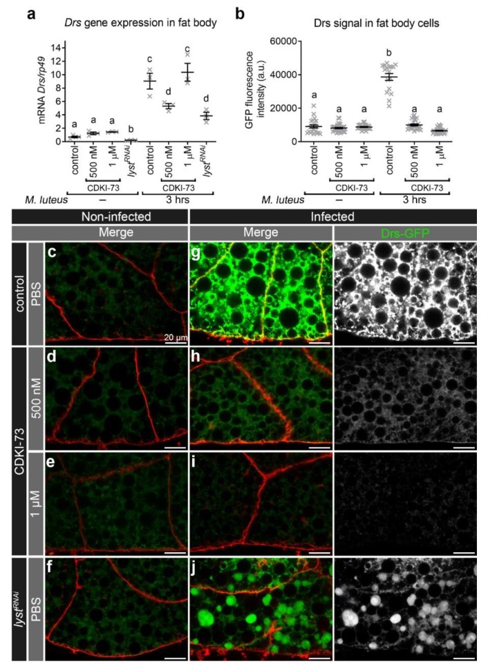
CDKI-73 depletes Drs in fat body cells after bacterial challenge. (a) The expression of Drs was characterised by qRT-PCR. (b) Histogram showing comparative analysis of intracellular Drs-GFP signal in fat body cells. One-way ANOVA and Tukey’s multiple comparison test showed significant differences between the means in designated groups (depicted by different letters on the bars in (a) and (b), p < 0.0001). Data are represented as mean ± SEM. (c–j) Confocal micrographs showing distribution of Drs-GFP (green) in fat body cells. The plasma membrane was outlined by CellMask™ Deep Red (red). Representative images were from fat body tissues treated for 30 min either with PBS (c,f,g,j), CDKI-73 at 500 nM (d, h) or 1 μM (e, i). Fat body tissues were from non-infected (c–f) and infected (Micrococcus luteus) larvae (g–j). Fat body tissues were from the following genotypes: CG-CAL4 > UAS-Rab11-GFP/+ (c–e, g–i) and UAS-lystRNAi/+; CG-CAL4 > UAS-Rab11-GFP/+ (f, j). Scale bars: 20 μm.

