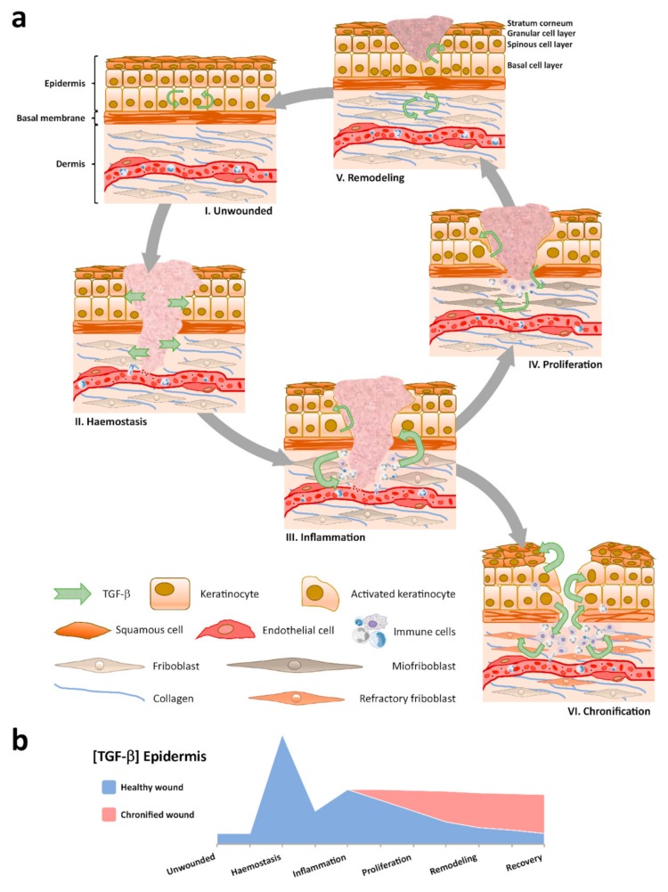
Figure 1.
Spatial and temporal patterns for TGF-β evolve during wound healing. (a) Distinct TGF-β signatures appear through the skin tissue compartments: i) Low TGF-β levels are characteristic of the homeostatic epidermis in unwounded skin; ii) After injury, a clot is constituted and TGF-β, from degranulated platelets, diffuses into neighboring skin tissue compartments; iii) Immune cells infiltrate and contribute to the regional cytokine milieu; iv) Molecular crosstalk between skin tissue compartments coordinates wound contraction and re-epithelization; v) Paracrine signaling drives dermal extracellular matrix (ECM) replacement and epidermal homeostasis recovery to a low TGF-β concentration environment; vi) Persistent immune infiltrates and aberrant cell behaviors establish, including dermal fibroblast becoming refractory to TGF-β signaling and the development of reactive hyperkeratosis and parakeratosis on exacerbated TGF-β expression in the epidermis. (b) The quantity of TGF-β varies in the epidermis through the sequential stages of wound healing.

