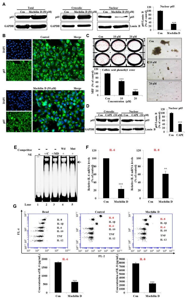
Figure 6.
Machilin D regulates the location of NF-κB p65 and secretion of IL-6 and IL-8. (A) The levels of p65 in the total, cytosolic, and nuclear proteins were measured in the MDA-MB-231 cells after treatment with machilin D for 24 h using western blot analyses. (B) Immunofluorescence (IF) analysis of p65 (green) expression and localization in the breast cancer cells under machilin D treatment. (C) The effect of caffeic acid phenethyl ester (CAPE), an inhibitor of NF-κB, on mammosphere formation. (D) The p65 levels in the cytosolic and nuclear proteins were measured in the MDA-MB-231 cells after treatment with CAPE for 24 h using western blot analyses. (E) Electrophoresis Mobility Shift Assays (EMSAs) of mammosphere nuclear proteins after treatment with machilin D. The nuclear extracts were reacted with the NF-κB probe and were analyzed by 6% native PAGE. Lane 1: NF-κB probe; lane 2: Nuclear extracts with the NF-κB probe; lane 3: Machilin D-treated nuclear proteins with the NF-κB probe; lane 4: Nuclear proteins incubated with the self-competitor (100×) oligo; lane 5: Nuclear extracts incubated with the mutated-NF-κB (100×) probe. The arrow indicates the DNA/NF-κB complex in the mammosphere nuclear lysates. (F) Transcriptional expression of the IL-6 and IL-8 genes was determined in the machilin D-treated mammospheres using specific primers. (G) Cytokine profile assay of the conditioned media and the machilin D-treated media using specific antibodies and cytokine beads. The data are presented as the mean ± SD of three independent experiments. ** p < 0.05; *** p < 0.01 versus the DMSO-treated control group indicated significant differences.

