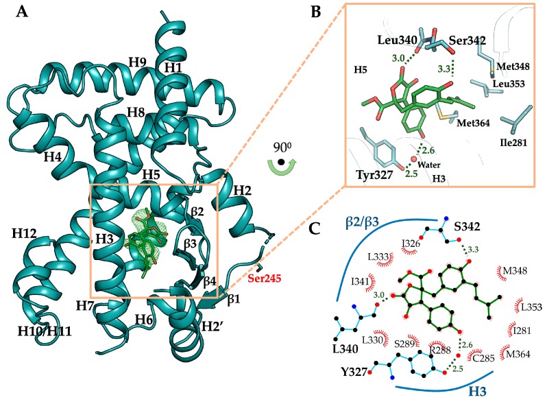
Figure 4.
Overall structure of butyrolactone I-bound PPARγ LBD and its binding mode. (A) Butyrolactone I-bound PPARγ LBD (chain B) displayed as cartoon representation colored in cyan. Ser245, a CDK5-mediated phosphorylation site, is shown as a stick model. Butyrolactone I occupying the ligand binding pocket is shown as green-colored stick model. The omit map (mFo–DFc, contoured at 2.0σ) of butyrolactone I is displayed in green-colored mesh representation. (B) Close-up view of the ligand binding pocket. Butyrolactone I shown as green-colored stick model occupies the ligand binding pocket forming three hydrogen bonds with PPARγ LBD residues Leu340, Ser342, and Tyr327 (shown as stick models), including a water-mediated hydrogen bond. The hydrogen bond-mediating water molecule is shown as red sphere. Residues involved in the interaction with 3-methyl-2-butenyl group of butyrolactone I are shown as stick models (Ile281, Met348, Leu353, and Met364 in the hydrophobic pocket of PPARγ LBD). (C) PPARγ-butyrolactone I interactions of the crystal structure were analyzed using LigPlot+ and presented in a two-dimensional scheme.

