Figure 8.
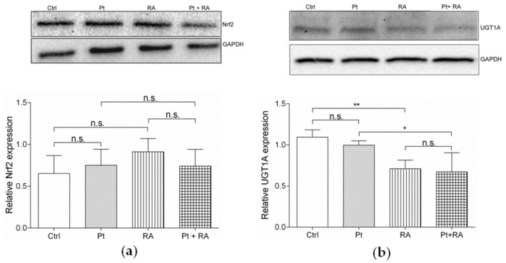
Representative Western blots and the corresponding densitometric quantification (mean ± SEM, n = 3) of (a) the relative Nrf2 expression and (b) the relative UGT1A expression in the transfected untreated SKOV-3 cells (Ctrl), after exposure to 15 µM cisplatin (Pt), to 20 µM retinoic acid (RA) and after co-incubation with 20 µM retinoic acid and 15 µM cisplatin (Pt + RA) for 24 h are shown. GAPDH was used as a loading control. *p < 0.05, ** p < 0.01, n.s. = not significant.

