Abstract
During a routine ovariohysterectomy on a 5-month-old ragdoll cat, right-sided segmental uterine aplasia and ipsilateral renal agenesis were discovered. The diagnosis was confirmed with histopathology. This condition is congenital and is a result of a failure of embryonic development of the paramesonephric ducts. Uterine aplasia and renal agenesis is a rare finding in cats but the prevalence in the ragdoll breed appears to be higher than in the general cat population.
Résumé
Aplasie utérine segmentaire et agénésie rénale ipsilatérale chez un chat ragdoll. Lors d’une ovario-hystérectomie de routine chez une chatte ragdoll âgée de 5 mois, une aplasie utérine segmentaire du côté droit et une agénésie rénale ipsilatérale furent découvertes. Le diagnostic fut confirmé par histopathologie. Cette condition est congénitale et est le résultat d’une défaillance de développement embryonnaire des conduits paramésonéphriques. L’aplasie utérine et l’agénésie rénale sont des trouvailles rares chez les chats mais la prévalence chez la race ragdoll semble être plus élevée que dans la population générale de chats.
(Traduit par Dr Serge Messier)
A 5-month-old female ragdoll cat was presented to the Brooklin Veterinary Hospital for routine vaccinations and consultation regarding ovariohysterectomy (OHE). At this time a date for the OHE was set but routine pre-surgical blood analysis was declined. As the final booster vaccinations in the routine kitten series would be completed within 2 wk of the date set for the OHE, the surgery was postponed and an alternative date was set. Prior to the newly scheduled date, the owner contacted the clinic and asked for the OHE to be completed promptly as the cat appeared to be entering her first heat cycle. On the day of the OHE procedure presurgical blood analysis was again declined.
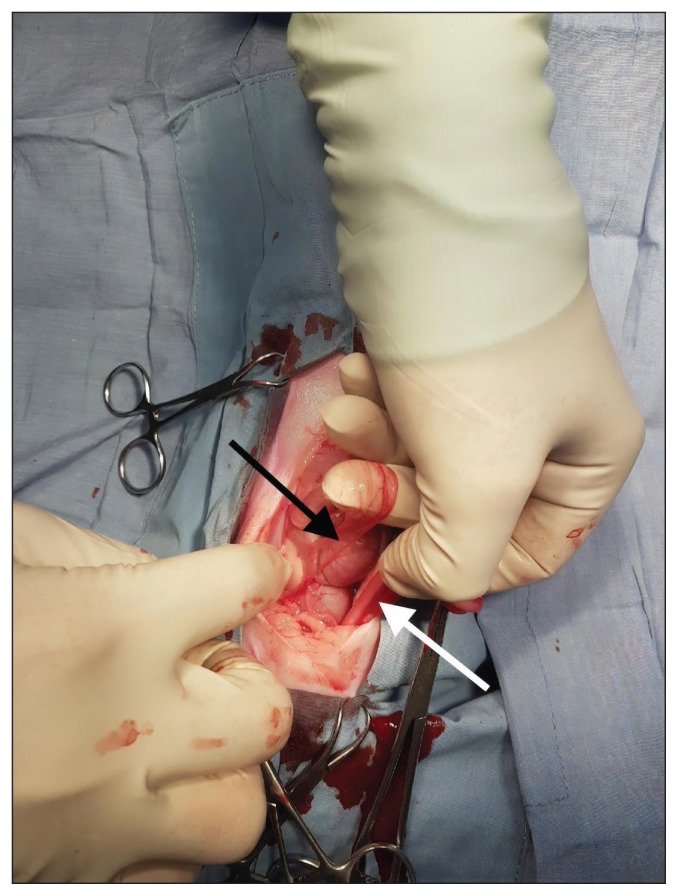
A midline incision was made just caudal to the umbilicus through the skin, subcutaneous tissue, and linea alba. The left ovarian horn was identified via the use of a spay hook. The left ovarian pedicle was ligated, and the uterine horn was followed to the uterine body. It was difficult to identify the bifurcation of the uterine body, and the right uterine horn could not be located using this method. The surgeon attempted to locate the right uterine horn with the spay hook but was unsuccessful. The midline incision through all 3 layers was extended caudally, but still the bifurcation and the right uterine horn could not be identified. At this time no palpable cervical structure could be identified. The midline incision was then extended through all 3 layers cranially to create an exploratory laparotomy incision. The surgeon then began to examine the contents of the abdominal cavity, and identified a right ovary attached to a ligamentous structure which terminated in the surrounding broad ligament before reaching the uterine body (Figure 1). The right ovarian pedicle was ligated and the ovary, as well as the abnormal structure, were removed. During this exploratory laparotomy, it was noted that the right kidney and ureter were absent.
Figure 1.
Normal left uterine horn (white arrow) compared with the aplastic right uterine horn (black arrow) in situ. The aplastic horn is shown to be terminating without attachment to the uterine body.
After completion of the OHE an abdominal ultrasound was performed to assess the abdominal organs and to confirm the absence of the right kidney. Routine post-operative pain management for cats following OHE at this clinic consists of meloxicam (Metacam; Boehringer Ingelheim, Burlington, Ontario), 0.05 mg/kg body weight (BW), SC; however, oral transmucosal buprenorphine, 0.02 mg/kg BW, compounded by Chiron Compounding Pharmacy, Guelph, Ontario was prescribed in this specific case because it is considered less nephrotoxic than meloxicam. At discharge the client was informed of the surgical findings and was cautioned that the cat may develop insufficient kidney function in the future. It was explained that cats with 2 kidneys frequently develop renal failure and it is likely that this condition may result in an earlier age of onset as the cat has only 1 functioning kidney doing the work of 2. Baseline blood analysis to assess current renal function was not elected at this time.
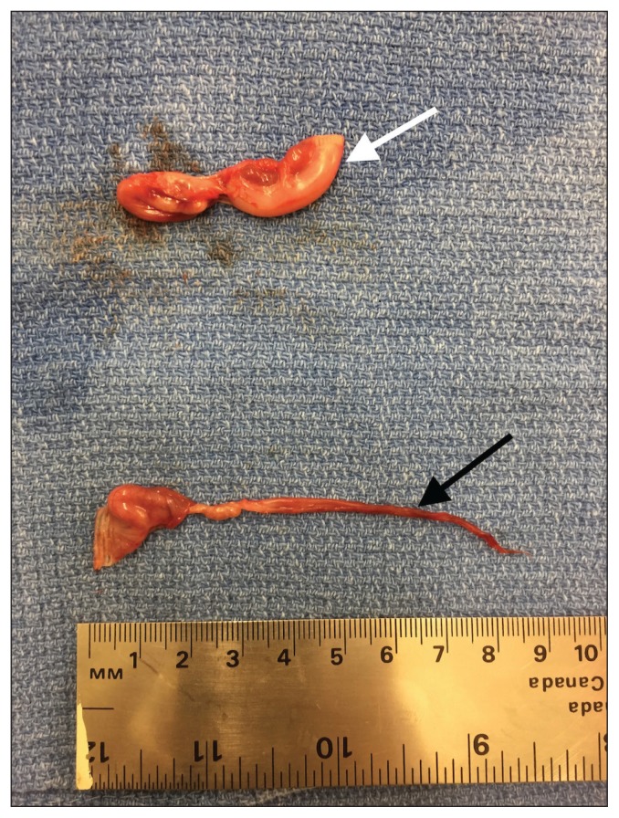
The excised left and right ovaries and associated uterine structures were fixed in formalin and sent for histopathology at the Animal Health Laboratory (AHL), University of Guelph (Figure 2). This investigation confirmed the diagnosis of segmental right uterine aplasia and ipsilateral renal agenesis. Results showed that the associated uterine tissue contained smooth muscle, collagen, and blood vessels but lacked any normal uterine tissue. In contrast to these abnormalities, the right ovarian structure was normal, as were the isthmus and infundibulum.
Figure 2.
Normal left uterine horn and ovary (white arrow) compared with the aplastic right uterine horn with normal ovary (black arrow) removed during surgery.
Discussion
Embryological development is a complex process that relies on a very specific series of events to occur to allow for complete development (1). When signals do not occur at the precise embryonic cell stage, development continues in an abnormal pattern (1). This may cause agenesis or dysgenesis, which results in complete absence of an organ or an abnormal organ, respectively (1). Congenital abnormalities are thus the result of the misfiring of signals during embryologic development, and result in alterations of structure and function of organs and physiologic systems (1). The urogenital tract arises due to complex interactions between 2 cell populations (2). The urinary tract and tubular uterine system develop from a set of cells originating in the intermediate mesoderm, while the ovaries develop from the gonadal ridge (2). This common ancestry of renal and uterine structures explains why renal and uterine abnormalities often occur in conjunction with one another, and the degree to which they are affected depends on the timing of the abnormal signals received by the tissues (2). The uterine tract itself forms from paired müllerian (or paramesonephric) ducts which elongate toward each other and fuse, thus uterine abnormalities can be seen on one or both sides of the tract (3,4). As the ovaries develop from a distinct cell population at the gonadal ridge, and respond to a different set of signals, the ovaries are often normal in the face of congenital urogenital abnormalities (2).
Segmental uterine aplasia (commonly referred to as uterus unicornis) is a condition in which portions of the tubular genital tract (uterus, cervix, and vagina) are not present (5,6). There are varying presentations of this anomaly which is caused by alterations in the timing of signaling mistakes (2,4). Signal misadventures occurring earlier in development lead to an anomaly which begins in a more proximal location of the tract and halts normal development of the rest of the organ, and thus a larger portion of the tract is affected than when the signal is disrupted at a later stage of development (2,4). Segmental uterine aplasia and ipsilateral renal agenesis are frequently identified together in animals with 2 normal ovaries, but the exact mechanism resulting in this failure is unknown (2). Mayer–Rokitansky– Küster–Hauser syndrome type 2 (MRKH t2), is a known human condition that has similarities in presentation to this condition in animals, which is inherited via an autosomal dominant mutation with incomplete penetrance and variable expressivity (7). However, MRKH t2 is not an exact replica for the condition seen in animals, and studies show low inheritance from affected female animals suggesting a more complex inheritance pattern likely involving more than 1 gene or signal (2,7).
While abnormalities can be seen in various species and body systems, urogenital abnormalities are rare in mammals and are seen most commonly in livestock animals (6). Segmental uterine aplasia with ipsilateral renal agenesis is most frequently presented in case report format, but case series investigations have been described as well, providing both qualitative and quantitative information (2–4,6,8). Uterine abnormalities are exceedingly rare in companion animals but are seen at almost twice the rate in cats (0.09%) compared with dogs (0.05%) (8). In cats with uterine abnormalities such as segmental uterine aplasia, ipsilateral renal agenesis is present in approximately 30% of cases (8). Ragdoll cats appear to be more commonly affected with this condition than are other cat breeds (5). Renal abnormalities such as chronic interstitial nephritis and polycystic kidney disease are commonly found in ragdoll cats as well and screening for such diseases can lead to the identification of segmental uterine aplasia with ipsilateral renal agenesis in otherwise healthy cats (9). Regardless of breed, this anomaly most commonly occurs on the right side of the body for reasons which are unknown (6,9). Cats presenting for OHE with this condition will almost always have 2 ovaries, and it is thus imperative that both are found and removed to successfully prevent heat cycles (4). It is also crucial that the extent of the condition is identified to take measures to preserve renal function of the sole kidney in future medical or surgical interventions (4).
Acknowledgments
The author thanks Dr. Hooman Afshari of Brooklin Veterinary Hospital for his contribution to this case, his surgical expertise leading to these findings and a successful OHE. Thank you to Dr. Rebecca Egan and the AHL for their contribution to the diagnostic information. CVJ
Footnotes
Use of this article is limited to a single copy for personal study. Anyone interested in obtaining reprints should contact the CVMA office (hbroughton@cvma-acmv.org) for additional copies or permission to use this material elsewhere.
References
- 1.Cuckow PM, Nyirady P, Winyard PJD. Normal and abnormal development of the urogenital tract. Prenat Diagn. 2001;21:908–916. doi: 10.1002/pd.214. [DOI] [PubMed] [Google Scholar]
- 2.Brookshire WC, Shivley J, Woodruff K, Cooley J. Uterus unicornis and pregnancy in two feline littermates. J Feline Med Surg. 2017;3:1–5. doi: 10.1177/2055116917743614. [DOI] [PMC free article] [PubMed] [Google Scholar]
- 3.Goo M-J, Williams BH, Hong I-H, et al. Multiple urogenital abnormalities in a Persian cat. J Feline Med Surg. 2009;11:153–155. doi: 10.1016/j.jfms.2008.04.007. [DOI] [PMC free article] [PubMed] [Google Scholar]
- 4.Carvallo FR, Wartluft AN, Melivilu RM. Unilateral uterine segmentary aplasia, papillary endometrial hyperplasia and ipsilateral renal agenesis in a cat. J Feline Med Surg. 2012;15:349–352. doi: 10.1177/1098612X12467786. [DOI] [PMC free article] [PubMed] [Google Scholar]
- 5.Little S. Feline reproduction problems and clinical challenges. J Feline Med Surg. 2011;13:508–515. doi: 10.1016/j.jfms.2011.05.008. [DOI] [PMC free article] [PubMed] [Google Scholar]
- 6.Chang J, Jung J-H, Yoon J, et al. Segmental aplasia of the uterine horn with ipsilateral renal agenesis in a cat. J Vet Med Sci. 2008;70:641–643. doi: 10.1292/jvms.70.641. [DOI] [PubMed] [Google Scholar]
- 7.Morcel K, Camborieux L. Programme de Recherches sur les Aplasies Müllériennes, Guerrier D. Mayer-Rokitansky-Küster-Hauser (MRKH) syndrome. Orphanet J Rare Dis. 2007;2:13. doi: 10.1186/1750-1172-2-13. [DOI] [PMC free article] [PubMed] [Google Scholar]
- 8.McIntyre RL, Levy JK, Roberts JF, Reep RL. Developmental uterine anomalies in cats and dogs undergoing elective ovariohysterectomy. J Am Vet Med Assoc. 2010;237:542–546. doi: 10.2460/javma.237.5.542. [DOI] [PubMed] [Google Scholar]
- 9.Paepe D, Saunders JH, Bavegems V, et al. Screening of ragdoll cats for kidney disease: A retrospective evaluation. J Small Anim Pract. 2012;53:572–577. doi: 10.1111/j.1748-5827.2012.01254.x. [DOI] [PubMed] [Google Scholar]


