
Some NLM-NCBI services and products are experiencing heavy traffic, which may affect performance and availability. We apologize for the inconvenience and appreciate your patience. For assistance, please contact our Help Desk at info@ncbi.nlm.nih.gov.

PLOS ONE logoLink to PLOS ONE
. 2020 Mar 16;15(3):e0230261. doi: 10.1371/journal.pone.0230261

Clinical implications of APOBEC3A and 3B expression in patients with breast cancer

Yong-seok Kim 1,#, Der Sheng Sun 2,#, Jung-sook Yoon 3, Yoon Ho Ko 4, Hye Sung Won 2,*, Jeong Soo Kim 5,*
Editor: Elizabeth Christie6
PMCID: PMC7075570  PMID: 32176735

Abstract

Background

We aimed to evaluate the expression of APOBEC3A (A3A), 3B (A3B) mRNA, and germline APOBEC3A/B deletion polymorphism in patients with breast cancers and to investigate the correlation between their expressions and clinicopathological characteristics.

Methods

RNA and DNA samples were extracted from 138 breast cancer tissues and adjacent normal breast tissues. The levels of A3A and A3B mRNA transcripts were determined using quantitative real-time polymerase chain reaction. Insertion and deletion PCR assays were performed to detect the A3B deletion allele. The serum concentrations of soluble programmed death-ligand 1 (sPD-L1) and interferon gamma were determined using enzyme-linked immunosorbent assays.

Results

A3B mRNA expression levels were significantly higher in triple-negative breast cancers compared to hormone receptor-positive, human epidermal growth factor receptor 2-negative breast cancers. Older age of the patient and high ki-67 expression were associated with increased expression levels of A3A and A3B mRNA. Advanced tumor stage, presence of lymph node involvement, and high histological grade were associated with increased expression levels of A3A mRNA. The APOBEC3A/B deletion allele was found in 77 (55.8%) patients. TP53 and PIK3CA mutations were detected in 62 (44.9%) and 31 (22.5%) patients, respectively. The presence of a PIK3CA mutation was associated with lower A3A mRNA expression levels. There was a weak positive relationship between A3A mRNA expression levels and serum sPD-L1 levels.

Conclusions

There was a difference in A3B mRNA expression levels according to breast cancer subtypes, and high levels of A3A and A3B mRNA expressions were associated with an aggressive phenotype. There was a high incidence of APOBEC3A/B deletion allele. Further studies are needed to identify the clinical significance of APOBEC in Asian patients with breast cancer.

Introduction

Genomic instability—a high frequency of mutations within the genome of cellular lineages—is known as the driving force of cancer development [1]. The mechanisms leading to genomic instability include inherited or acquired defects in pathways that maintain genomic integrity, such as DNA repair, DNA replication or cell cycle control [2]. DNA damage as a source of genomic instability can be caused by exogenous agents such as environmental chemicals and endogenous mutagenic events. Alexandrov LB et al. found that the catalogue of somatic mutations from a cancer genome bears the signatures of the mutational processes and a signature attributed to the Apolipoprotein B mRNA-editing enzyme, catalytic polypeptide-like (APOBEC) family is notably present in many cancer types [3]. APOBEC family is known to function in the innate immune system that protects against retroviruses by deaminating cytosine to uracil in single-stranded DNA. APOBEC has attracted much attention as an endogenous mutagen in various types of cancers. APOBEC can induce base substitutions in tumor genomes. Cytosine deamination by APOBEC in single-strand DNA results in an uracil residue. Excision of this uracil residue by uracil DNA glycosylase generates an abasic site, and insertion of adenine opposite the abasic site results in a C to T transition in a preferential motif [4]. Analyses using data from The Cancer Genome Atlas (TCGA) have revealed that the APOBEC mutation signature is particularly enriched in cancers of the bladder, cervix, head and neck, breast, and lung. Among the four intrinsic subtypes of breast cancer, it occurred much more frequently in the human epidermal growth factor receptor 2 (HER2) enriched type of breast cancer [5]. In addition, APOBEC mRNA levels correlated positively with the number of APOBEC mutation signature [5].

Humans have at least 11 APOBEC genes: activation-induced cytidinedeaminase, APOBEC1, APOBEC2 and APOBECs 3A–3H [6]. Although the roles of each APOBEC member are not yet established, previous studies have suggested that APOBEC3B (A3B) might be a major candidate inducing the APOBEC mutation signature in various cancers [7, 8]. In breast cancers, APOBEC3A (A3A) and A3B showed positive correlations between their expression levels and the APOBEC mutation signature [810].

With the expression of APOBEC, a germline copy number polymorphism involving A3A and A3B, which effectively deletes A3B, is known to have a role in carcinogenesis. Nik-Zainal S et al. reported that breast cancers in carriers of APOBEC3A/B deletion polymorphism showed more mutations of the putative APOBEC-dependent genome-wide signatures than cancers in non-carriers [11]. This deletion is more common in East Asians and case-control studies in Asian populations have found that the APOBEC3A/B deletion allele was associated with increased risk for breast cancer [1214]. However, this finding was not reproduced in Western populations [15, 16].

Previous studies on the role of APOBEC in breast cancer have been conducted mainly in Western patients. We aimed to evaluate the expression of A3A and A3B mRNA according to subtypes and the incidence of APOBEC3A/B deletion polymorphisms in Korean patients with breast cancers. We investigated correlations between their expression levels and clinicopathological characteristics and their impact on the immune response.

Methods

Patients

The patients who underwent surgery for breast cancer in our hospital between January 2013 and December 2016 were evaluated. Finally, this study included 138 cases with similar proportions of each subtype based on the clinicopathological data. Tissue samples were frozen immediately after surgery and stored at –70°C until DNA and RNA isolation. Venous blood samples were taken from patients at the time of surgery and centrifuged at 3,000 rpm for 10 min. The serum obtained was divided into aliquots and stored at –20°C. The following clinical data were collected: age, sex, body mass index, smoking and alcohol history, any family history of breast cancer, comorbidity, tumor stage, surgery type, surgery date, pathology reports, the use of chemotherapy, radiotherapy or endocrine therapy, tumor recurrence, and survival. Hormone receptor (HR) positivity was defined as ≥ 10% of tumor cells staining for estrogen receptor and/or progesterone receptor. HER2 positivity was defined as a HER2/CEP17 fluorescence in situ hybridization ratio of ≥ 2.0 and/or an immunohistochemical staining score of 3+. This study was approved by the institutional review board of the Catholic Medical Center (IRB No. UC18SEI0099) and was conducted in accordance with the Declaration of Helsinki. The written informed consent was obtained from all participants in this study.

Quantitative real-time polymerase chain reaction (qPCR) assays for APOBEC3A and 3B mRNA

Total RNA was extracted from 138 breast cancer tissues and 10 adjacent normal breast tissues using RNeasy mini kits (Qiagen, Hilden, Germany) according to the manufacturer’s instructions. The cDNA was synthesized from RNA using RevertAid First Strand cDNA Synthesis kits with a random hexamer primer (Fermentas International, Inc., Burlington, ON, Canada). For quantification of A3A and A3B mRNA, qPCR was performed with a CFX96 Real-time qPCR detection system (Bio-Rad, Hercules, CA, USA) using an iTaqUniversal SYBR Green Supermix (Bio-Rad). After an initial incubation at 95°C for 3 min, the reactions were followed by 40 cycles at 95°C for 10 sec and at 55°C for 30 sec. The primers used for the A3A and A3B genes were: A3A_F,5′–GAGAAGGGACAAGCACATGG–3′; A3A_R,5′–TGGATCCATCAAGTGTCTGG–3′; A3B_F,5′–GACCCTTTGGTCCTTCGAC–3′; and A3B_R,5′–GCACAGCCCCAGGAGAAG–3′, respectively [17]. The relative mRNA expression level was normalized to the β-actin gene expression, and then expressed as the log2 fold change of the delta-delta Ct values. To validate the primers for qPCR, we checked A3A and A3B mRNA levels in SKBR3 (A3Bnull) and MCF-7 breast cancer cell line, and confirmed that A3B mRNA level was very low in SKBR3 breast cancer cell line (S1 Fig) [10].

Genotyping assay of germline APOBEC3A/B deletion polymorphism

Genomic DNA was extracted from 138 matched normal breast tissues using DNeasy blood & tissue kits (Qiagen) according to the manufacturer’s instructions. We designed PCR using oligonucleotide primer sequences as described previously [17]: Deletion_F,5′–TAGGTGCCACCCCGAT–3′; Deletion_R,5′–TTGAGCATAATCTTACTCTTGTAC–3′; Insertion1_F,5′–TTGGTGCTGCCCCCTC–3′; Insertion1_R,5′–TAGAGACTGAGGCCCAT–3′; and Insertion2_F,5′–TGTCCCTTTTCAGAGTTTGAGTA–3′; and Insertion2_R,5′–TGGAGCCAATTAATCACTTCAT–3′. Insertion and deletion PCR reactions were performed separately. The PCR products for Deletion and Insertion2 primers were pooled and loaded into a standard 1.5% agarose gel. In addition, samples that appeared homozygous for the deletion, were subjected to amplifications with primer Insertion1. The following cycling conditions were used: 5 min at 95°C, followed by 40 cycles at 95°C for 1 min, 60°C for 1 min, and 72°C for 1 min. The final extension step was for 7 min at 72°C.

Mutations of TP53 and PIK3CA

The entire coding sequence of the TP53 gene (exons 2–11) was analyzed for mutations by direct sequencing of the PCR products. The PCR reactions for the TP53 gene were performed using Platinum Taq DNA polymerase (Thermo Fisher, Walthman, MA, USA), 10 mM dNTPs (Invitrogen Life Technologies, Carlsbad, CA, USA) and Betaine solution (Sigma-Aldrich, St. Louis, MO, USA) with the cDNA. After an initial incubation at 97°C for 2 min, the reaction was followed by 35 cycles at 95°C for 1 min, 67°C for 30 sec, and 72°C for 1 min. The hotspot mutations in exons 9 and 20 of PIK3CA were assessed by direct sequencing of the PCR products: E542K (exon 9), E545K (exon 9), H1047L (exon 20), and H1047R (exon 20) [18, 19]. The PCR reactions for the PIK3CA gene were using Ex-Taq (TaKaRa Bio Inc., Otsu, Shiga, Japan) with the cDNA. After an initial incubation at 95°C for 5 min, the reaction was followed by 35 cycles at 95°C for 1 min, 60°C for 30 sec and 72°C for 1 min. The final extension step was at 72°C for 7 min. The primers used for the TP53 gene were: TP53_F,5′–GAGCCGCAGTCAGATCCTAG–3′ and TP53_R,5′–GTCTGAGTCAGGCCCTTCTG–3′. The primers used for the PIK3CA gene were: exon 9_F,5′–TGGCCAGTACCTCATGGATTAGAA–3′; exon 9_R,5′–GAGGCCAATCTTTTACCAAGCA–3′; exon 20_F,5′–AATGCACAAAGACAAGAGAATTTGAG–3′; and exon 20_R,5′–AATTCCTATGCAATCGGTCTTTGC–3′.

Determination of soluble programmed death-ligand 1 (sPD-L1) and interferon gamma (IFN-γ) concentrations

The serum concentrations of sPD-L1 and IFN-γ were determined using commercially available enzyme-linked immunosorbent assay kits in accordance with the manufacturers’ instructions (Cloud-Clone Corp., Houston, TX, USA and R&D Systems Inc., Minneapolis, MN, USA). These assays use the quantitative sandwich enzyme immunoassay technique and serum levels were calculated according to standard curves. The minimum detectable levels of sPD-L1 and IFN-γ were 0.056 ng/mL and 8.0pg/mL, respectively.

Statistical analysis

A3A and A3B mRNA levels are expressed as the median and interquartile range. A log power of 2 or fold changes in expression was used to generate box and whisker plots based on the median and interquartile range. Boxplots show the log2 fold change values for the relative expression of the APOBEC mRNA levels. Boxplots show the median (center bar) and the third and first quartiles (upper and lower edge of box, respectively). The Mann-Whitney nonparametric U test was used for comparisons of differences between continuous variables in two groups. The significance between three or more groups was tested using Kruskal-Wallis test. Dunn-Bonferroni post hoc test was employed to correct the significance for between-group comparisons. Categorical variables were compared using chi-squared or Fisher’s exact tests. To determine the significant correlations between two continuous variables, we used Spearman’s rank correlation coefficient. All statistical analyses were performed using SPSS program and p < 0.05 was considered significant.

Results

APOBEC3A and 3B mRNA expression according to breast cancer subtypes and characteristics

We measured A3A and A3B mRNA levels from 138 breast cancer samples and 10 adjacent normal breast tissues. A3A mRNA levels tended to be higher in the breast cancer tissues than in the normal breast tissues, but the differences were not statistically significant (median 0.20 vs. 0.16; p = 0.396). A3B mRNA levels in the breast cancer tissues were significantly higher than in the normal breast tissues (median 0.55 vs. 0.13; p = 0.013). The median mRNA levels for A3A was lower than that of A3B in the breast cancer tissues.

We classified breast cancers into three subtypes based on the expression of the estrogen receptor, progesterone receptor, and HER2: 1) HR-positive and HER2-negative breast cancer, 2) HER2-positive breast cancer, and 3) triple-negative breast cancer. The numbers of patients in each subtype were 56 (40.6%), 49 (35.5%), and 33 (23.9%), respectively. Among 49 patients with HER2-positive breast cancer, 34 (69.3%) patients were also HR-positive. With respect to breast cancer subtypes, A3A mRNA expression showed a tendency to be higher in HER2-positive breast cancers, and A3B mRNA expression was significantly higher in triple-negative breast cancers compared to HR-positive, HER2-negative breast cancers (p = 0.004) (Fig 1). In post hoc test, p-values between HR-positive breast cancers and HER2-positive breast cancers and between HER2-positive breast cancers and triple-negative breast cancers and between HR-positive breast cancers and triple-negative breast cancers were 0.853, 0.061, and 0.003, respectively. Table 1 shows the mRNA expression levels of A3A and A3B according to clinicopathological characteristics of the 138 patients. Both A3A and A3B mRNA levels were higher in older patients and Ki-67 > 20%. Advanced tumor stage, presence of lymph node involvement, and high histological grade showed significant associations with higher A3A mRNA levels.

Fig 1. Comparative quantification of APOBEC mRNA expression according to breast cancer subtypes.

Fig 1

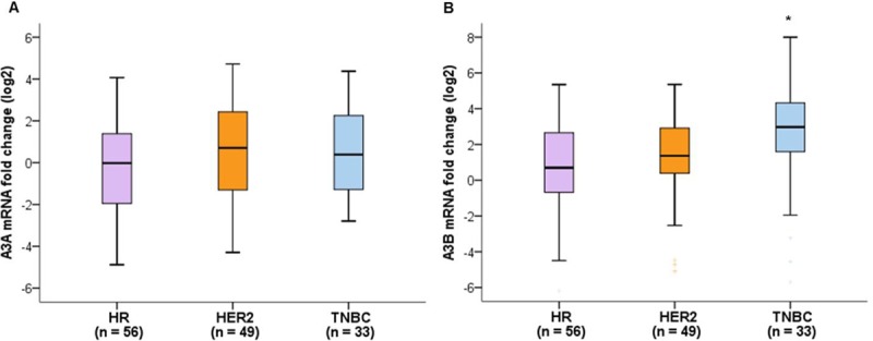
(A) A3A mRNA expression levels showed a tendency to be higher in HER2-positive breast cancer (p = 0.158). (B) A3B mRNA expression levels were significantly higher in triple-negative breast cancers compared to HR-negative, HER2-positive breast cancers (p = 0.004). The significance between groups was tested using Kruskal-Wallis test followed by Dunn’s multiple comparison post hoc test. Global p-values are indicated.*p < 0.05.

Table 1. APOBEC3A and APOBEC3B mRNA expression levels according to patient characteristics.

Characteristics No. of patients (%) APOBEC3A median (interquartile range) p-value APOBEC3B median (interquartile range) p-value
Age (median: 52 years) 0.012 0.036
    ≤ 52 72 (52.2) 0.16 (0.62) 0.37 (1.00)
    > 52 66 (47.8) 0.32 (0.63) 0.92 (1.89)
BMI (kg/m2) 0.113 0.028
    Normal (< 23.0) 44 (31.9) 0.12 (0.76) 0.29 (0.87)
    Overweight (≥ 23.0) 94 (68.1) 0.24 (0.59) 0.85 (1.69)
Menopausal status 0.189 0.852
    Premenopausal 59 (43.1) 0.16 (0.75) 0.54 (1.28)
    Postmenopausal 78 (56.9) 0.24 (0.59) 0.61 (1.68)
Smoking 0.933 0.622
    Current/Ex-smoker 14 (10.1) 0.16 (0.66) 0.47 (1.81)
    Non-smoker 124 (89.9) 0.21 (0.64) 0.57 (1.61)
Alcohol 0.980 0.421
    Yes 10 (7.8) 0.19 (0.55) 1.28 (2.6)
    No 128 (92.8) 0.21 (0.64) 0.54 (1.52)
Family history 0.483 0.178
    Yes 8 (5.8) 0.72 (1.24) 1.38 (4.91)
    No 130 (94.2) 0.19 (0.61) 0.54 (1.44)
Diabetes 0.203 0.326
    Yes 22 (15.9) 0.24 (0.74) 0.45 (0.83)
    No 116 (84.1) 0.19 (0.64) 0.62 (1.73)
Hypertension 0.161 0.279
    Yes 44 (31.9) 0.22 (0.69) 0.90 (1.67)
    No 94 (68.1) 0.19 (0.60) 0.44 (1.36)
Site 0.091 0.711
    Right 50 (36.8) 0.17 (0.42) 0.43 (1.78)
    Left 86 (63.2) 0.26 (0.73) 0.57 (1.40)
Tumor size 0.071 0.901
    ≤ 2 cm (pT1) 50 (36.2) 0.18 (0.39) 0.41 (1.64)
    > 2 cm (pT2/3) 88 (63.8) 0.29 (0.75) 0.63 (1.52)
No. of involved LNs 0.004 0.974
    0 79 (57.2) 0.16 (0.34) 0.50 (1.66)
    1–3 36 (26.1) 0.42 (1.01) 0.62 (1.19)
    ≥ 4 23 (16.7) 0.28 (0.73) 0.60 (1.51)
pStage 0.007 0.811
    I 37 (26.8) 0.10 (0.24) 0.37 (1.77)
    II 75 (54.4) 0.29 (0.73) 0.62 (1.22)
    III/IV 26 (18.8) 0.35 (1.02) 0.63 (1.69)
Grade 0.001 0.067
    Well/ moderate 72 (52.2) 0.18 (0.34) 0.36 (1.33)
    Poor 66 (47.8) 0.39 (1.05) 0.71 (1.78)
Ki-67 0.001 0.027
    ≤ 20% 57 (41.3) 0.11 (0.46) 0.35 (0.92)
    > 20% 81 (58.7) 0.30 (0.81) 0.90 (1.83)
Lymphatic invasion 0.093 0.142
    Yes 58 (43.0) 0.26 (0.76) 0.91 (1.73)
    No 77 (57.0) 0.18 (0.49) 0.40 (1.39)
Subtype 0.158 0.004
    HR+/ HER2- 56 (40.6) 0.02 (0.75) 0.70 (1.96)
    HER2+ 49 (35.5) 0.70 (1.13) 1.37 (2.57)
    HR-/ HER2- 33 (23.9) 0.39 (0.97) 2.97 (2.90)

The mRNA levels are presented as the log2 fold change of the delta-delta Ct values.

The significance between groups was tested using Mann-Whitney U test and Kruskal-Wallis test followed by Dunn’s multiple comparison post hoc test. Global p-values are indicated.

BMI, body mass index; No., number, LNs: lymph nodes.

Relationships between APOBEC3A/B deletion polymorphism and tumor characteristics

We performed genotyping assays for APOBEC3A/B deletion polymorphism (A3B deletion) from 138 normal breast tissues. The samples were classified as A3BD/D (two-copy deletion), A3BD/I (one-copy deletion), and A3BI/I (no deletion). Among these patients, 77 had A3BD/I (55.8%) and there was no instance of A3BD/D. There was no association between A3A mRNA levels and A3B deletions, but A3B mRNA levels showed a tendency to be lower in breast cancers with A3B deletion (p = 0.299 and 0.075, respectively) (Fig 2). There was no difference in the frequency of A3B deletion according to breast cancer subtypes. The association of A3B deletion with clinicopathological features is summarized in Table 2. We observed no significant associations between A3B deletion and any clinicopathological features of these breast cancers except for histological grade. The incidence of tumors with poor differentiation was significantly higher in breast cancers with A3B deletion.

Fig 2. Comparative quantification of APOBEC mRNA expression according to the presence of the APOBEC3A/B deletion polymorphism.

Fig 2

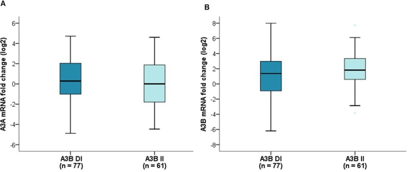
(A) There was no association between A3A mRNA expression levels and A3B deletion (p = 0.299). (B) A3B mRNA expression levels showed a tendency to be lower in breast cancers with A3B deletion (p = 0.075). A3B DI, one-copy deletion; A3B II, no deletion. Mann-Whitney U test.

Table 2. Association of APOBEC3A/B deletion polymorphism with clinicopathological features in patients with breast cancers.

Characteristics A3BD/I (n = 77) No.(%) A3BI/I (n = 61) No.(%) p-value
Age (year) 0.696
Mean±SD 53.7±10.3 54.4±12.1
BMI (kg/m2) 0.368
Normal (< 23.0) 27 (35.1) 17 (27.9)
Overweight (≥ 23.0) 50 (64.9) 44 (72.1)
Menopausal status 0.668
Premenopausal 33 (42.8) 26 (42.6)
Postmenopausal 43 (57.2) 35 (57.4)
Smoking 0.214
Current/Ex-smoker 10 (13.0) 4 (6.6)
Non-smoker 67 (87.0) 57 (93.4)
Alcohol 0.348
Yes 7 (9.1) 3 (4.9)
No 70 (90.9) 58 (95.1)
Family history 0.283
Yes 3 (3.8) 5 (8.2)
No 74 (96.2) 56 (91.8)
Diabetes 0.550
Yes 11 (14.3) 11 (18.0)
No 66 (85.7) 50 (82.0)
Hypertension 0.869
Yes 25 (32.5) 19 (31.1)
No 52 (67.5) 42 (68.9)
Site 0.231
Right 30 (39.0) 20 (32.8)
Left 47 (61.0) 39 (63.9)
Both 0 2 (3.3)
Tumor size 0.391
≤ 2 cm (pT1) 31 (40.3) 29 (47.5)
> 2 cm (pT2/3) 46 (59.7) 32 (52.5)
No. of involved LNs 0.694
0 45 (58.4) 34 (55.7)
1–3 21 (27.3) 15 (24.6)
≥ 4 11 (14.3) 12 (19.7)
pStage 0.094
I 18 (23.4) 19 (31.1)
II 48 (62.3) 27 (44.3)
III/IV 11 (14.3) 15 (24.6)
Grade 0.014
Well/ moderate 33 (42.9) 39 (63.9)
Poor 44 (57.1) 22 (36.1)
Ki-67 0.094
≤ 20% 27 (35.1) 30 (49.2)
> 20% 50 (64.9) 31 (50.8)
Lymphatic invasion 0.848
Yes 31 (41.3) 27 (45.0)
No 44 (58.7) 33 (55.0)
Subtype 0.614
HR+/ HER2- 33 (42.8) 23 (37.7)
HER2+ 28 (36.4) 21 (34.4)
HR-/ HER2- 16 (20.8) 17 (27.9)

A3BD/I, one-copy deletion; A3BI/I, no deletion; SD, standard deviation; BMI, body mass index; HR, hormone receptor; HER2, human epidermal growth factor receptor 2; No., number; LNs, lymph nodes.

Analysis of TP53 and PIK3CA mutations

Among 138 patients with breast cancers, TP53 and PIK3CA mutations were detected in 62 (44.9%) and 31 (22.5%) patients, respectively. We evaluated the association between TP53 and PIK3CA mutations and A3A and A3B mRNA expression levels. The A3A and A3B mRNA expression levels did not differ significantly according to the presence of TP53 mutations. With respect to PIK3CA mutations, A3A mRNA expression levels were significantly lower in tumors carrying mutant PIK3CA than in tumors carrying wild-type PIK3CA (p = 0.038, Fig 3A), but there was no difference in A3B mRNA expression levels according to PIK3CA mutation status (p = 0.127, Fig 3B). There were no significant associations between A3B deletion and TP53, PIK3CA mutations.

Fig 3. Comparative quantification of APOBEC mRNA expression according to PIK3CA mutation status.

Fig 3

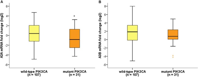
(A) A3A mRNA expression levels were significantly lower in breast cancers with PIK3CA mutations (p = 0.038). (B) There was no association between A3B mRNA expression levels and PIK3CA mutation status (p = 0.127). *p < 0.05, Mann-Whitney U test.

Analysis of serum concentrations of sPD-L1 and IFN-γ

We measured serum sPD-L1 and IFN-γ levels from blood samples obtained at diagnosis in 81 of the 138 patients. For sPD-L1, it was possible to measure serum concentrations in all cases, but only 47 cases of IFN-γ were available because the serum levels were too low to be detected. The median levels of sPD-L1 and IFN-γ were 1.69 ng/mL (range, 0.174–9.757) and 1.78 pg/mL (range, 0.018–51.940), respectively. We analyzed the relationship between the serum levels of sPD-L1 and A3A, and A3B mRNA expression levels. There was a significant weak positive correlation between the serum levels of sPD-L1 and A3A mRNA expression levels (Spearman’s r = 0.264, p = 0.017) (Fig 4A). The serum levels of sPD-L1 in tumors with high A3A mRNA expression levels (≥ median) and low A3A mRNA levels (< median) were 1.99 ng/mL and 1.34 ng/mL, respectively (p = 0.038) (Fig 4B). There was no significant correlation between the serum levels of sPD-L1 and A3B mRNA expression levels or between the serum levels of IFN-γ and A3A mRNA expression levels, or those of IFN-γ and A3B mRNA expression levels. There were no significant differences in serum sPD-L1 and IFN-γ levels according to A3B deletion.

Fig 4. Correlation of serum sPDL-1 levels and APOBEC 3A mRNA expression levels.

Fig 4

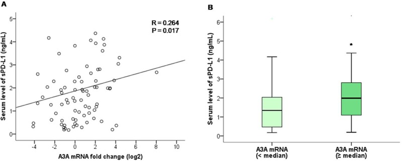
(A) Scatter plot graph showing a positive correlation between serum sPDL-1 levels and A3A mRNA expression levels. (B) Serum levels of sPD-L1 was higher in tumors with high A3A mRNA expression levels (≥ median) than in tumors with low A3A mRNA levels (< median) (p = 0.038). *p < 0.05, Mann-Whitney U test.

Discussion

We quantified mRNA levels of A3A and A3B in 138 breast cancer specimens and in 10 normal tissues from an adjacent area. We found that A3B mRNA expression levels were higher in breast cancer tissues than in adjacent normal breast tissues. According to breast cancer subtypes, A3B mRNA levels were significantly higher in triple-negative breast cancers compared to HR-positive, HER2-negative breast cancers. Robert SA et al. showed that APOBEC mutagenesis was the highest in HER2-enriched subtype, followed by basal-like, and luminal subtype [5]. Our studies showed that A3B mRNA levels was the highest in triple-negative breast cancers, followed by HER2-positive breast cancers, and the lowest in HR-positive, HER2-negative breast cancers. It is consistent that APOBEC mutagenesis is relatively low in HR-positive breast cancer, but there is a difference in the subtype that has the highest values. The cause of this difference is unclear, but we can consider a few things: discrepancy between immunohistochemistry-based clinical subtypes and intrinsic molecular subtypes, small sample size, and ethnic difference etc. Further studies with larger sample size of Asian population are warranted. The association of APOBEC mutagenesis with breast cancer subtypes is in line with tumor mutation burden and immunogenicity according to breast cancer subtypes. There is a higher mean mutational load in HR-negative breast cancers than in HR-positive breast cancers, and triple-negative breast cancers appear to be more immunogenic compared to other subtypes [20].

APOBEC overexpression as a potential source of genomic instability might accelerate cancer progression and be associated with poor prognostic factors. Tsuboi et al. reported that high A3B expression was associated with lymph node metastasis, lymphatic invasion, venous invasion, and high nuclear grade in Japanese patients with breast cancers [17]. Sieuwerts et al. also reported that the expression of A3B mRNA was positively correlated with nodal status, tumor size, and grade [21]. Here we found that both A3A and A3B mRNA levels were higher in patients with old age and Ki-67 > 20%. Advanced tumor stage, the presence of lymph node involvement, and a high histological grade showed significant associations with higher A3A mRNA levels. This suggests that APOBEC mutagenesis is linked with an aggressive phenotype in breast cancers.

TP53 and PIK3CA mutations are the most commonly altered genes in breast cancers and the association between these mutations and APOBEC expression has been studied. Based on the TCGA dataset, the overall prevalence rates of TP53 mutations are 75%, 84%, 14%, and 31% in HER2-enriched, basal-like, luminal A, and B subtypes, respectively [22]. The overall frequency of PIK3CA mutations in breast cancers has been reported as 20–40% and they are more prevalent in HR-positive and HER2-positive breast cancers than in triple-negative breast cancers [22]. The majority of PIK3CA mutations clustered in two ‘hotspot’ regions in exons 9 and 20, corresponding to the helical and catalytic domains of PIK3CA, respectively. Previous evidence has shown that exon 20 mutations predominate in breast cancers [18]. A previous study from Korean breast cancer cohort reported that the prevalence of TP53 and PIK3CA mutations was 47.9% and 28.5%, respectively [23]. In our study, TP53 and PIK3CA mutations were identified in 44.9% and 22.5%, respectively. All patients with PIK3CA mutations except for four were identified as having exon 20 mutations. The METABRIC and TCGA datasets showed that A3B expression was increased in tumors with TP53 mutations and the presence of a PIK3CA alteration was associated with lower A3B expression [13]. In our study, no correlations were found between A3A or A3B mRNA expression levels and TP53 mutation status. The presence of PIK3CA mutations was associated with low A3A mRNA expression in breast cancer. The causes and clinical significance of the relationship between PIK3CA mutations and low APOBEC expression in breast cancers are not yet clear. One possible explanation is the association with the predominant type of PIK3CA mutations in breast cancer. Henderson et al. reported that tumors with enrichment for APOBEC signature mutations were associated with PIK3CA helical domain mutations (exon 9), but not with catalytic domain mutations (exon 20) [19].

APOBEC-mediated hypermutation can induce immunogenicity, leading to increased immune-related markers. Here we evaluated serum sPD-L1 and IFN-γ levels as immune-related markers. IFN-γ is a well-known proinflammatory cytokine that is important for innate and adaptive immunity. It is produced by natural killer cells and CD4 and CD8 cytotoxic T lymphocyte effector T cells, once antigen-specific immunity develops [24]. PD-L1 is an inhibitory ligand that binds to PD-1 to suppress T cell activation. The expression of PD-L1 is induced by multiple proinflammatory molecules including IFN-γ. sPD-L1 is a soluble form of PD-L1, which can be released or shed from PD-L1-positive tumor cells or immune cells. Previous studies showed that high sPD-L1 levels were associated with a poor prognosis in various types of cancers [25, 26]. Unfortunately, the baseline levels of IFN-γ in many patients in our study were too low to make a proper analysis. However, we found that the serum levels of sPD-L1 were positively correlated with A3A mRNA expression levels. This result may suggest a possible association between APOBEC mutagenesis and tumor immunogenicity. It is obviously insufficient to draw clear conclusions. This correlation was statistically significant but weak. The roles and function of sPD-L1 in immune regulation are not fully understood. It is not yet clear whether sPD-L1 plays a significant role like membranous PD-L1, and there are questions to be answered about the clinical significance of sPD-L1. Further studies are warranted to clarify associations between APOBEC mRNA levels and tumor immunogenicity including immune cell infiltration.

Previous studies reported that germlineAPOBEC3A/B deletion polymorphism were not associated with the clinicopathological features of breast cancers, but were linked to a hypermutation phenotype [13]. The prevalence of deletion polymorphisms differs greatly between groups. There was a higher incidence (40–90%) of deletion allele in East Asian and Pacific Island populations than in African and European populations (0.9–6%) [12]. Wen et al. reported that A3B expression was significantly lower in A3B deletion carriers, but A3A expression was not observed to be significantly higher in A3B deletion carriers [27]. To detect germline APOBEC3A/B deletion polymorphism, we performed genotyping assays from 138 matched normal breast tissues. We found that 77 (55.8%) patients carried a single-copy deletion of APOBEC3A/B, and A3B mRNA expression levels showed a tendency to be lower in breast cancers with A3B deletion allele. We learned that some points should be considered when analyzing the APOBEC in Asian patients with a high frequency of A3B deletions. Recently, Starrett et al. reported that APOBEC3H haplotype I (A3H-I) associated with the APOBEC mutation signature in breast tumors lacking A3B. They suggested that combination of A3B and A3H-I explained the full APOBEC signature mutation, especially in Asia with high incidence of A3B deletion allele [28]. An analysis of A3H-I was not included in the current study, and it was a limitation of this study. Second, A3A mRNA levels can reflect values from a deletion allele as well as a non-deletion allele in case of using the common primer that used in Western studies. It is unclear whether there are differences in characteristics and levels of A3A mRNA between A3B deletion carriers and non-carriers. However, considering the possibility of the effect of A3B deletion allele on A3A mRNA, it may be more accurate analysis to measure each one separately. Further studies are necessary to clarify the effect of A3B deletion allele on A3A mRNA levels and the best method to measure A3A mRNA levels in A3B deletion carriers.

In conclusion, A3B mRNA expression levels were significantly higher in triple-negative breast cancers compared to HR-positive, HER2-negative breast cancers. A3B and A3A mRNA expression levels were associated with aggressive phenotypes such as a high mitotic index. Tumors with PIK3CA mutations showed a relatively low A3A mRNA expression levels. There was a weak positive relationship between A3A mRNA expression and sPD-L1 as an immune-related marker. Taken together, we suggest that a combination of the mRNA expression levels of A3A and A3B might provide better clinical significance and prediction of APOBEC mutagenesis than each of these variables alone in patients with breast cancers. Further studies of including analysis of A3H-I are needed to identify more clearly the clinical significance of APOBEC in Asian patients with breast cancer.

Supporting information

S1 Fig. A3A and A3B mRNA expression in breast cancer cell lines.

(TIF)

S1 Data

(XLSX)

S2 Data

(XLSX)

Data Availability

All relevant data are within the manuscript and its Supporting Information files.

Funding Statement

This work was supported by the the Basic Science Research Program through the National Research Foundation of Korea (NRF) funded by the Ministry of Education (NRF-2016R1D1A1B03933245, W.H.S) and by The Catholic University of Korea, Uijeongbu St. Mary’s Hospital Clinical Research Laboratory Foundation made in the program year of 2018 (W.H.S). The funders had no role in study design, data collection and analysis, decision to publish, or preparation of the manuscript

References

  • 1.Shen Z. Genomic instability and cancer: an introduction. J Mol Cell Biol. 2011;3(1):1–3. 10.1093/jmcb/mjq057 [DOI] [PubMed] [Google Scholar]
  • 2.Yao Y, Dai W. Genomic Instability and Cancer. J Carcinog Mutagen. 2014;5. [DOI] [PMC free article] [PubMed] [Google Scholar]
  • 3.Alexandrov LB, Nik-Zainal S, Wedge DC, Aparicio SA, Behjati S, Biankin AV, et al. Signatures of mutational processes in human cancer. Nature. 2013;500(7463):415–21. 10.1038/nature12477 [DOI] [PMC free article] [PubMed] [Google Scholar]
  • 4.Roberts SA, Gordenin DA. Hypermutation in human cancer genomes: footprints and mechanisms. Nat Rev Cancer. 2014;14(12):786–800. 10.1038/nrc3816 [DOI] [PMC free article] [PubMed] [Google Scholar]
  • 5.Roberts SA, Lawrence MS, Klimczak LJ, Grimm SA, Fargo D, Stojanov P, et al. An APOBEC cytidine deaminase mutagenesis pattern is widespread in human cancers. Nat Genet. 2013;45(9):970–6. 10.1038/ng.2702 [DOI] [PMC free article] [PubMed] [Google Scholar]
  • 6.Smith HC, Bennett RP, Kizilyer A, McDougall WM, Prohaska KM. Functions and regulation of the APOBEC family of proteins. Semin Cell Dev Biol. 2012;23(3):258–68. 10.1016/j.semcdb.2011.10.004 [DOI] [PMC free article] [PubMed] [Google Scholar]
  • 7.Kuong KJ, Loeb LA. APOBEC3B mutagenesis in cancer. Nat Genet. 2013;45(9):964–5. 10.1038/ng.2736 [DOI] [PMC free article] [PubMed] [Google Scholar]
  • 8.Burns MB, Temiz NA, Harris RS. Evidence for APOBEC3B mutagenesis in multiple human cancers. Nat Genet. 2013;45(9):977–83. 10.1038/ng.2701 [DOI] [PMC free article] [PubMed] [Google Scholar]
  • 9.Chan K, Roberts SA, Klimczak LJ, Sterling JF, Saini N, Malc EP, et al. An APOBEC3A hypermutation signature is distinguishable from the signature of background mutagenesis by APOBEC3B in human cancers. Nat Genet. 2015;47(9):1067–72. 10.1038/ng.3378 [DOI] [PMC free article] [PubMed] [Google Scholar]
  • 10.Burns MB, Lackey L, Carpenter MA, Rathore A, Land AM, Leonard B, et al. APOBEC3B is an enzymatic source of mutation in breast cancer. Nature. 2013;494(7437):366–70. 10.1038/nature11881 [DOI] [PMC free article] [PubMed] [Google Scholar]
  • 11.Nik-Zainal S, Wedge DC, Alexandrov LB, Petljak M, Butler AP, Bolli N, et al. Association of a germline copy number polymorphism of APOBEC3A and APOBEC3B with burden of putative APOBEC-dependent mutations in breast cancer. Nat Genet. 2014;46(5):487–91. 10.1038/ng.2955 [DOI] [PMC free article] [PubMed] [Google Scholar]
  • 12.Kidd JM, Newman TL, Tuzun E, Kaul R, Eichler EE. Population stratification of a common APOBEC gene deletion polymorphism. PLoS Genet. 2007;3(4):e63 10.1371/journal.pgen.0030063 [DOI] [PMC free article] [PubMed] [Google Scholar]
  • 13.Cescon DW, Haibe-Kains B, Mak TW. APOBEC3B expression in breast cancer reflects cellular proliferation, while a deletion polymorphism is associated with immune activation. Proc Natl Acad Sci U S A. 2015;112(9):2841–6. 10.1073/pnas.1424869112 [DOI] [PMC free article] [PubMed] [Google Scholar]
  • 14.Long J, Delahanty RJ, Li G, Gao YT, Lu W, Cai Q, et al. A common deletion in the APOBEC3 genes and breast cancer risk. J Natl Cancer Inst. 2013;105(8):573–9. 10.1093/jnci/djt018 [DOI] [PMC free article] [PubMed] [Google Scholar]
  • 15.Gohler S, Da Silva Filho MI, Johansson R, Enquist-Olsson K, Henriksson R, Hemminki K, et al. Impact of functional germline variants and a deletion polymorphism in APOBEC3A and APOBEC3B on breast cancer risk and survival in a Swedish study population. J Cancer Res Clin Oncol. 2016;142(1):273–6. 10.1007/s00432-015-2038-7 [DOI] [PubMed] [Google Scholar]
  • 16.Gansmo LB, Romundstad P, Hveem K, Vatten L, Nik-Zainal S, Lonning PE, et al. APOBEC3A/B deletion polymorphism and cancer risk. Carcinogenesis. 2018;39(2):118–24. 10.1093/carcin/bgx131 [DOI] [PMC free article] [PubMed] [Google Scholar]
  • 17.Tsuboi M, Yamane A, Horiguchi J, Yokobori T, Kawabata-Iwakawa R, Yoshiyama S, et al. APOBEC3B high expression status is associated with aggressive phenotype in Japanese breast cancers. Breast Cancer. 2016;23(5):780–8. 10.1007/s12282-015-0641-8 [DOI] [PubMed] [Google Scholar]
  • 18.Saal LH, Holm K, Maurer M, Memeo L, Su T, Wang X, et al. PIK3CA mutations correlate with hormone receptors, node metastasis, and ERBB2, and are mutually exclusive with PTEN loss in human breast carcinoma. Cancer Res. 2005;65(7):2554–9. 10.1158/0008-5472-CAN-04-3913 [DOI] [PubMed] [Google Scholar]
  • 19.Henderson S, Chakravarthy A, Su X, Boshoff C, Fenton TR. APOBEC-mediated cytosine deamination links PIK3CA helical domain mutations to human papillomavirus-driven tumor development. Cell Rep. 2014;7(6):1833–41. 10.1016/j.celrep.2014.05.012 [DOI] [PubMed] [Google Scholar]
  • 20.Luen S, Virassamy B, Savas P, Salgado R, Loi S. The genomic landscape of breast cancer and its interaction with host immunity. Breast. 2016;29:241–50. 10.1016/j.breast.2016.07.015 [DOI] [PubMed] [Google Scholar]
  • 21.Sieuwerts AM, Willis S, Burns MB, Look MP, Meijer-Van Gelder ME, Schlicker A, et al. Elevated APOBEC3B correlates with poor outcomes for estrogen-receptor-positive breast cancers. Horm Cancer. 2014;5(6):405–13. 10.1007/s12672-014-0196-8 [DOI] [PMC free article] [PubMed] [Google Scholar]
  • 22.Cancer Genome Atlas N. Comprehensive molecular portraits of human breast tumours. Nature. 2012;490(7418):61–70. 10.1038/nature11412 [DOI] [PMC free article] [PubMed] [Google Scholar]
  • 23.Kan Z, Ding Y, Kim J, Jung HH, Chung W, Lal S, et al. Multi-omics profiling of younger Asian breast cancers reveals distinctive molecular signatures. Nat Commun. 2018;9(1):1725 10.1038/s41467-018-04129-4 [DOI] [PMC free article] [PubMed] [Google Scholar]
  • 24.Lee S, Margolin K. Cytokines in cancer immunotherapy. Cancers (Basel). 2011;3(4):3856–93. [DOI] [PMC free article] [PubMed] [Google Scholar]
  • 25.Zhang J, Gao J, Li Y, Nie J, Dai L, Hu W, et al. Circulating PD-L1 in NSCLC patients and the correlation between the level of PD-L1 expression and the clinical characteristics. Thorac Cancer. 2015;6(4):534–8. 10.1111/1759-7714.12247 [DOI] [PMC free article] [PubMed] [Google Scholar]
  • 26.Zhou J, Mahoney KM, Giobbie-Hurder A, Zhao F, Lee S, Liao X, et al. Soluble PD-L1 as a Biomarker in Malignant Melanoma Treated with Checkpoint Blockade. Cancer Immunol Res. 2017;5(6):480–92. 10.1158/2326-6066.CIR-16-0329 [DOI] [PMC free article] [PubMed] [Google Scholar]
  • 27.Wen WX, Soo JS, Kwan PY, Hong E, Khang TF, Mariapun S, et al. Germline APOBEC3B deletion is associated with breast cancer risk in an Asian multi-ethnic cohort and with immune cell presentation. Breast Cancer Res. 2016;18(1):56 10.1186/s13058-016-0717-1 [DOI] [PMC free article] [PubMed] [Google Scholar]
  • 28.Starrett GJ, Luengas EM, McCann JL, Ebrahimi D, Temiz NA, Love RP, et al. The DNA cytosine deaminase APOBEC3H haplotype I likely contributes to breast and lung cancer mutagenesis. Nat Commun. 2016;7:12918 10.1038/ncomms12918 [DOI] [PMC free article] [PubMed] [Google Scholar]

Decision Letter 0

Elizabeth Christie

31 Oct 2019

PONE-D-19-26098

Clinical implications of APOBEC3A and 3B expression in patients with breast cancer

PLOS ONE

Dear Dr. Won,

Thank you for submitting your manuscript to PLOS ONE. After careful consideration, we feel that it has merit but does not fully meet PLOS ONE’s publication criteria as it currently stands. Therefore, we invite you to submit a revised version of the manuscript that addresses the points raised during the review process. Please ensure all of the major comments from both reviewers are addressed, particularly providing evidence that the qPCR assays are specific.

We would appreciate receiving your revised manuscript by Dec 15 2019 11:59PM. When you are ready to submit your revision, log on to https://www.editorialmanager.com/pone/ and select the 'Submissions Needing Revision' folder to locate your manuscript file.

If you would like to make changes to your financial disclosure, please include your updated statement in your cover letter.

To enhance the reproducibility of your results, we recommend that if applicable you deposit your laboratory protocols in protocols.io, where a protocol can be assigned its own identifier (DOI) such that it can be cited independently in the future. For instructions see: http://journals.plos.org/plosone/s/submission-guidelines#loc-laboratory-protocols

Please include the following items when submitting your revised manuscript:

  • A rebuttal letter that responds to each point raised by the academic editor and reviewer(s). This letter should be uploaded as separate file and labeled 'Response to Reviewers'.

  • A marked-up copy of your manuscript that highlights changes made to the original version. This file should be uploaded as separate file and labeled 'Revised Manuscript with Track Changes'.

  • An unmarked version of your revised paper without tracked changes. This file should be uploaded as separate file and labeled 'Manuscript'.

Please note while forming your response, if your article is accepted, you may have the opportunity to make the peer review history publicly available. The record will include editor decision letters (with reviews) and your responses to reviewer comments. If eligible, we will contact you to opt in or out.

We look forward to receiving your revised manuscript.

Kind regards,

Elizabeth Christie

Academic Editor

PLOS ONE

Journal Requirements:

When submitting your revision, we need you to address these additional requirements.

Please ensure that your manuscript meets PLOS ONE's style requirements, including those for file naming. The PLOS ONE style templates can be found at

http://www.journals.plos.org/plosone/s/file?id=wjVg/PLOSOne_formatting_sample_main_body.pdf and http://www.journals.plos.org/plosone/s/file?id=ba62/PLOSOne_formatting_sample_title_authors_affiliations.pdf

[Note: HTML markup is below. Please do not edit.]

Reviewers' comments:

Reviewer's Responses to Questions

Comments to the Author

1. Is the manuscript technically sound, and do the data support the conclusions?

The manuscript must describe a technically sound piece of scientific research with data that supports the conclusions. Experiments must have been conducted rigorously, with appropriate controls, replication, and sample sizes. The conclusions must be drawn appropriately based on the data presented.

Reviewer #1: No

Reviewer #2: Partly

**********

2. Has the statistical analysis been performed appropriately and rigorously?

Reviewer #1: Yes

Reviewer #2: I Don't Know

**********

3. Have the authors made all data underlying the findings in their manuscript fully available?

The PLOS Data policy requires authors to make all data underlying the findings described in their manuscript fully available without restriction, with rare exception (please refer to the Data Availability Statement in the manuscript PDF file). The data should be provided as part of the manuscript or its supporting information, or deposited to a public repository. For example, in addition to summary statistics, the data points behind means, medians and variance measures should be available. If there are restrictions on publicly sharing data—e.g. participant privacy or use of data from a third party—those must be specified.

Reviewer #1: Yes

Reviewer #2: No

**********

4. Is the manuscript presented in an intelligible fashion and written in standard English?

PLOS ONE does not copyedit accepted manuscripts, so the language in submitted articles must be clear, correct, and unambiguous. Any typographical or grammatical errors should be corrected at revision, so please note any specific errors here.

Reviewer #1: Yes

Reviewer #2: Yes

**********

5. Review Comments to the Author

Please use the space provided to explain your answers to the questions above. You may also include additional comments for the author, including concerns about dual publication, research ethics, or publication ethics. (Please upload your review as an attachment if it exceeds 20,000 characters)

Reviewer #1: Kim and coworkers study a cohort of 138 Korean breast cancer patients and attempt to associate A3A and A3B mRNA levels with various clinical and genetic features. The results are inconclusive due to a lack of validation studies for the RTqPCR assays. Some of the rationale is also unclear.

Major:

1) Because the authors are focused an Asian populations (Korean), where an A3A-B deletion allele is common, they should also consider A3H haplotypes which Starrett and coworkers implicated in causing APOBEC signature mutations in the absence of A3B (PMID 27650891). In other words, A3H could also be clinically relevant.

2) The RTqPCR assays for A3A and A3B may not be specific due to extremely high homology between these genes. What tests have the authors done to validate these assays and ensure specificity?

3) Lines 202-207 – what are the units for A3A and A3B mRNA levels? This is unclear. All raw values should be provided in a supporting table (ie. not just medians).

4) It is hard to believe that there are no patients completely lacking A3B (ie. homozygous null) given the 77/(138x2) allele frequency of the deletion in this population. Can the deletion allele be verified using an independent assay such as with different PCR primers and sequencing?

5) The rationale for correlating serum sPD-L1 and tumor A3A and A3B mRNA levels is not explained. This analysis makes no sense at all.

Minor:

- Line 100 should also cite PMID:23389445, which is the first paper to implicate A3B in cancer

- Line 161 – dNTPs

Reviewer #2: Clinical implications of APOBEC3A and 3B expression 1 in patients with breast cancer

In this study the investigators have performed 4 main analyses of a Korean breast cancer cohort, firstly, they quantitated the mRNA levels of A3A and A3B by qRTPCR. They correlate these values with various clinical features and also the results of the subsequent analyses. Secondly, they quantified the prevalence of a germline deletion that leads to the 3’UTR of A3B being spliced onto the 3’ end of the A3A 3’ UTR, which also leads to the loss of one copy of the A3B gene in their patient cohort. Thirdly, they examined the prevalence of TP53 and PIK3CA mutations in their patient cohort by “direct” sequencing. Fourth, they correlated serum sPDL1 and IFNg protein levels with A3A and A3B mRNA expression levels. These studies found a number of statistically significant associations with either A3A or A3B levels and clinical features. Finally, they state that they performed survival analyses on the cohort by dividing the patients up based on high and low A3A and A3B expression levels. However, they found no statistically significant associations with survival. This is the first investigation of A3A and A3B in an all Korean population of breast cancer patients, and this maybe important as it is known that east Asian populations have a higher incidence of the deletion allele than western cohorts.

Of this study I have the following major concerns:

A. The methods are not described in sufficient detail to completely understand how the data was generated:

1. How were ER expression and Her2 expression measured and what were the cut-offs for being called ER positive and Her2 positive? What proportion of Her2 patients are also ER positive?

2. Does the RTPCR for A3A also amplify the deletion allele?

3. Are the mRNA levels reported in the tables deltadeltaCT values, this is not completely clear from the methods?

4. What is meant by “direct” sequencing – was this Sanger sequencing? Why was cDNA analysed and not genomic DNA? Furthermore, the exact mutations detected for each sample should be tabulated and provided as supplementary data.

5. Which statistical tests were used to generate the p values in the tables? Due to the large interquartile range I’m surprised at some of the p values. I would like to note however that as I am not familiar with all the statistical tests outlined in the methods and am unable to say whether they are the most appropriate statistical test for this data. In addition, without the full data being available (the authors only provide summary statistics) it is hard to test the veracity of the statistics.

6. What two variables are being compared to generate the statistics in fig 1? My understanding is that the K-W test that was used can demonstrate that one sample significantly dominates the other samples in the analysis, but it does not identify which sample is the dominant one and this needs to be determined using a post-hoc test?

B. The high prevalence of the deletion phenotype in this cohort (56%) makes the mRNA analysis of A3A problematic. I presume that the A3A RTPCR used will amplify mRNA from both the normal A3A allele and the deletion allele, while the A3B RTPCR would only amplify mRNA from the normal A3B allele? If this is not the case this should be clarified in the methods. If it does amplify both alleles this means that for a large number of patients the read out from the A3A assay will be measuring a combination of A3A and mutant gene expression. Thus, from the authors analysis they cannot categorically state the associations are due to A3A mRNA levels, rather than the deletion allele mRNA. While the deletion allele is identical in coding sequence the change in the 3’ UTR could change the stability of the mRNA and also the rate of translation of mRNA to protein. As the mutant allele has been associated with increased risk of breast cancer by a number of other groups the prevalence of this allele is important to the understanding of A3A biology (there is a meta-analysis of this in this publication: 10.18632/oncotarget.19400). This issue could affect a large number of the subsequent analyses that compare other clinical parameters to supposed A3A mRNA levels. Ideally the investigators would redo this analysis with an assay which can separately measure the mutant allele and normal allele.

C. The rationale for studying the link between A3A and A3B expression levels and immune related parameters such as serum IFN-gamma and sPDL1 seems tenuous. In this cohort they have not shown that the level of A3A associates with mutational burden or immune cell infiltration into the tumour. Furthermore, as they were unable to measure IFN-gamma in a large number of samples and the association of sPDL1 with A3A is marginal (R2=0.03) I think this data is not very useful/informative. Particularly as the association may or may not be confounded by the relative abundance of the deletion allele.

D. As the investigators show that the levels of A3B associate with the TNBC subtype then subsequent associations of A3B with clinical parameters need to have the caveat that the association may be due to differences in breast cancer subtype proportions within the groups studied rather than A3B directly.

E. I do not think that the survival analyses are informative and should be removed. As they only finished collecting samples from this cohort in December 2016 there is insufficient follow-up time to draw conclusions about survival. If they do want to include survival data then the Kaplan-Meier curves need to be included, and these should also state the numbers of censored events over time. In addition, it should be noted that the survival data could also be influenced by other factors such as that the population analysed contains all the different subtypes of breast cancer that would each have been treated differently (hormone therapy for ER pos, Herceptin for Her2, and chemo for TNBC). Although the numbers would be quite small they could do separate analyses for each subtype of breast cancer.

Minor points:

This cohort seems unusual with only 40% luminal breast cancer patients (most cohorts have >70% luminal subtype), the authors should comment on why this is the case. Is this normal for a Korean population or did the selection criteria skew the relative abundance of luminal patients?

The authors should also clarify whether the deletion allele is associated with any particular subtype of breast cancer (this is not completely clear from table 2 as they do not include breast cancer subtypes).

**********

6. PLOS authors have the option to publish the peer review history of their article (what does this mean?). If published, this will include your full peer review and any attached files.

If you choose “no”, your identity will remain anonymous but your review may still be made public.

Do you want your identity to be public for this peer review? For information about this choice, including consent withdrawal, please see our Privacy Policy.

Reviewer #1: No

Reviewer #2: No

[NOTE: If reviewer comments were submitted as an attachment file, they will be attached to this email and accessible via the submission site. Please log into your account, locate the manuscript record, and check for the action link "View Attachments". If this link does not appear, there are no attachment files to be viewed.]

While revising your submission, please upload your figure files to the Preflight Analysis and Conversion Engine (PACE) digital diagnostic tool, https://pacev2.apexcovantage.com/. PACE helps ensure that figures meet PLOS requirements. To use PACE, you must first register as a user. Registration is free. Then, login and navigate to the UPLOAD tab, where you will find detailed instructions on how to use the tool. If you encounter any issues or have any questions when using PACE, please email us at figures@plos.org. Please note that Supporting Information files do not need this step.

PLoS One. 2020 Mar 16;15(3):e0230261. doi: 10.1371/journal.pone.0230261.r002

Author response to Decision Letter 0


23 Nov 2019

Dear Editor,

We’re very grateful for the constructive comments of the reviewers. It is clear that comments make our paper better. We revised our manuscript in response to reviewers’ comment. We have highlighted the major changes with a blue color. We hope that the revision satisfies reviewers’ requirement. Thank you very much.

Reviewer 1

1) Because the authors are focused an Asian populations (Korean), where an A3A-B deletion allele is common, they should also consider A3H haplotypes which Starrett and coworkers implicated in causing APOBEC signature mutations in the absence of A3B (PMID 27650891). In other words, A3H could also be clinically relevant.

� We appreciate your good comments. New findings on APOBEC mutation signature in cancer have been reported in recent years. We planned to investigate the most studied APOBEC3A and 3B in our patients, because there was not much data on other APOBEC family including APOBEC3H in cancer. We added the A3H haplotype you mentioned as a limitation of our study in discussion.

2) The RTqPCR assays for A3A and A3B may not be specific due to extremely high homology between these genes. What tests have the authors done to validate these assays and ensure specificity?

� We agree with reviewer’s opinion. We confirmed that using Primer-BLAST-NCBI. We attached the captured image of the results.

3) Lines 202-207 – what are the units for A3A and A3B mRNA levels? This is unclear. All raw values should be provided in a supporting table (ie. not just medians).

� Thank you for your comments. The relative mRNA expression level was normalized to the β-actin gene expression, and then expressed as thelog2 fold change of the delta-delta Ct values. We added it in Method. We submit all raw values as supplement file (S1).

4) It is hard to believe that there are no patients completely lacking A3B (ie. homozygous null) given the 77/(138x2) allele frequency of the deletion in this population. Can the deletion allele be verified using an independent assay such as with different PCR primers and sequencing?

� Yes, the deletion allele was verified using different PCR primers. We rechecked for heterozygous deletion/ insertion using insertion primer in samples that showed homozygous deletion in genotyping. (Reference: Kidd JM et al. Population stratification of a common APOBEC gene deletion polymorphism. PLoS Genet 3(4): e63)

5) The rationale for correlating serum sPD-L1 and tumor A3A and A3B mRNA levels is not explained. This analysis makes no sense at all.

� Thank you for your comment. APOBEC is well known as an endogenous mutagen in various cancers. High tumor mutational burden and APOBEC mutagenesis signature are associated with immune signatures and with increased expression of immune-related genes. Therefore, we wanted to evaluate the relationships between APOBEC mRNA levels and serum levels of sPD-L1 and IFN-γ as immune-related markers. Obviously, there are better ways to evaluate cancer immunogenicity including immune cell infiltration. Unfortunately, we were not able to evaluate it properly. We described this as a limitation of our research in the discussion.

Minor

- Line 100 should also cite PMID:23389445, which is the first paper to implicate A3B in cancer.

� We added that paper according to your comment. Thank you.

- Line 161 – dNTPs

� We revised it. Thank you.

Reviewer 2

1. How were ER expression and Her2 expression measured and what were the cut-offs for being called ER positive and Her2 positive? What proportion of Her2 patients are also ER positive?

� Thank you for your comments. We added definitions of hormone receptor positivity and HER2 positivity in method. Among 49 patients with HER2-positive breast cancer, 34 patients were also HR-positive. We added it in result.

2. Does the RTPCR for A3A also amplify the deletion allele?

� With reference to other papers measuring A3A mRNA levels, we used the A3A primer that was amplified from both the normal A3A allele and the deletion allele.

3. Are the mRNA levels reported in the tables deltadeltaCT values, this is not completely clear from the methods?

� Thank you for your comments. The relative mRNA expression level was normalized to the β-actin gene expression, and then expressed as the log2 fold change of the delta-delta Ct values. We added it in Method.

4. What is meant by “direct” sequencing – was this Sanger sequencing? Why was cDNA analysed and not genomic DNA? Furthermore, the exact mutations detected for each sample should be tabulated and provided as supplementary data.

� To evaluate mutations in exons, cDNA was amplified by PCR with each primer of TP53 and PIK3CA, and then we did Sanger sequencing for the products. Finally, we analyzed the mutations by comparing the sequencing results with reference sequence. We submit all raw values as supplement file (S2).

5. Which statistical tests were used to generate the p values in the tables? Due to the large interquartile range I’m surprised at some of the p values. I would like to note however that as I am not familiar with all the statistical tests outlined in the methods and am unable to say whether they are the most appropriate statistical test for this data. In addition, without the full data being available (the authors only provide summary statistics) it is hard to test the veracity of the statistics.

� To compare the differences of continuous variables between two or more groups, we performed Mann-Whitney U test and Kruskal-Wallis test as nonparametric tests because our data isn’t normally distributed. We added the statistical methods below the table and figure.

6. What two variables are being compared to generate the statistics in fig 1? My understanding is that the K-W test that was used can demonstrate that one sample significantly dominates the other samples in the analysis, but it does not identify which sample is the dominant one and this needs to be determined using a post-hoc test?

� We appreciate your good comment. Dunn-Bonferroni post hoc testing is carried out on each pair of groups after a Kruskal-Wallis test. We added detailed information and results about this.

B. The high prevalence of the deletion phenotype in this cohort (56%) makes the mRNA analysis of A3A problematic. I presume that the A3A RTPCR used will amplify mRNA from both the normal A3A allele and the deletion allele, while the A3B RTPCR would only amplify mRNA from the normal A3B allele? If this is not the case this should be clarified in the methods. If it does amplify both alleles this means that for a large number of patients the read out from the A3A assay will be measuring a combination of A3A and mutant gene expression. Thus, from the authors analysis they cannot categorically state the associations are due to A3A mRNA levels, rather than the deletion allele mRNA. While the deletion allele is identical in coding sequence the change in the 3’ UTR could change the stability of the mRNA and also the rate of translation of mRNA to protein. As the mutant allele has been associated with increased risk of breast cancer by a number of other groups the prevalence of this allele is important to the understanding of A3A biology (there is a meta-analysis of this in this publication: 10.18632/oncotarget.19400). This issue could affect a large number of the subsequent analyses that compare other clinical parameters to supposed A3A mRNA levels. Ideally the investigators would redo this analysis with an assay which can separately measure the mutant allele and normal allele.

� We appreciate your comment and understand your suggestion. We used the primer mentioned in previous studies. It measured the A3A mRNA levels from both deletion allele and non-deletion allele. As you mentioned, there may be differences between A3A mRNA from deletion allele and A3A mRNA from non-deletion allele. Some study reported that APOBEC3B expression was significantly lower in A3B deletion carriers, but APOBEC3A expression was not observed to be significantly higher in A3B deletion carriers. Some study mentioned that A3A mRNA from deletion allele was more stable, resulting in higher intracellular A3A levels. We think that further studies are needed to clarify the effect of A3B deletion allele on A3A mRNA levels and the best method to measure A3A mRNA levels in A3B deletion carriers. We added this in discussion. Thank you for your important advice.

C. The rationale for studying the link between A3A and A3B expression levels and immune related parameters such as serum IFN-gamma and sPDL1 seems tenuous. In this cohort they have not shown that the level of A3A associates with mutational burden or immune cell infiltration into the tumour. Furthermore, as they were unable to measure IFN-gamma in a large number of samples and the association of sPDL1 with A3A is marginal (R2=0.03) I think this data is not very useful/informative. Particularly as the association may or may not be confounded by the relative abundance of the deletion allele.

� Thank you for your comment. We agree with your opinion. We describe this as a limitation of our research in the discussion.

D. As the investigators show that the levels of A3B associate with the TNBC subtype then subsequent associations of A3B with clinical parameters need to have the caveat that the association may be due to differences in breast cancer subtype proportions within the groups studied rather than A3B directly.

� We understand your concerns. However, correlation with clinical parameters showed more apparent in A3A than in A3B. Actually, there is no big difference in A3A mRNA levels between subtypes. Therefore, we think that it’s hard to say that associations of A3B with clinical parameters are only caused by differences in breast cancer subtype proportions.

E. I do not think that the survival analyses are informative and should be removed. As they only finished collecting samples from this cohort in December 2016 there is insufficient follow-up time to draw conclusions about survival. If they do want to include survival data then the Kaplan-Meier curves need to be included, and these should also state the numbers of censored events over time. In addition, it should be noted that the survival data could also be influenced by other factors such as that the population analysed contains all the different subtypes of breast cancer that would each have been treated differently (hormone therapy for ER pos, Herceptin for Her2, and chemo for TNBC). Although the numbers would be quite small they could do separate analyses for each subtype of breast cancer.

� We agreed with your opinion. Survival analyses have some limitations due to small number of patients and short follow-up time. We decided to remove the survival data according to your recommendation.

Minor points:

This cohort seems unusual with only 40% luminal breast cancer patients (most cohorts have >70% luminal subtype), the authors should comment on why this is the case. Is this normal for a Korean population or did the selection criteria skew the relative abundance of luminal patients?

� We agreed with your opinion. Luminal subtype accounts for about 60-70% of all patients with breast cancer. Based on the clinicopathological data, we chose samples with similar proportions for each subtype to prevent the number of one subtype from becoming too small. We added this comments in method. Thank you.

The authors should also clarify whether the deletion allele is associated with any particular subtype of breast cancer (this is not completely clear from table 2 as they do not include breast cancer subtypes).

� We described that in result (Line 241-242):There was no difference in the frequency of A3B deletion according to breast cancer subtypes.

Attachment

Submitted filename: APOBEC_Response.docx

Decision Letter 1

Elizabeth Christie

20 Jan 2020

PONE-D-19-26098R1

Clinical implications of APOBEC3A and 3B expression in patients with breast cancer

PLOS ONE

Dear Dr. Won,

Thank you for submitting your manuscript to PLOS ONE. After careful consideration, we feel that it has merit but does not fully meet PLOS ONE’s publication criteria as it currently stands. Therefore, we invite you to submit a revised version of the manuscript that addresses the points raised during the review process.

Please address all of the reviewers comments - both the new issues that have been identified and previous comments from Reviewer 1.

We would appreciate receiving your revised manuscript by Mar 05 2020 11:59PM. When you are ready to submit your revision, log on to https://www.editorialmanager.com/pone/ and select the 'Submissions Needing Revision' folder to locate your manuscript file.

If you would like to make changes to your financial disclosure, please include your updated statement in your cover letter.

To enhance the reproducibility of your results, we recommend that if applicable you deposit your laboratory protocols in protocols.io, where a protocol can be assigned its own identifier (DOI) such that it can be cited independently in the future. For instructions see: http://journals.plos.org/plosone/s/submission-guidelines#loc-laboratory-protocols

Please include the following items when submitting your revised manuscript:

  • A rebuttal letter that responds to each point raised by the academic editor and reviewer(s). This letter should be uploaded as separate file and labeled 'Response to Reviewers'.

  • A marked-up copy of your manuscript that highlights changes made to the original version. This file should be uploaded as separate file and labeled 'Revised Manuscript with Track Changes'.

  • An unmarked version of your revised paper without tracked changes. This file should be uploaded as separate file and labeled 'Manuscript'.

Please note while forming your response, if your article is accepted, you may have the opportunity to make the peer review history publicly available. The record will include editor decision letters (with reviews) and your responses to reviewer comments. If eligible, we will contact you to opt in or out.

We look forward to receiving your revised manuscript.

Kind regards,

Elizabeth Christie

Academic Editor

PLOS ONE

[Note: HTML markup is below. Please do not edit.]

Reviewers' comments:

Reviewer's Responses to Questions

Comments to the Author

1. If the authors have adequately addressed your comments raised in a previous round of review and you feel that this manuscript is now acceptable for publication, you may indicate that here to bypass the “Comments to the Author” section, enter your conflict of interest statement in the “Confidential to Editor” section, and submit your "Accept" recommendation.

Reviewer #1: (No Response)

Reviewer #2: (No Response)

**********

2. Is the manuscript technically sound, and do the data support the conclusions?

The manuscript must describe a technically sound piece of scientific research with data that supports the conclusions. Experiments must have been conducted rigorously, with appropriate controls, replication, and sample sizes. The conclusions must be drawn appropriately based on the data presented.

Reviewer #1: No

Reviewer #2: Partly

**********

3. Has the statistical analysis been performed appropriately and rigorously?

Reviewer #1: N/A

Reviewer #2: I Don't Know

**********

4. Have the authors made all data underlying the findings in their manuscript fully available?

The PLOS Data policy requires authors to make all data underlying the findings described in their manuscript fully available without restriction, with rare exception (please refer to the Data Availability Statement in the manuscript PDF file). The data should be provided as part of the manuscript or its supporting information, or deposited to a public repository. For example, in addition to summary statistics, the data points behind means, medians and variance measures should be available. If there are restrictions on publicly sharing data—e.g. participant privacy or use of data from a third party—those must be specified.

Reviewer #1: Yes

Reviewer #2: No

**********

5. Is the manuscript presented in an intelligible fashion and written in standard English?

PLOS ONE does not copyedit accepted manuscripts, so the language in submitted articles must be clear, correct, and unambiguous. Any typographical or grammatical errors should be corrected at revision, so please note any specific errors here.

Reviewer #1: Yes

Reviewer #2: No

**********

6. Review Comments to the Author

Please use the space provided to explain your answers to the questions above. You may also include additional comments for the author, including concerns about dual publication, research ethics, or publication ethics. (Please upload your review as an attachment if it exceeds 20,000 characters)

Reviewer #1: The authors have not yet addressed my most important comments. Just because they are using published PCR assays does not mean they are excused from doing proper controls for assay specificity (prior work could be incorrect). For instance, both RTqPCR and PCR assay specificity could be validated using breast cancer cell lines with known APOBEC genotypes. SKBR3, for instance, is A3B null (PMID 23389445). MCF10A is wildtype (as far as anyone knows) and A3B is inducible with PKC agonists and A3A with interferon-alpha (PMID 26420215).

Reviewer #2: Clinical implications of APOBEC3A and 3B expression in patients with breast cancer

(Revised version)

In this study the investigators have performed 4 main analyses of a Korean breast cancer cohort, firstly, they quantitated the mRNA levels of A3A and A3B by qRTPCR. They correlate these values with various clinical features and also the results of the subsequent analyses. Secondly, they quantified the prevalence of a germline deletion that leads to the 3’UTR of A3B being spliced onto the 3’ end of the A3A 3’ UTR, which also leads to the loss of one copy of the A3B gene in their patient cohort. Thirdly, they examined the prevalence of TP53 and PIK3CA mutations in their patient cohort by Sanger sequencing. Fourth, they correlated serum sPDL1 and IFNg protein levels with A3A and A3B mRNA expression levels. These studies found a number of statistically significant associations with either A3A or A3B levels and clinical features. This is the first investigation of A3A and A3B in an all Korean population of breast cancer patients, and this maybe important as it is known that east Asian populations have a higher incidence of the deletion allele than western cohorts.

The authors have addressed and clarified a number of my issues with the original manuscript. With the exception that the raw data they provided, as this was not annotated with information such as breast cancer subtype it was not very helpful. In addition, supplementary table 2 only roughly summarises the mutations discovered and does not list the exact mutations. Unfortunately, upon re-reading the manuscript a number of additional issues came up these are listed below.

Major points:

1. In the discussion they state they use matched adjacent normal tissue in their analyses but in the results section they state only 10 normal tissue samples were used and from the supplementary table 1 it appears they have used an average normal value that is not matched to the patient tumour samples. This needs to be clarified. In addition, did they include a similar distribution of mutant allele carriers in the normal tissue as in the tumour samples? Does the deletion allele affect expression of A3A and A3B in normal tissue? If this could be a confounding factor it should be discussed.

2. In both the results and discussion, it is stated that A3A and A3B are higher in younger patients, however, the results that are in table 1 show the opposite, with higher median levels of both A3A and A3B in the older (>52) patients. This needs to be clarified and corrected.

Minor points:

1. The full breast cancer subtype data and associations with A3A, A3B and the mutation allele should be included in Tables 1 and 2 for completeness.

2. Were outliers included or excluded in the generation of the line of best fit for the R-squared analysis of sPDL1 and A3A levels? While there is a significant association between sPDL1 and A3A levels the R squared value is very low, this should be noted in the results or discussion.

3. In the introduction it is stated that previous studies have shown that the Apobec mutation profile is highest in the Her2 subtype, this should be commented on in relation to the results from this study in the discussion.

4. A number of times it is stated that A3B levels are significantly higher in TNBC, this should be qualified with the fact that this is only in comparison to HR positive cancers not Her2 positive.

5. The number of Her2 positive patients in Table 2 equals 48 whereas the text states there were 49 Her2 patients.

6. The initial manuscript was written with good English however a number of the revisions/edits added to the manuscript following the initial review need to be checked for grammar.

**********

7. PLOS authors have the option to publish the peer review history of their article (what does this mean?). If published, this will include your full peer review and any attached files.

If you choose “no”, your identity will remain anonymous but your review may still be made public.

Do you want your identity to be public for this peer review? For information about this choice, including consent withdrawal, please see our Privacy Policy.

Reviewer #1: No

Reviewer #2: No

[NOTE: If reviewer comments were submitted as an attachment file, they will be attached to this email and accessible via the submission site. Please log into your account, locate the manuscript record, and check for the action link "View Attachments". If this link does not appear, there are no attachment files to be viewed.]

While revising your submission, please upload your figure files to the Preflight Analysis and Conversion Engine (PACE) digital diagnostic tool, https://pacev2.apexcovantage.com/. PACE helps ensure that figures meet PLOS requirements. To use PACE, you must first register as a user. Registration is free. Then, login and navigate to the UPLOAD tab, where you will find detailed instructions on how to use the tool. If you encounter any issues or have any questions when using PACE, please email us at figures@plos.org. Please note that Supporting Information files do not need this step.

PLoS One. 2020 Mar 16;15(3):e0230261. doi: 10.1371/journal.pone.0230261.r004

Author response to Decision Letter 1


5 Feb 2020

Dear Editor,

We once again appreciate good comments of the reviewers and it is definitely helpful. We revised our manuscript in response to reviewers’ comments. We have highlighted the major changes with a blue color. We hope that the revision satisfies reviewers’ requirement. Thank you very much.

Reviewer 1

1) The authors have not yet addressed my most important comments. Just because they are using published PCR assays does not mean they are excused from doing proper controls for assay specificity (prior work could be incorrect). For instance, both RTqPCR and PCR assay specificity could be validated using breast cancer cell lines with known APOBEC genotypes. SKBR3, for instance, is A3B null (PMID 23389445). MCF10A is wildtype (as far as anyone knows) and A3B is inducible with PKC agonists and A3A with interferon-alpha (PMID 26420215).

� We appreciate your good comment. According to your recommendation, we checked the A3A and A3B mRNA expression levels in SKBR3 and MCF7 breast cancer cell lines. We confirmed that A3B mRNA level in SKBR3 (A3B null type) cells was relatively very low (undetectable level). We submited the result as a supporting figure (S1 Fig). We described that in method.

Reviewer 2

Major points:

1) In the discussion they state they use matched adjacent normal tissue in their analyses but in the results section they state only 10 normal tissue samples were used and from the supplementary table 1 it appears they have used an average normal value that is not matched to the patient tumour samples. This needs to be clarified. In addition, did they include a similar distribution of mutant allele carriers in the normal tissue as in the tumour samples? Does the deletion allele affect expression of A3A and A3B in normal tissue? If this could be a confounding factor it should be discussed.

� Thank you for your comment. First, to make sure that A3A and A3B mRNA levels are increased in tumor tissues rather than normal breast tissues, we got the mRNA samples from 138 breast tumor tissues and 10 adjacent normal breast tissues of 138 patients, and compared the mRNA levels between tumor tissues and normal tissues. Second, in the case of genotyping assay of germline APOBEC3A/B deletion polymorphism, it is not a somatic mutation but a germline mutation. Therefore, we got the genomic DNA from 138 matched normal breast tissues, not tumor tissues. We’re sorry for causing confusion. To make this clear, we added the number of cases in each method, results and discussion.

2) In both the results and discussion, it is stated that A3A and A3B are higher in younger patients, however, the results that are in table 1 show the opposite, with higher median levels of both A3A and A3B in the older (>52) patients. This needs to be clarified and corrected.

� Thank you for finding our mistake. We confirmed that the result in Table was correct. We revised the sentence in results and discussion.

Minor points:

1) The full breast cancer subtype data and associations with A3A, A3B and the mutation allele should be included in Tables 1 and 2 for completeness.

� We appreciate your good comment. We added and revised the breast cancer subtype data in Table 1 and 2 according to your recommendation.

2) Were outliers included or excluded in the generation of the line of best fit for the R-squared analysis of sPDL1 and A3A levels? While there is a significant association between sPDL1 and A3A levels the R squared value is very low, this should be noted in the results or discussion.

� We appreciate your good comment. We included all the values in correlation analysis. We changed coefficient of determination (r2) to correlation coefficient (r). The data has showed that there is a significant weak positive correlation (r = 0.264, p = 0.017). We mentioned this in results and discussion.

3) In the introduction it is stated that previous studies have shown that the Apobec mutation profile is highest in the Her2 subtype, this should be commented on in relation to the results from this study in the discussion.

� Thank you for your comment. We added this in the discussion according to your recommendation.

4) A number of times it is stated that A3B levels are significantly higher in TNBC, this should be qualified with the fact that this is only in comparison to HR positive cancers not Her2 positive.

� Thank you for your comment. We agreed with your opinion. We revised the sentence in results and discussion.

5) The number of Her2 positive patients in Table 2 equals 48 whereas the text states there were 49 Her2 patients.

� Thank you for your comment. We revised the breast cancer subtype data according to your previous recommendation (No.1).

6) The initial manuscript was written with good English however a number of the revisions/edits added to the manuscript following the initial review need to be checked for grammar.

� We agree with your opinion. We will correct the final version again via professional editing service web site (Online English -http://www.oleng.com.au/).

Attachment

Submitted filename: 2ndRevision_response.docx

Decision Letter 2

Elizabeth Christie

26 Feb 2020

Clinical implications of APOBEC3A and 3B expression in patients with breast cancer

PONE-D-19-26098R2

Dear Dr. Won,

We are pleased to inform you that your manuscript has been judged scientifically suitable for publication and will be formally accepted for publication once it complies with all outstanding technical requirements.

Within one week, you will receive an e-mail containing information on the amendments required prior to publication. When all required modifications have been addressed, you will receive a formal acceptance letter and your manuscript will proceed to our production department and be scheduled for publication.

Shortly after the formal acceptance letter is sent, an invoice for payment will follow. To ensure an efficient production and billing process, please log into Editorial Manager at https://www.editorialmanager.com/pone/, click the "Update My Information" link at the top of the page, and update your user information. If you have any billing related questions, please contact our Author Billing department directly at authorbilling@plos.org.

If your institution or institutions have a press office, please notify them about your upcoming paper to enable them to help maximize its impact. If they will be preparing press materials for this manuscript, you must inform our press team as soon as possible and no later than 48 hours after receiving the formal acceptance. Your manuscript will remain under strict press embargo until 2 pm Eastern Time on the date of publication. For more information, please contact onepress@plos.org.

With kind regards,

Elizabeth Christie

Academic Editor

PLOS ONE

Additional Editor Comments (optional):

Reviewers' comments:

Reviewer's Responses to Questions

Comments to the Author

1. If the authors have adequately addressed your comments raised in a previous round of review and you feel that this manuscript is now acceptable for publication, you may indicate that here to bypass the “Comments to the Author” section, enter your conflict of interest statement in the “Confidential to Editor” section, and submit your "Accept" recommendation.

Reviewer #1: All comments have been addressed

Reviewer #2: All comments have been addressed

**********

2. Is the manuscript technically sound, and do the data support the conclusions?

The manuscript must describe a technically sound piece of scientific research with data that supports the conclusions. Experiments must have been conducted rigorously, with appropriate controls, replication, and sample sizes. The conclusions must be drawn appropriately based on the data presented.

Reviewer #1: Yes

Reviewer #2: Yes

**********

3. Has the statistical analysis been performed appropriately and rigorously?

Reviewer #1: Yes

Reviewer #2: Yes

**********

4. Have the authors made all data underlying the findings in their manuscript fully available?

The PLOS Data policy requires authors to make all data underlying the findings described in their manuscript fully available without restriction, with rare exception (please refer to the Data Availability Statement in the manuscript PDF file). The data should be provided as part of the manuscript or its supporting information, or deposited to a public repository. For example, in addition to summary statistics, the data points behind means, medians and variance measures should be available. If there are restrictions on publicly sharing data—e.g. participant privacy or use of data from a third party—those must be specified.

Reviewer #1: Yes

Reviewer #2: Yes

**********

5. Is the manuscript presented in an intelligible fashion and written in standard English?

PLOS ONE does not copyedit accepted manuscripts, so the language in submitted articles must be clear, correct, and unambiguous. Any typographical or grammatical errors should be corrected at revision, so please note any specific errors here.

Reviewer #1: Yes

Reviewer #2: Yes

**********

6. Review Comments to the Author

Please use the space provided to explain your answers to the questions above. You may also include additional comments for the author, including concerns about dual publication, research ethics, or publication ethics. (Please upload your review as an attachment if it exceeds 20,000 characters)

Reviewer #1: The authors have addressed my comments; however, fig 4 should be expanded to show both A3A and A3B correlations with PDL1 (even if the A3B results are negative).

Reviewer #2: The authors have now addressed my concerns and i'm happy for their paper to be published in PLOSone.

**********

7. PLOS authors have the option to publish the peer review history of their article (what does this mean?). If published, this will include your full peer review and any attached files.

If you choose “no”, your identity will remain anonymous but your review may still be made public.

Do you want your identity to be public for this peer review? For information about this choice, including consent withdrawal, please see our Privacy Policy.

Reviewer #1: No

Reviewer #2: No

Acceptance letter

Elizabeth Christie

3 Mar 2020

PONE-D-19-26098R2

Clinical implications of APOBEC3A and 3B expression in patients with breast cancer

Dear Dr. Won:

I am pleased to inform you that your manuscript has been deemed suitable for publication in PLOS ONE. Congratulations! Your manuscript is now with our production department.

If your institution or institutions have a press office, please notify them about your upcoming paper at this point, to enable them to help maximize its impact. If they will be preparing press materials for this manuscript, please inform our press team within the next 48 hours. Your manuscript will remain under strict press embargo until 2 pm Eastern Time on the date of publication. For more information please contact onepress@plos.org.

For any other questions or concerns, please email plosone@plos.org.

Thank you for submitting your work to PLOS ONE.

With kind regards,

PLOS ONE Editorial Office Staff

on behalf of

Dr. Elizabeth Christie

Academic Editor

PLOS ONE

Associated Data

    This section collects any data citations, data availability statements, or supplementary materials included in this article.

    Supplementary Materials

    S1 Fig. A3A and A3B mRNA expression in breast cancer cell lines.

    (TIF)

    S1 Data

    (XLSX)

    S2 Data

    (XLSX)

    Attachment

    Submitted filename: APOBEC_Response.docx

    Attachment

    Submitted filename: 2ndRevision_response.docx

    Data Availability Statement

    All relevant data are within the manuscript and its Supporting Information files.


    Articles from PLoS ONE are provided here courtesy of PLOS

    RESOURCES