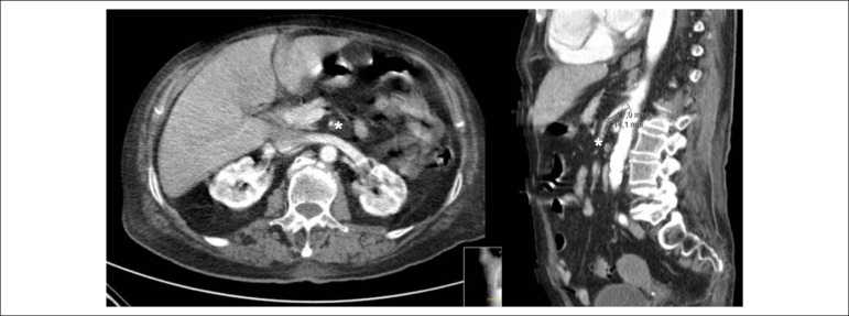
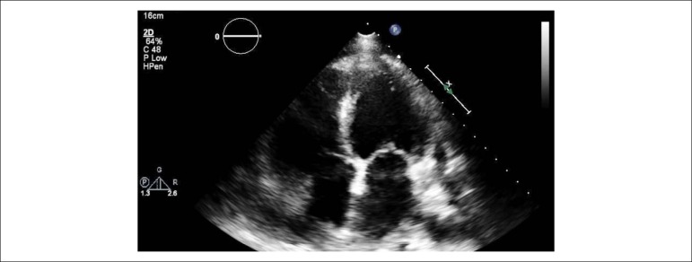
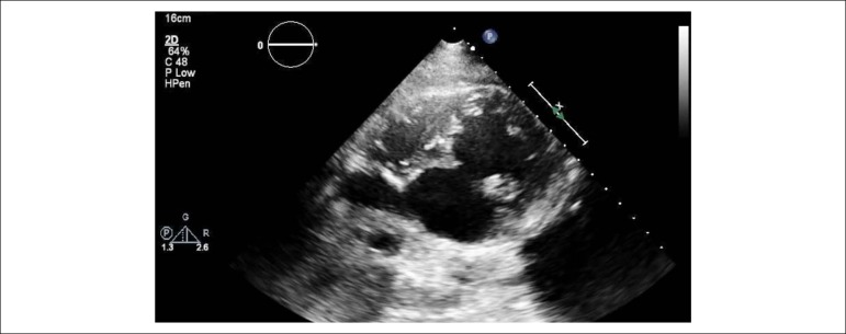
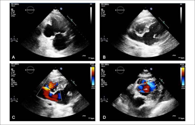
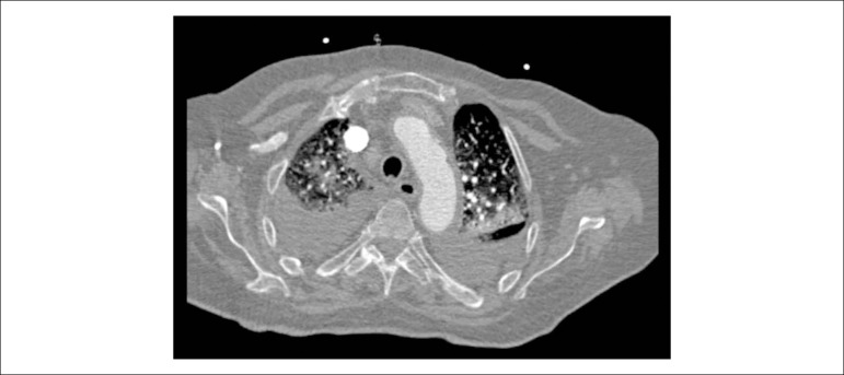
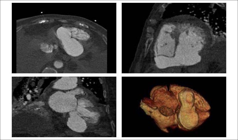
An 84-years-old woman with hypertension and dyslipidemia was admitted in the emergency room with acute abdominal pain; patient complained of chest pain three weeks before hospital admission. On physical examination, she was hypotensive, tachycardic (with arrhythmic pulse), tachypneic and with diffuse abdominal pain. Electrocardiogram showed atrial fibrillation with rapid ventricular response, complete left bundle branch block and inferior Q waves. Abdominal computed tomography (CT) revealed a thrombus in the superior mesenteric artery (Figure 1, white asterisk). The patient showed a clinical course with congestive heart failure and low cardiac output. Transthoracic echocardiogram (Videos 1-2) showed mild left ventricular dilatation with a mild dysfunction and a pseudoaneurysm of the basal half of the posterior and inferior walls with left-to-right shunt, confirmed by color Doppler imaging (Figure 2 A-D). Cardiac CT (Video 3) revealed contained myocardial rupture, located at the basal segments of the inferior and posterior septal walls, extending to the free wall of the right ventricle, forming a pseudo-cavity, which communicates with the true cavity of the right ventricle (Figure 3). Despite vasopressor and inotropic support and proposal for cardiac surgery, the patient had an unfavorable course.
Figure 1.
Abdominal computed tomography showing a thrombus in the superior mesenteric artery (white asterisk).
Video 1.
Transthoracic echocardiogram - apical windows. Link: http://publicacoes.cardiol.br/portal/abc/portugues/2020/v11402/dor-abdominal-uma-apresentacaoincomum-de-ruptura-miocardica.asp
Video 2.
Transthoracic echocardiogram - modified subcostal window. Link: http://publicacoes.cardiol.br/portal/abc/portugues/2020/v11402/dor-abdominal-umaapresentacao-incomum-de-ruptura-miocardica.asp
Figure 2.
Pseudoaneurysm of left ventricular inferior wall on transthoracic echocardiography (TTE), apical two-chamber view (A). Left-right shunt in the basal segment of interventricular septum (B, C and D).
Video 3.
Thoracic CT scan .Link: http://publicacoes.cardiol.br/portal/abc/portugues/2020/v11402/dor-abdominal-uma-apresentacao-incomum-de-ruptura-miocardica.asp
Figure 3.
Cardiac computerized tomography showing contained myocardial rupture forming a pseudocavity that communicates with the true cavity of the right ventricle
Myocardial rupture demands a prompt diagnosis.1 Occurrence of late myocardial infarction should raise suspicion and clinical signs may be atypical.2
This case illustrates an interesting entity - pseudoaneurysm, with left-to-right shunt and contained myocardial rupture, which extended to the right ventricle, leading to a dismal prognosis.
Footnotes
Sources of Funding
There were no external funding sources for this study.
Study Association
This study is not associated with any thesis or dissertation work.
Ethics approval and consent to participate
This article does not contain any studies with human participants or animals performed by any of the authors.
Contribuição dos autores
Conception and design of the research: Seabra D; Acquisition of data, analysis and interpretation of the data and writing of the manuscript: Seabra D, Neto A, Oliveira I; Critical revision of the manuscript for intellectual content: Seabra D, Santos RP, Azevedo J, Pinto P.
Potential Conflict of Interest
No potential conflict of interest relevant to this article was reported.
References
- 1.Durko A, Budde R, Geleijnse M, Kappetein A. Recognition, assessment and management of the mechanical complications of acute myocardial infarction. Heart. 2018;104(14):1216–1223. doi: 10.1136/heartjnl-2017-311473. [DOI] [PubMed] [Google Scholar]
- 2.Helmy TA, Nicholson WJ, Lick S, Uretsky BF. Contained myocardial rupture: a variant linking complete and incomplete rupture. Heart. 2005;91(2):e13. doi: 10.1136/hrt.2004.048082. [DOI] [PMC free article] [PubMed] [Google Scholar]


