Dear Sir,
Recently, a type of pneumonia caused by a novel Betacoronavirus, the 2019 novel coronavirus (2019-nCoV), has spread in China, mainly in Wuhan [1]. It is known that cases of this disease have also been identified in other provinces of China [2].
A 53-year-old man has lived in Wuhan for many years, and recently came to Guangzhou, China. He was admitted to the hospital with fever and cough in the last 2 days, and his highest body temperature was 38.3 °C before admission. The laboratory examinations showed decreased white blood cell count (2.50 × 10⁹/L) and elevated C-reactive protein (16.2 mg/L; normal range 0–10 mg/L).
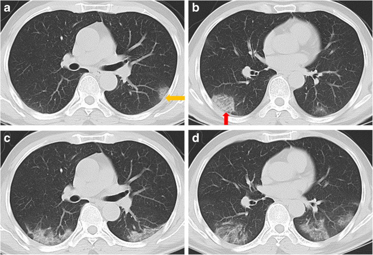
Non-contrast-enhanced chest computed tomography (CT) (Fig. 1a, b) showed multiple, bilateral, peripheral, patchy ground glass opacities, lobular, and subsegmental, with fading margins. The lesions had mainly a subpleural distribution.
Fig. 1.
CT scan of the patient. CT demonstrated slight thickening of the adjacent pleura (yellow arrow) and interlobular septa in the lesion (red arrow)
CT also demonstrated slight thickening of the adjacent pleura (yellow arrow) and interlobular septa in the lesion (red arrow). Subsequently, the patients had laboratory-confirmed 2019-nCoV infection by real-time polymerase chain reaction (PCR). After 5 days, CT scan (Fig. 1c, d) showed enlarged lesions, compared with previous images, and progressive ground glass opacities in both lower lobes, indicating disease progression.
Infection with 2019-nCoV was confirmed based on the patient’s epidemiological history, clinical manifestations, imaging characteristics, and laboratory tests.
Bilateral, multiple, patchy, or ground glass opacities, with subpleural distribution, were the lung CT appearances of this patient, affected by 2019-nCoV pneumonia. These characteristics should be recognized by professionals working in imaging departments.
Authors’ contributions
JL and LL designed the study. XX was a major contributor in writing the manuscript. LZ and CY revised it critically for important intellectual content. All authors have read and approved the manuscript.
Compliance with ethical standards
Conflict of interest
The authors declare that they have no conflicts of interest.
Ethics approval
Patient’s consent for publication of fully anonymized images was waived.
Footnotes
Please note that due to the time sensitive nature of the work presented in this article, standard peer-review has been bypassed to ensure rapid publication. The article has been directly assessed by the Editor-in-Chief.
This article is part of the Topical Collection on The Infection and Inflammation
Publisher’s note
Springer Nature remains neutral with regard to jurisdictional claims in published maps and institutional affiliations.
Contributor Information
Xi Xu, Email: 736461654@qq.com.
Chengcheng Yu, Email: 1515185140@qq.com.
Lieguang Zhang, Email: zhlieguang@126.com.
Liangping Luo, Email: tluolp@jnu.edu.cn.
Jinxin Liu, Email: Liujx83710378@126.com.
References
- 1.Zhu N, Zhang D, Wang W, Li X, Yang B, Song J, et al. A novel coronavirus from patients with pneumonia in China, 2019 [J]. N Engl J Med. 2020. 10.1056/NEJMoa2001017.
- 2.Huang C, Wang Y, Li X, Ren L, Zhao J, Hu Y, et al. Clinical features of patients infected with 2019 novel coronavirus in Wuhan, China [J]. Lancet. 2020. 10.1016/S0140-6736(20)30183-5. [DOI] [PMC free article] [PubMed]


