Abstract
Given that local cell-mediated immunity (CMI) against the human papillomavirus type 16 E6 (HPV16 E6) protein is important for eradication of HPV16 E6-expressing cancer cells in the cervical mucosa, the HPV16 E6 protein may be a target for the mucosal immunotherapy of cervical cancer. Here, we expressed the HPV16 E6 antigen on Lactobacillus casei (L. casei) and investigated E6-specific CMI following oral administration of the L. casei-PgsA-E6 to mice. Surface expression of HPV16 E6 antigens was confirmed and mice were orally inoculated with the L. casei-PgsA or the L. casei-PgsA-E6. Compared to the L. casei-PgsA-treated mice, significantly higher levels of serum IgG and mucosal IgA were observed in L. casei-PgsA-E6-immunized mice; these differences were significantly enhanced after boost. Consistent with this, systemic and local CMI were significantly increased after the boost, as shown by increased counts of IFN-γ-secreting cells in splenocytes, mesenteric lymph nodes (MLN), and vaginal samples. Furthermore, in the TC-1 tumor model, animals receiving the orally administered L. casei-PgsA-E6 showed reduced tumor size and increased survival rate versus mice receiving control (L. casei-PgsA) immunization. We also found that L. casei-PgsA-E6-induced antitumor effect was decreased by in vivo depletion of CD4+ or CD8+ T cells. Collectively, these results indicate that the oral administration of lactobacilli bearing the surface-displayed E6 protein induces T cell-mediated cellular immunity and antitumor effects in mice.
Keywords: HPV16 E6, Lactobacillus casei, Antitumor effect, PgsA, Cell-mediated immunity
Introduction
Cervical cancer is the second leading cause of cancer death among women worldwide [1], and human papillomavirus (HPV) infection is closely associated with cervical cancer [2–4]. More than 100 HPV genotypes have been identified [5], but the HPV type 16 (HPV16) has been associated with more than 50% of the HPV-related cervical cancer cases studied [6–8]. The E6 protein physically interacts with p53, leading to its dysfunction, while the E7 protein binds to the retinoblastoma protein (pRb) and activates cell cycle-associated genes [9–11]. Because the E6 and E7 proteins are consistently expressed in cervical cancer cells [12], these two oncoproteins represent attractive targets for developing vaccines and immunotherapeutic strategies against HPV-associated diseases. Recently, Nakagawa et al. [13–15] demonstrated the presence of E6-specific memory T cells in women whose HPV infection had been cleared and concluded that the cytotoxic T lymphocyte (CTL) response to HPV16 E6 antigens was more important than that to E7 antigens for controlling HPV16 infection. Thus, E6-specific CTL activity may be a major immune response for controlling persistent HPV16 infection.
Because HPV infects the epithelium of the lower female genital tract, where it can lead to cervical carcinoma [16], one strategy against cervical cancer might be to induce effective mucosal immunity with an aim toward inhibiting genital HPV transmission/infection and eradicating HPV-infected cells. Several studies have demonstrated that oral immunization with viral antigens can induce antigen-specific humoral and cellular immunity [17–21]. Oral administration is not only the way to increase mucosal immunity, but it is generally more convenient than parenteral administration. Lactic acid bacteria (LAB), which are relatively safe and exhibit an adjuvant effect in modulating the immune responses, may be a good delivery vehicle for mucosal immunization [22–24]. In addition, surface-displayed antigens on lactobacilli were reportedly efficient for inducing antigen presentation in dendritic cells [23], and surface localization of HPV16 antigens on lactococci was reported to induce greater cellular immunity compared to the intracellular or secreted forms [25]. Furthermore, we previously demonstrated that oral administration of viral antigens displayed on Lactobacillus casei (L. casei), such as the severe acute respiratory syndrome-associated coronavirus S protein and the HPV16 E7 protein, effectively induces antigen-specific neutralizing antibodies [18] and antitumor responses in mice [19]. In this study, we used the pgsA gene product, a transmembrane protein derived from the poly-γ-glutamic acid synthetase complex (the Pgs-BCA system) of Bacillus subtilis [26, 27], as a surface anchoring motif for surface display of antigens on L. casei. Here, we examined whether oral administration of E6 antigens expressed on L. casei could elicit E6-specific cellular immunity. The induction of effective immunity was assessed by production of antigen-specific serum IgG and mucosal IgA, and by quantification of antigen-specific IFN-γ-secreting CD8+ cells in splenocytes or local lymphocytes by the enzyme-linked immunospot (ELISPOT) assay. Finally, we tested the in vivo tumor regression effect of HPV16 E6-expressing L. casei in a mouse tumor model seeded with the E6-expressing tumor cell line (TC-1).
Materials and methods
Construction of HPV16 E6-expressing L. casei
We generated the expression vector, pKV:PgsA-E6 (Fig. 1a), encoding PgsA fused to the E6 protein of HPV type 16. The HCE promoter of pHAT:PgsA-E7 [20] was replaced with a promoter of the fructose-bisphosphate aldolase (Pald) gene originating from L. casei, and the E7 protein-encoding gene downstream of the poly-γ-glutamic acid synthetase A (PgsA) gene was replaced with that encoding the E6 protein of HPV16. Briefly, the gene encoding the E6 protein of HPV16, was amplified by reverse transcription-PCR from the CaSki cell line [28], using forward (5′-CAG GGA TCC ATG CAC CAA AAG AGA ACT GC-3′) and reverse (5′-CGC TCT AGA TCA TTA CAG CTG GGT TTC TCT ACG TG -3′) primers that contained BamHI and XbaI sites, respectively. The E7 gene of pHAT:PgsA-E7 was then replaced with the HPV16 E6-encoding fragment using these restriction enzyme sites. The plasmids were first established in E. coli JM83 and then transformed into L. casei BLS by electroporation. E. coli JM83 cells were grown in Luria–Bertani medium with erythromycin (150 μg/ml) at 37°C. Recombinant and expression host L. casei BLS cells were grown in MRS medium with erythromycin (16 μg/ml) at 30°C. The collected cells were washed with PBS and killed by boiling for 10 min followed by lyophilization.
Fig. 1.
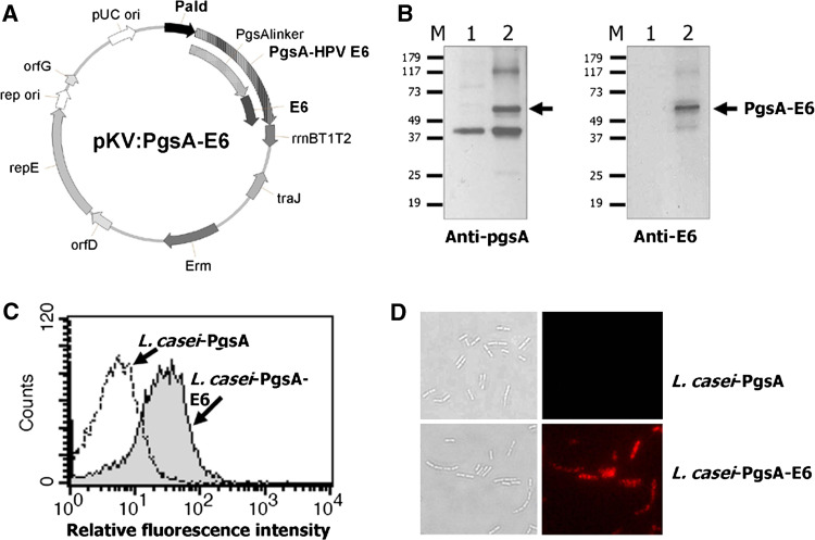
Surface expression of HPV16 E6 on L. casei. a A schematic diagram of the pKV-HPV16E6 plasmid used for HPV16 E6 expression on the surface of L. casei. b E6 protein expression in L. casei, as demonstrated by Western blot analysis using anti-pgsA3D (left) and anti-HPV16 E6 (right) polyclonal antibodies. M molecular weight markers, lane 1 L. casei-PgsA, lane 2 L. casei-PgsA-E6. A ~61-kDa protein band corresponding to the expected size of the PgsA-E6 fusion protein is present in lane 2. c FACS histogram of L. casei-PgsA (open) and L. casei-PgsA-E6 (filled) cells probed with polyclonal anti-HPV16 E6 antibodies, followed by an Alexa Fluor 594-conjugated anti-goat IgG antibody. d Representative immunofluorescence images of L. casei-PgsA and L. casei-PgsA-E6 (right) and corresponding differential interference contrast images (DIC) (left) are shown (×1,000)
Western blot analysis
Lactobacillus casei cells (2 × 10⁹ cells) were washed thrice with 700 μl of PBS and resuspended and sonicated in 500 μl of PBS containing 1 mM phenylmethylsulfonyl fluoride (PMSF). The samples were placed on ice (all subsequent steps were performed on ice), and 100 μl of cell lysate was mixed with 30 μl of 5× sample buffer and boiled for 10 min. The samples were centrifuged at 13,000 rpm for 3 min at 4°C, and the supernatants were resolved by 12% SDS-PAGE and transferred onto polyvinylidene fluoride (PVDF) membranes (Millipore, MA, USA). HPV16 E6 protein bands were detected by labeling with a polyclonal anti-pgsA antibody or a polyclonal goat anti-HPV16 E6 antibody (Santa Cruz Biotechnology, CA, USA), followed by the addition of horseradish peroxidase (HRP)-conjugated anti-goat IgG antibody (Cell Signaling Technology, MA, USA) diluted in 5% skim milk in PBS. The blots were visualized by chemiluminescence using an ECL Detection Kit (GE Healthcare, Uppsala, Sweden).
Flow cytometry and immunofluorescence microscopy
Lactobacillus casei cells (1 × 10⁸ cells) were harvested, washed twice with PBS, and resuspended in TE buffer containing polyclonal anti-HPV16 E6 antibody. The cell suspensions were incubated for 16 h at 4°C, washed twice with PBS and then incubated on ice for 2 h with Alexa Fluor 594-conjugated anti-goat IgG antibody (Molecular Probes, OR, USA) in TE buffer. The cells were then washed twice with PBS. After centrifugation, the cell pellets were resuspended in PBS and examined either with a BD FACSCalibur flow cytometer and analyzed by the CELLQuest software (Becton–Dickinson, CA, USA) or with a fluorescence microscope (Carl Zeiss MicroImaging Inc., NY, USA) and photographed with an Axiocam HRC (Carl Zeiss MicroImaging Inc.).
Immunization and sample collection
Female 6- to 8-week-old C57BL/6 mice were purchased from Dae Han BioLink (Chung-ju, Korea) and housed in the specific pathogen-free animal facility at the Korea Research Institute of Bioscience and Biotechnology (Daejeon, Korea). Recombinant L. casei cells constitutively expressing fusion proteins (PgsA or PgsA-E6) were harvested, washed thrice with PBS and resuspended in PBS at 109 cells/ml. Oral doses of 5 × 10⁹ of recombinant lactobacilli diluted in PBS (200 μl of the cell suspension) were administered daily via the intra-gastric route on days 0–4, 7–11, 21–25, and 35–39. Immune sera were taken at 14, 28, and 42 days and stored at −20°C until use. Intestinal lavages were obtained by washing intestine with 0.5 ml of ice-cold saline containing protease inhibitors; these samples were centrifuged at 13,000 rpm for 20 min at 4°C, and the supernatants were stored at −20°C until analysis. Blood was collected by eye bleeding, and spleens and mesenteric lymph nodes (MLNs) were harvested aseptically. Vaginal secretions were collected by washing the vaginal tracts with 250 μl of sterile PBS containing 1 mM PMSF; these samples were cleared by centrifugation at 13,000 rpm for removal of tissue and cellular debris and stored at −20°C until use. Vaginal lymphocytes were isolated as described by Dupuy et al. [29]. Briefly, mouse vaginas were excised, the cervix was removed, and the vaginal tissue was minced in Hank’s buffered salt solution (HBSS), washed 4 times with 1 mM EDTA in HBSS and digested for 1 h 37°C in RPMI 1640 (Invitrogen-GIBCO, CA, USA) supplemented with 5% (vol/vol) FBS, 1 mg/ml collagenase type 4, and 1 mg/ml dispase. The remaining cells were filtered through a sterile gauze mesh and washed with RPMI 1640 containing 10% FBS. Lymphocytes were purified using Ficoll-histopaque (Sigma–Aldrich, MO, USA). Purified vaginal lymphocytes from five mice were pooled for the IFN-γ ELISPOT analysis.
ELISA
For quantification of antigen-specific IgG and IgA antibody levels, a MaxiSorp 96-well plate (Nunc, Denmark) was incubated overnight at 4°C with purified His-HPV16 E6 (200 ng/well) in coating buffer (0.1 M sodium carbonate, 0.02% sodium azide, pH 9.6). For use as an ELISA coating antigen, the recombinant His-E6 protein was produced in E. coli. Briefly, the HPV16 E6 gene was inserted into the pET-28 vector (Novagen, Darmstadt, Germany) and transformed into E. coli. The induced His-E6 protein was isolated from cell lysates, purified using Ni-NTA affinity columns (QIAGEN, CA, USA) and used for coating the plate. The coated plate was blocked with 5% skim milk in PBS for 1 h at 37°C, washed twice with PBS containing 0.01% Tween-20, loaded with serially diluted serum samples for detecting IgG and tissue wash samples for detecting local IgA and incubated for 2 h at 37°C. HRP-conjugated goat anti-mouse IgG or IgA (Sigma–Aldrich) (1:5,000 in 3% skim milk in PBS) was added for detection of antigen-specific antibodies. Following incubation, plates were washed and TMB substrate reagent (Sigma–Aldrich) was added to the wells. After incubation for 20 min at room temperature in the dark, the reaction was stopped with 1 M sulfuric acid. Finally, OD450 was measured using an ELISA reader (Victor3, Perkin Elmer, USA).
IFN-γ ELISPOT
The IFN-γ ELISPOT assay was performed using a mouse IFN-γ ELISPOT kit (BD Biosciences, CA, USA), according to the manufacturer’s instructions. Briefly, 1 day before single cell preparation, anti-mouse IFN-γ capture antibody (5 μg/ml) in PBS was distributed (100 μl/well) to a BDTM ELISPOT 96-well plate, and the plate was incubated at 4°C overnight. After washing, 200 μl/well of blocking solution containing complete RPMI 1640 media was added, and the plate was incubated for 2 h. Freshly isolated lymphocytes were added at 2 × 10⁵ cells per well in a medium containing 10 μg/ml HPV16 E6 peptide (amino acid 48–57; containing the MHC class I epitope). The plate was incubated for 3 days, the cells were discarded, and the plate was sequentially treated with biotinylated anti-mouse IFN-γ antibody, streptavidin-HRP, and substrate solution. The plate was washed with deionized water and dried for at least 2 h in the dark. The spots were automatically counted using an ImunoScan Entry analyzer (Cellular Technology Ltd., OH, USA). As a negative control, the number of spot-forming cells (SFC) per 106 cells was counted in the absence of the peptides which was less than 50 SFC/106 cells.
Intracellular cytokine staining
Single cell suspensions were prepared from spleen and MLN. For experiments, 5 × 10⁶ cells were incubated in media containing 1 μg/ml of E6 48–57 peptide (EVYDFAFRDL) and 1 μl/ml GolgiPlug (BD Biosciences) at 37°C overnight. Cells were then washed and stained with phycoerythrin (PE)-conjugated monoclonal rat anti-mouse CD8 antibody (BD Biosciences), followed by the intracellular cytokine staining with FITC-conjugated IFN-γ antibody (BD Biosciences) using the Cytofix/Cytoperm kit (BD Biosciences), according to the manufacturer’s instructions. The fluorescence intensities were measured by the FACSCalibur flow cytometry and analyzed using the CELLQuest software (Becton–Dickinson).
In vivo tumor challenge experiment
For preventive experiments, C57BL/6 mice (eight per group) were orally administered with Lactobacillus expressing the HPV16 E6 antigen as described above. One week after first administration, C57BL/6 mice were challenged with TC-1 tumor cells (2 × 10⁴ cells per mouse) that had been co-transformed with the HPV16 E6 and E7 and activated by the ras oncogene [30]. The tumor challenge was performed by subcutaneous injection in the left inguinal region. To measure the therapeutic efficacy, oral administration of the recombinant L. casei-PgsA-E6 or L. casei-PgsA was initiated 3 days after TC-1 challenge on days 0–4 and 7–11, and the boost was performed on days 21–25 and 35–39. The TC-1 cells (kindly provided by T.C. Wu, Johns Hopkins University, Baltimore, MD) were cultured in RPMI-1640 supplemented with 10% (vol/vol) FBS (Hyclone, Logan, UT, USA), 1% antibiotics–antimycotics, and 2 mM nonessential amino acids at 37°C with 5% CO2. Tumor size was expressed as the tumor area (mm2) determined by monitoring 2 perpendicular diameters of the tumors 3 times a week using a caliper.
In vivo antibody depletion experiment
To examine the role of CD4+ and CD8+ T cells in the antitumor effects of L. casei-PgsA-E6, we performed in vivo antibody depletion as previously described [31], followed by tumor challenge. The anti-CD4 monoclonal antibody (MAb), GK1.5, and the anti-CD8 MAb, 2.43, were used for CD4+ and CD8+ T cell depletion, respectively. The initial depletion was performed by intraperitoneal injection with 100 μg of the MAb per day for 3 days. To check the role of T cells in the preventive antitumor model, the depletion was initiated simultaneously with the first oral administration of recombinant L. casei, which was a week before tumor challenge and the depletion continued to be done once a week for 5 weeks. For the therapeutic antitumor model, the initial depletion started 3 days before tumor challenge and the depletion continued to be done once a week for 5 weeks.
Statistical analysis
Statistical analyses were performed using Student’s t tests for data. A p value of <0.05 was taken as indicating a statistically significant difference.
Results
HPV16 E6 protein was expressed on L. casei-PgsA-E6
We first constructed a Lactobacillus expression vector, pKV:PgsA-E6, where PgsA was used as an anchoring protein for direct surface expression of the HPV16 E6 protein on L. casei (Fig. 1a). After transformation, L. casei cells harboring the pKV:PgsA or the pKV:PgsA-E6 were grown overnight at 30°C for induction of recombinant protein expression. To confirm proper expression of the HPV16 E6 protein, equal amounts of whole cell lysates from the recombinant L. casei harboring the pKV-PgsA or the pKV-PgsA-E6 were resolved by SDS-PAGE and subjected to Western blot analysis using both anti-HPV16 E6 and anti-PgsA antibodies (Fig. 1b). The lane containing the L. casei-PgsA-E6 (Fig. 1b, lane 2) contained a ~61 kDa immunoreactive band that most likely represents the fusion protein comprising the 17-kDa E6 protein and the 44-kDa PgsA surface display motif. A smaller band representing expression of the PgsA anchor protein was detected in L. casei-PgsA lysates (Fig. 1b, lane 1). The surface localization of the HPV16 E6 protein on L. casei was confirmed by flow cytometry and immunofluorescence microscopy, using a polyclonal anti-HPV16 E6 antibody and Alexa Fluor 594-conjugated anti-goat IgG antibody. FACS analysis revealed a significant increase in fluorescence intensity of the L. casei-PgsA-E6 [mean fluorescence intensity (MFI) = 33.42] compared to that of L. casei-PgsA (MFI = 4.16) (Fig. 1c). The immunofluorescence microscopy analysis showed that the L. casei-PgsA-E6 was immunostained positive for HPV16 E6, while the control L. casei-PgsA was not (Fig. 1d). These data demonstrate that our recombinant HPV16 E6 protein was efficiently expressed on the surface of L. casei-PgsA-E6.
Oral immunization with L. casei-PgsA-E6 induces E6-specific humoral immunity
To determine whether recombinant Lactobacilli possess the ability to induce antibody production to surface-displayed E6 antigens, female C57BL/6 mice were orally immunized with the L. casei-PgsA-E6 or the L. casei-PgsA (5 × 10⁹ CFU) on days 0–4 and 7–11 and then boosted on days 21–25 and 35–39. E6-specific serum IgG and mucosal IgA levels were determined on days 14, 28, and 42 by ELISA using the purified His-E6 protein as the coating antigen (Fig. 2). The mean E6-specific IgG log10 titer in sera from mice immunized with the L. casei-PgsA-E6 was 2.33 ± 0.21, compared with 1.14 ± 0.56 in serum samples from the L. casei-PgsA-immunized control group. On days 28 and 42, the mean log10 titers of the serum IgG in mice immunized orally with the L. casei-PgsA-E6 were 3.20 ± 0.43 and 3.67 ± 0.42, respectively, indicating that the mean titer of serum IgG in L. casei-PgsA-E6-immunized mice was increased ~7- and ~22-fold after the first and second boosts, respectively. In contrast, the IgG titers of L. casei-PgsA-immunized mice were not significantly increased on days 28 or 42 (Fig. 2a). These results suggest that a significant E6-specific humoral response was induced by oral immunization with the L. casei-PgsA-E6.
Fig. 2.
HPV16 E6-specific serum IgG and mucosal IgA production in mice following oral immunization with L. casei-PgsA-E6. a E6-specific serum IgG collected from mice orally immunized with L. casei-PgsA or L. casei-PgsA-E6 was determined by ELISA using recombinant His-E6 as a coating antigen. b E6-specific intestinal IgA and c vaginal IgA were determined in lavage fluids. Asterisks represent statistically significant differences relative to the L. casei-PgsA control (*p < 0.05; **p < 0.01). Data are given as mean ± SD of duplicate experiments
To examine E6-specific mucosal immune responses, we detected the production of mucosal IgA antibodies in intestinal and vaginal lavage fluids collected from orally immunized mice on days 14, 28, and 42 post-immunization (PI). The results revealed that mice immunized with the L. casei-PgsA-E6 developed significant IgA antibody levels in both the intestinal and vaginal mucosa (Fig. 2b, c). Compared with the mean optical densities (ODs) of intestinal IgA in the L. casei-PgsA-immunized mice, those in the L. casei-PgsA-E6-immunized mice were significantly higher (p < 0.01). The mean OD value of the intestinal fluids after immunization with the L. casei-PgsA-E6 was 0.28 ± 0.05, and it was significantly enhanced after the first and second boosts (0.41 ± 0.09 and 0.49 ± 0.18, respectively). In contrast, the level of intestinal IgA in the L. casei-PgsA-immunized mice was not significantly different following the initial immunization or the first and second boosts (Fig. 2b). On day 14 PI, the mean ODs of vaginal washes from L. casei-PgsA- or L. casei-PgsA-E6-immunized mice were similar (0.13 ± 0.02 vs. 0.18 ± 0.01, respectively). In contrast, compared to the L. casei-PgsA-immunized mice, those immunized with the L. casei-PgsA-E6 demonstrated higher E6-specific vaginal IgA responses following both the first (0.26 ± 0.05 vs. 0.14 ± 0.01) and second (0.34 ± 0.07 vs. 0.20 ± 0.03) boosts (Fig. 2c). The vaginal IgA levels in the L. casei-PgsA-E6-immunized mice were significantly increased after each boost (p < 0.05), but this response was not observed in the L. casei-PgsA-treated mice. These data indicate that both systemic (serum IgG) and mucosal (intestinal/vaginal IgA) E6-specific humoral immune responses were induced by oral administration of the L. casei-PgsA-E6.
Oral administration of L. casei-PgsA-E6 induces E6-specific cellular immune responses in spleen
To investigate E6-specific CMI responses generated by oral administration of the L. casei-PgsA-E6 in C57BL/6 mice, we performed flow cytometric analysis and ELISPOT assay. After vaccination with the L. casei-PgsA or the L. casei-PgsA-E6, murine splenocytes were prepared on days 14, 28, and 42 PI. The E6-specific IFN-γ-expressing CD8+ T cell response was observed after stimulation with the class I-restricted E6 48-57 peptide (EVYDFAFRDL). As shown in Fig. 3a, we observed a significantly higher number of E6-specific IFN-γ-expressing CD8+ T cells in mice immunized with the L. casei-PgsA-E6 compared to mice vaccinated with the same dose of the L. casei-PgsA. In the L. casei-PgsA-E6 vaccinated mice, the E6-specific IFN-γ-expressing CD8+ T cell response was increased approximately threefold after the first boost and fivefold after the second boost (p < 0.05); such effects were not seen in the L. casei-PgsA-immunized mice. As previously reported, the population of IFN-γ-expressing CD8 negative T cells was observed in response to E6 CTL peptide (Fig. 3a) [32, 33]. Next, E6-specific IFN-γ-secreting cells were evaluated in splenocytes from the L. casei-PgsA or the L. casei-PgsA-E6-immunized mice, using the ELSPOT assay. Consistent with the above findings, the L. casei-PgsA-E6-treated mice showed a significant CMI response (589.41 ± 114.59 spot-forming cells (SFC)/106 cells) after second boost (p < 0.01). In contrast, mice orally dosed with the L. casei-PgsA showed a moderate response (103.23 ± 57.32 SFC/106 cells) (Fig. 3b) that was similar to that seen in preimmunized mice (data not shown). Taken together, these results indicate that E6-specific CMI responses were induced in the splenocytes of mice orally immunized with the L. casei-PgsA-E6.
Fig. 3.
Systemic CMI response induced by oral administration of L. casei-PgsA-E6. a To evaluate the E6-specific IFN-γ+ CD8+ T cell response, splenocytes were collected from mice orally dosed with the L. casei-PgsA or the L. casei-PgsA-E6 and stimulated with 1 μg/ml of the class I-restricted E6 peptide (EVYDFAFRDL) and 1 μl/ml GolgiPlug. The fluorescence intensities were measured by FACSCalibur flow cytometry. b E6-specific IFN-γ-secreting splenic T cells stimulated with 1 μg/ml of the E6 peptide were measured by ELISPOT assay. Asterisks represent statistically significant differences relative to the L. casei-PgsA control (*p < 0.05; **p < 0.01). Data are given as mean ± SD of triplicate experiments
Oral administration of L. casei-PgsA-E6 induces mucosal E6-specific cellular immune responses
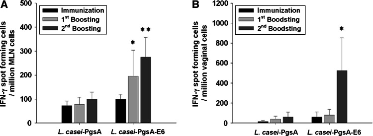
As the local E6-specific cellular response is important for elimination of HPV16-infected cells located in mucosa, we investigated local E6-specific CMI responses in mesenteric lymph nodes (MLN) and vaginal lymphocytes from mice orally treated with the L. casei-PgsA or the L. casei-PgsA-E6 (Fig. 4). By the ELISPOT assay, the number of E6-specific IFN-γ-secreting cells in MLN cells was increased approximately 2.6-fold after the second boost in the L. casei-PgsA-E6-vaccinated mice compared to the control mice (p < 0.01) (Fig. 4a). In contrast, no such effect was seen in the L. casei-PgsA-immunized mice. In vaginal lymphocytes, the number of E6-specific IFN-γ-secreting cells was increased approximately sevenfold after the second boost in the L. casei-PgsA-E6-vaccinated mice (p < 0.05) (Fig. 4b). These results indicate that oral immunization with the L. casei-PgsA-E6 appears to be a very effective method for inducing local CMI responses in the MLN and vagina of mice.
Fig. 4.
Mucosal CMI response induced by oral administration of L. casei-PgsA-E6. MLN cells and vaginal lymphocytes were isolated from L. casei-PgsA- or L. casei-PgsA-E6-immunized mice and examined for local E6-specific cellular responses. a MLN cells and b vaginal lymphocytes were analyzed for the presence of IFN-γ-secreting cells following stimulation with the E6 peptide, using an IFN-γ ELISPOT assay. Asterisks represent statistically significant differences relative to the control L. casei-PgsA (*p < 0.05; **p < 0.01)
Preventive effect of orally administered L. casei-PgsA-E6 on TC-1 tumor
To determine whether the orally administered L. casei-PgsA-E6 elicits an effective protective immunity against progressive tumor cells, C57BL/6 mice were orally treated with the L. casei-PgsA or the L. casei-PgsA-E6 on days 0–4 and 7–11, and boosted on days 21–25 and 35–39. On day 6, mice were challenged with TC-1 tumor cells (2 × 10⁴ cells per mouse) by subcutaneous injection in the left inguinal region (Fig. 5). All mice that had been orally dosed with the L. casei-PgsA developed tumors within 3 weeks after tumor injection. In contrast, 50% of mice immunized with the L. casei-PgsA-E6 remained tumor free at 5 weeks post-challenge, and 37.5% of this group remained tumor free at 40 days post-challenge (Fig. 5a). Thirty-seven days after tumor cell injection, the average tumor size (48.08 mm2) in the L. casei-PgsA-E6-immunized mice was significantly (p < 0.05) smaller than that (227.83 mm2) in the L. casei-PgsA-immunized mice (Fig. 5b). Consistent with this finding, all mice immunized with the L. casei-PgsA succumbed to their tumors within 62 days after tumor inoculation, whereas mice immunized with L. casei-PgsA-E6 exhibited 62.5% survival over the same time period and 12.5% survival 90 days after tumor inoculation (Fig. 5c). These results indicate that oral administration of the L. casei-PgsA-E6 elicits antitumor effects against E6-expressing TC-1 tumor cells in C57BL/6 mice.
Fig. 5.

Preventive effect of orally administered L. casei-PgsA-E6 on TC-1 tumor. C57BL/6 mice (eight per group) were orally treated with L. casei-PgsA or L. casei-PgsA-E6 and then injected s.c. with 2 × 10⁴ TC-1 tumor cells in the left inguinal region. a The percent rate of tumor-free mice, and b tumor size, and c survival rate of TC-1 tumor-bearing mice were checked. Tumor size was periodically monitored by measurement of two perpendicular diameters of the tumors, and the tumor size was expressed as the tumor area (mm2). Asterisks represent statistically significant differences relative to the L. casei-PgsA control (*p < 0.05; **p < 0.01)
Therapeutic effect of orally administered L. casei-PgsA-E6 on established TC-1 tumor
We further analyzed the therapeutic potential of the orally administered L. casei-PgsA-E6 in mice with established tumor. C57BL/6 mice were orally treated with the L. casei-PgsA or the L. casei-PgsA-E6 on day 3 after subcutaneous inoculation of TC-1 tumor cells. All mice that had been orally dosed with the L. casei-PgsA developed tumors within 3 weeks after tumor injection. In contrast, 40% of mice immunized with the L. casei-PgsA-E6 remained tumor free at 3 weeks after tumor injection and 10% of this group was still tumor free at 27 days after TC-1 tumor challenge (Fig. 6a). Thirty-four days after tumor cell injection, the average tumor size (289.1 mm2) in the L. casei-PgsA-E6-immunized mice was significantly (p < 0.01) smaller than that (494.4 mm2) in the L. casei-PgsA-immunized mice (Fig. 6b). Finally, none of mice immunized with the L. casei-PgsA survived at 48 days after tumor injection, whereas half of the mice immunized with the L. casei-PgsA-E6 survived; the remaining mice survived until 75 days after inoculation of the TC-1 tumor (Fig. 6c). Thus, in the therapy experiments, we observed that oral administration of the L. casei-PgsA-E6 elicits antitumor effects against E6-expressing TC-1 tumor cells in C57BL/6 mice.
Fig. 6.

Therapeutic effect of orally administered L. casei-PgsA-E6 on TC-1 tumor. TC-1 tumor cells (2 × 10⁴ cells) were injected subcutaneously into C57BL/6 mice (ten per group) 3 days before the oral immunization of the L. casei-PgsA or the L. casei-PgsA-E6. a The percent rate of tumor-free mice, and b tumor size, and c survival rate of TC-1 tumor-bearing mice were checked. Tumor size was periodically monitored by measurement of two perpendicular diameters of the tumor and expressed as the tumor area (mm2). Asterisks represent statistically significant differences relative to the L. casei-PgsA control (*p < 0.05; **p < 0.01)
The antitumor effects of L. casei-PgsA-E6 are dependent on CD4+ and CD8+ T cells
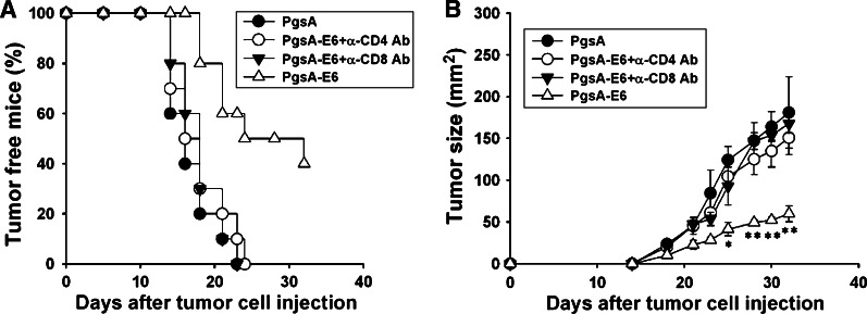
The cellular immunity is mediated by both CD4+ and CD8+ T cells, often through the action of secreted cytokines and cytolytic activity, respectively. To determine the subsets of T lymphocytes that are important for the antitumor effect against E6-expressing tumor cells, we performed in vivo antibody depletion experiments. Depletion of CD4+ or CD8+ T cells was accomplished by intraperitoneal injection of GK1.5 or 2.43 antibodies, respectively. As shown in Fig. 7, preventive antitumor effects were abrogated through either CD4+ or CD8+ T cells depletion in the L. casei-PgsA-E6-treated mice. In addition, in the therapeutic model, the L. casei-PgsA-E6-treated mice that had been depleted of CD4+ or CD8+ T cells prior to tumor challenge consistently developed tumor regardless of L. casei-PgsA-E6 treatment (Fig. 8). Taken together, these results indicated that both CD4+ and CD8+ T cells were essential for the antitumor effect generated by oral administration of the recombinant L. casei-PgsA-E6 against TC-1.
Fig. 7.
The depletion of CD4+ or CD8+ T cells eliminates the preventive antitumor effect of L. casei-PgsA-E6. In vivo antibody depletion experiments were performed to determine the effects of T lymphocyte subsets on the tumor protection of the L. casei-PgsA-E6. MAb GK1.5 was used for CD4 depletion, MAb 2.43 was used for CD8 depletion. CD4 or CD8 depletions were initiated a week before tumor challenge. a The percent rate of tumor-free mice, and b tumor size was checked. Tumor size was periodically monitored by measurement of two perpendicular diameters of the tumors, and expressed as the tumor area (mm2). Asterisks represent statistically significant differences relative to the control (*p < 0.05; **p < 0.01)
Fig. 8.

The depletion of CD4+ or CD8+ T cells eliminates the therapeutic antitumor effect of L. casei-PgsA-E6. In vivo depletion of CD4+ or CD8+ T cells (3 days before tumor challenge) was performed as mentioned in “Materials and methods”. a The percent rate of tumor-free mice, and b tumor size, and c survival rate of TC-1 tumor-bearing mice were examined. Tumor size was periodically monitored by measurement of two perpendicular diameters of the tumor and expressed as the tumor area (mm2). Asterisk represent statistically significant differences relative to the L. casei-PgsA control (*p < 0.05)
Discussion
Considering that cervical cancer occurs in the vaginal mucosa and has been highly associated with HPV16, a vaccine capable of inducing CMI against the HPV16 E6 antigen might prove effective for eradicating cervical tumor cells. Here, we developed an effective recombinant Lactobacillus-based oral vaccine and tested its ability to induce systemic and local immune responses and therapeutic effects against HPV16 E6-expressing tumor.
Mucosal (in this case, oral) immunization has several advantages over other routes of antigen delivery, including convenience, cost-effectiveness, and more importantly, induction of both local and systemic immune responses, as opposed to parenteral vaccines that only elicit systemic immune responses [34, 35]. For mucosal vaccination purposes, lactic acid bacteria are attractive delivery vehicles because they are considered safer than many other live vaccine carriers (e.g. Salmonella, E. coli, Vaccinia) [23]. We chose L. casei because it is a good agent for oral vaccination due to its high intrinsic adjuvant effect and ability to increase innate immunity [36, 37]. The L. casei strain we used as the vaccine vehicle in this study can activate immature human bone marrow dendritic cells through upregulation of IL-12 secretion and surface expression of costimulatory molecules (CD40) (unpublished data).
Cell-mediated immune responses play a crucial role in protecting the host from invading pathogens. In this study, we investigated the feasibility of targeting the HPV16 E6 oncoprotein for vaccine development to control HPV-associated lesions. We showed that oral treatment of mice with HPV16 E6 proteins displayed on L. casei could induce E6-specific serum IgG and mucosal IgA production, and E6-specific T cell responses in both the systemic and mucosal immune systems. There was a significant increase of E6-specific IFN-γ-secreting cells in vaginal lymphocytes from L. casei-PgsA-E6-immunized mice only after the second boost. The mucosal immune system has both inductive and effector sites [35]. This vaccine was orally administered, meaning that its inductive site was mainly in the intestinal regions, which is somewhat distant from the effector site (vaginal region). Therefore, it is possible that the CMI response at this distant effector site could be delayed. However, we found that the number of E6-specific IFN-γ-secreting cells in the L. casei-PgsA-E6-immunized mice was significantly higher than that in the L. casei-PgsA-immunized mice. More importantly, in an in vivo mouse tumor model, the orally administered L. casei-PgsA-E6 showed antitumor effects against HPV16 E6-expressing murine tumors, as evidenced by reduced tumor size and increased survival rate versus mice receiving control (L. casei-PgsA) immunizations. In accordance with the observation, in vivo depletion of CD4+ or CD8+ T cells by MAb GK1.5 or MAb 2.43 completely abrogated anti-TC1 tumor activity in mice orally administration with the L. casei-PgsA-E6. These results showed that both CD4+ and CD8+ T cells were essential for antitumor immunity against E6-expressing tumor. The human clinical study using L. casei expressing HPV16 E7 on patients of cervical intraepithelial neoplasia 3 (CIN3) to check the safety and efficacy is currently in progress. The additional administration of recombinant L. casei expressing HPV16 E6 may enhance its therapeutic efficacy. Collectively, our findings suggest that recombinant L. casei expressing HPV16 E6 antigens may be a promising therapeutic vaccine candidate against cervical cancer. In addition, our surface display system could be used to design vaccines against other mucosally transmitted pathogens.
Acknowledgments
This work was supported by grants of the Korea Health 21 R&D Project (A050562) and National R&D Program for Cancer Control (0720510), Ministry of Health and Welfare, Republic of Korea and a grant from KRIBB Initiative program to H. Poo.
Abbreviations
- HPV
Human papillomavirus
- PgsA
Poly-gamma-glutamic acid synthetase complex A
- IFN-γ
Interferon gamma
- ELISA
Enzyme-linked immunosorbent assay
- ELISPOT
Enzyme-linked immunospot
- SD
Standard deviation
References
- 1.Walboomers JM, Jacobs MV, Manos MM, Bosch FX, Kummer JA, Shah KV, Snijders PJ, Peto J, Meijer CJ, Munoz N. Human papillomavirus is a necessary cause of invasive cervical cancer worldwide. J Pathol. 1999;189:12–19. doi: 10.1002/(SICI)1096-9896(199909)189:1<12::AID-PATH431>3.0.CO;2-F. [DOI] [PubMed] [Google Scholar]
- 2.zur Hausen H. Papillomavirus infections—a major cause of human cancers. Biochim Biophys Acta. 1996;1288:F55–F78. doi: 10.1016/0304-419x(96)00020-0. [DOI] [PubMed] [Google Scholar]
- 3.zur Hausen H. Papillomaviruses causing cancer: evasion from host-cell control in early events in carcinogenesis. J Natl Cancer Inst. 2000;92:690–698. doi: 10.1093/jnci/92.9.690. [DOI] [PubMed] [Google Scholar]
- 4.Parkin DM, Bray F, Ferlay J, Pisani P. Estimating the world cancer burden: Globocan 2000. Int J Cancer. 2001;94:153–156. doi: 10.1002/ijc.1440. [DOI] [PubMed] [Google Scholar]
- 5.de Villiers EM. Papillomavirus and HPV typing. Clin Dermatol. 1997;15:199–206. doi: 10.1016/S0738-081X(96)00164-2. [DOI] [PubMed] [Google Scholar]
- 6.Bosch FX, Manos MM, Munoz N, Sherman M, Jansen AM, Peto J, Schiffman MH, Moreno V, Kurman R, Shah KV. Prevalence of human papillomavirus in cervical cancer: a worldwide perspective. International biological study on cervical cancer (IBSCC) Study Group. J Natl Cancer Inst. 1995;87:796–802. doi: 10.1093/jnci/87.11.796. [DOI] [PubMed] [Google Scholar]
- 7.Munoz N, Bosch FX, de Sanjose S, Herrero R, Castellsague X, Shah KV, Snijders PJ, Meijer CJ. Epidemiologic classification of human papillomavirus types associated with cervical cancer. N Engl J Med. 2003;348:518–527. doi: 10.1056/NEJMoa021641. [DOI] [PubMed] [Google Scholar]
- 8.Matsukura T, Sugase M. Identification of genital human papillomaviruses in cervical biopsy specimens: segregation of specific virus types in specific clinicopathologic lesions. Int J Cancer. 1995;61:13–22. doi: 10.1002/ijc.2910610104. [DOI] [PubMed] [Google Scholar]
- 9.Crook T, Tidy JA, Vousden KH. Degradation of p53 can be targeted by HPV E6 sequences distinct from those required for p53 binding and trans-activation. Cell. 1991;67:547–556. doi: 10.1016/0092-8674(91)90529-8. [DOI] [PubMed] [Google Scholar]
- 10.Heck DV, Yee CL, Howley PM, Munger K. Efficiency of binding the retinoblastoma protein correlates with the transforming capacity of the E7 oncoproteins of the human papillomaviruses. Proc Natl Acad Sci USA. 1992;89:4442–4446. doi: 10.1073/pnas.89.10.4442. [DOI] [PMC free article] [PubMed] [Google Scholar]
- 11.Doorbar J. The papillomavirus life cycle. J Clin Virol. 2005;32(Suppl 1):S7–S15. doi: 10.1016/j.jcv.2004.12.006. [DOI] [PubMed] [Google Scholar]
- 12.Ling M, Kanayama M, Roden R, Wu TC. Preventive and therapeutic vaccines for human papillomavirus-associated cervical cancers. J Biomed Sci. 2000;7:341–356. doi: 10.1007/BF02255810. [DOI] [PubMed] [Google Scholar]
- 13.Nakagawa M, Stites DP, Patel S, Farhat S, Scott M, Hills NK, Palefsky JM, Moscicki AB. Persistence of human papillomavirus type 16 infection is associated with lack of cytotoxic T lymphocyte response to the E6 antigens. J Infect Dis. 2000;182:595–598. doi: 10.1086/315706. [DOI] [PubMed] [Google Scholar]
- 14.Nakagawa M, Kim KH, Moscicki AB. Patterns of CD8 T-cell epitopes within the human papillomavirus type 16 (HPV 16) E6 protein among young women whose HPV 16 infection has become undetectable. Clin Diagn Lab Immunol. 2005;12:1003–1005. doi: 10.1128/CDLI.12.8.1003-1005.2005. [DOI] [PMC free article] [PubMed] [Google Scholar]
- 15.Wang X, Moscicki AB, Tsang L, Brockman A, Nakagawa M. Memory T cells specific for novel human papillomavirus type 16 (HPV16) E6 epitopes in women whose HPV16 infection has become undetectable. Clin Vaccine Immunol. 2008;15:937–945. doi: 10.1128/CVI.00404-07. [DOI] [PMC free article] [PubMed] [Google Scholar]
- 16.Tjiong MY, Out TA, Ter Schegget J, Burger MP, Van Der Vange N. Epidemiologic and mucosal immunologic aspects of HPV infection and HPV-related cervical neoplasia in the lower female genital tract: a review. Int J Gynecol Cancer. 2001;11:9–17. doi: 10.1046/j.1525-1438.2001.011001009.x. [DOI] [PubMed] [Google Scholar]
- 17.Lin CW, Lee JY, Tsao YP, Shen CP, Lai HC, Chen SL. Oral vaccination with recombinant Listeria monocytogenes expressing human papillomavirus type 16 E7 can cause tumor growth in mice to regress. Int J Cancer. 2002;102:629–637. doi: 10.1002/ijc.10759. [DOI] [PubMed] [Google Scholar]
- 18.Nardelli-Haefliger D, Roden RB, Benyacoub J, Sahli R, Kraehenbuhl JP, Schiller JT, Lachat P, Potts A, De Grandi P. Human papillomavirus type 16 virus-like particles expressed in attenuated Salmonella typhimurium elicit mucosal and systemic neutralizing antibodies in mice. Infect Immun. 1997;65:3328–3336. doi: 10.1128/iai.65.8.3328-3336.1997. [DOI] [PMC free article] [PubMed] [Google Scholar]
- 19.Lee JS, Poo H, Han DP, Hong SP, Kim K, Cho MW, Kim E, Sung MH, Kim CJ. Mucosal immunization with surface-displayed severe acute respiratory syndrome coronavirus spike protein on Lactobacillus casei induces neutralizing antibodies in mice. J Virol. 2006;80:4079–4087. doi: 10.1128/JVI.80.8.4079-4087.2006. [DOI] [PMC free article] [PubMed] [Google Scholar]
- 20.Poo H, Pyo HM, Lee TY, Yoon SW, Lee JS, Kim CJ, Sung MH, Lee SH. Oral administration of human papillomavirus type 16 E7 displayed on Lactobacillus casei induces E7-specific antitumor effects in C57BL/6 mice. Int J Cancer. 2006;119:1702–1709. doi: 10.1002/ijc.22035. [DOI] [PubMed] [Google Scholar]
- 21.Santi L, Batchelor L, Huang Z, Hjelm B, Kilbourne J, Arntzen CJ, Chen Q, Mason HS. An efficient plant viral expression system generating orally immunogenic Norwalk virus-like particles. Vaccine. 2008;26:1846–1854. doi: 10.1016/j.vaccine.2008.01.053. [DOI] [PMC free article] [PubMed] [Google Scholar]
- 22.Thones N, Muller M. Oral immunization with different assembly forms of the HPV 16 major capsid protein L1 induces neutralizing antibodies and cytotoxic T-lymphocytes. Virology. 2007;369:375–388. doi: 10.1016/j.virol.2007.08.004. [DOI] [PubMed] [Google Scholar]
- 23.Hanniffy S, Wiedermann U, Repa A, Mercenier A, Daniel C, Fioramonti J, Tlaskolova H, Kozakova H, Israelsen H, Madsen S, Vrang A, Hols P, Delcour J, Bron P, Kleerebezem M, Wells J. Potential and opportunities for use of recombinant lactic acid bacteria in human health. Adv Appl Microbiol. 2004;56:1–64. doi: 10.1016/S0065-2164(04)56001-X. [DOI] [PubMed] [Google Scholar]
- 24.Seegers JF. Lactobacilli as live vaccine delivery vectors: progress and prospects. Trends Biotechnol. 2002;20:508–515. doi: 10.1016/S0167-7799(02)02075-9. [DOI] [PubMed] [Google Scholar]
- 25.Perdigon G, Maldonado Galdeano C, Valdez JC, Medici M. Interaction of lactic acid bacteria with the gut immune system. Eur J Clin Nutr. 2002;56(Suppl 4):S21–S26. doi: 10.1038/sj.ejcn.1601658. [DOI] [PubMed] [Google Scholar]
- 26.Ashiuchi M, Nawa C, Kamei T, Song JJ, Hong SP, Sung MH, Soda K, Misono H. Physiological and biochemical characteristics of poly gamma-glutamate synthetase complex of Bacillus subtilis . Eur J Biochem. 2001;268:5321–5328. doi: 10.1046/j.0014-2956.2001.02475.x. [DOI] [PubMed] [Google Scholar]
- 27.Ashiuchi M, Soda K, Misono H. A poly-gamma-glutamate synthetic system of Bacillus subtilis IFO 3336: gene cloning and biochemical analysis of poly-gamma-glutamate produced by Escherichia coli clone cells. Biochem Biophys Res Commun. 1999;263:6–12. doi: 10.1006/bbrc.1999.1298. [DOI] [PubMed] [Google Scholar]
- 28.Kim TY, Myoung HJ, Kim JH, Moon IS, Kim TG, Ahn WS, Sin JI. Both E7 and CpG-oligodeoxynucleotide are required for protective immunity against challenge with human papillomavirus 16 (E6/E7) immortalized tumor cells: involvement of CD4+ and CD8+ T cells in protection. Cancer Res. 2002;62:7234–7240. [PubMed] [Google Scholar]
- 29.Dupuy C, Buzoni-Gatel D, Touze A, Bout D, Coursaget P. Nasal immunization of mice with human papillomavirus type 16 (HPV-16) virus-like particles or with the HPV-16 L1 gene elicits specific cytotoxic T lymphocytes in vaginal draining lymph nodes. J Virol. 1999;73:9063–9071. doi: 10.1128/jvi.73.11.9063-9071.1999. [DOI] [PMC free article] [PubMed] [Google Scholar]
- 30.Lin KY, Guarnieri FG, Staveley-O’Carroll KF, Levitsky HI, August JT, Pardoll DM, Wu TC. Treatment of established tumors with a novel vaccine that enhances major histocompatibility class II presentation of tumor antigen. Cancer Res. 1996;56:21–26. [PubMed] [Google Scholar]
- 31.Kim TW, Lee TY, Bae HC, Hahm JH, Kim YH, Park C, Kang TH, Kim CJ, Sung MH, Poo H. Oral administration of high molecular mass poly-gamma-glutamate induces NK cell-mediated antitumor immunity. J Immunol. 2007;179:775–780. doi: 10.4049/jimmunol.179.2.775. [DOI] [PubMed] [Google Scholar]
- 32.Peng S, Ji H, Trimble C, He L, Tsai YC, Yeatermeyer J, Boyd DA, Hung CF, Wu TC. Development of a DNA vaccine targeting human papillomavirus type 16 oncoprotein E6. J Virol. 2004;78:8468–8476. doi: 10.1128/JVI.78.16.8468-8476.2004. [DOI] [PMC free article] [PubMed] [Google Scholar]
- 33.Manuri PR, Nehete B, Nehete PN, Reisenauer R, Wardell S, Courtney AN, Gambhira R, Lomada D, Chopra AK, Sastry KJ. Intranasal immunization with synthetic peptides corresponding to the E6 and E7 oncoproteins of human papillomavirus type 16 induces systemic and mucosal cellular immune responses and tumor protection. Vaccine. 2007;25:3302–3310. doi: 10.1016/j.vaccine.2007.01.010. [DOI] [PMC free article] [PubMed] [Google Scholar]
- 34.Frazer I. Vaccines for papillomavirus infection. Virus Res. 2002;89:271–274. doi: 10.1016/S0168-1702(02)00195-8. [DOI] [PubMed] [Google Scholar]
- 35.Neutra MR, Kozlowski PA. Mucosal vaccines: the promise and the challenge. Nat Rev Immunol. 2006;6:148–158. doi: 10.1038/nri1777. [DOI] [PubMed] [Google Scholar]
- 36.Vitini E, Alvarez S, Medina M, Medici M, de Budeguer MV, Perdigon G. Gut mucosal immunostimulation by lactic acid bacteria. Biocell. 2000;24:223–232. [PubMed] [Google Scholar]
- 37.Galdeano CM, Perdigon G. The probiotic bacterium Lactobacillus casei induces activation of the gut mucosal immune system through innate immunity. Clin Vaccine Immunol. 2006;13:219–226. doi: 10.1128/CVI.13.2.219-226.2006. [DOI] [PMC free article] [PubMed] [Google Scholar]


