Currently, there is no commercially available vaccine against African swine fever. Outbreaks of this disease are devastating the swine industry from Central Europe to East Asia, and they are being caused by circulating strains of African swine fever virus derived from the Georgia 2007 isolate. Here, we report the discovery of a previously uncharacterized virus gene, which when deleted completely attenuates the Georgia isolate. Importantly, animals infected with this genetically modified virus were protected from developing ASF after challenge with the virulent parental virus. Interestingly, ASFV-G-ΔI177L confers protection even at low doses (102 HAD50) and remains completely attenuated when inoculated at high doses (106 HAD50), demonstrating its potential as a safe vaccine candidate. At medium or higher doses (104 HAD50), sterile immunity is achieved. Therefore, ASFV-G-ΔI177L is a novel efficacious experimental ASF vaccine protecting pigs from the epidemiologically relevant ASFV Georgia isolate.
KEYWORDS: ASF, ASFV, African swine fever virus, vaccine
ABSTRACT
African swine fever virus (ASFV) is the etiological agent of a contagious and often lethal disease of domestic pigs that has significant economic consequences for the swine industry. The disease is devastating the swine industry in Central Europe and East Asia, with current outbreaks caused by circulating strains of ASFV derived from the 2007 Georgia isolate (ASFV-G), a genotype II ASFV. In the absence of any available vaccines, African swine fever (ASF) outbreak containment relies on the control and culling of infected animals. Limited cross-protection studies suggest that in order to ensure a vaccine is effective, it must be derived from the current outbreak strain or at the very least from an isolate with the same genotype. Here, we report the discovery that the deletion of a previously uncharacterized gene, I177L, from the highly virulent ASFV-G produces complete virus attenuation in swine. Animals inoculated intramuscularly with the virus lacking the I177L gene, ASFV-G-ΔI177L, at a dose range of 102 to 106 50% hemadsorbing doses (HAD50), remained clinically normal during the 28-day observational period. All ASFV-G-ΔI177L-infected animals had low viremia titers, showed no virus shedding, and developed a strong virus-specific antibody response; importantly, they were protected when challenged with the virulent parental strain ASFV-G. ASFV-G-ΔI177L is one of the few experimental vaccine candidate virus strains reported to be able to induce protection against the ASFV Georgia isolate, and it is the first vaccine capable of inducing sterile immunity against the current ASFV strain responsible for recent outbreaks.
IMPORTANCE Currently, there is no commercially available vaccine against African swine fever. Outbreaks of this disease are devastating the swine industry from Central Europe to East Asia, and they are being caused by circulating strains of African swine fever virus derived from the Georgia 2007 isolate. Here, we report the discovery of a previously uncharacterized virus gene, which when deleted completely attenuates the Georgia isolate. Importantly, animals infected with this genetically modified virus were protected from developing ASF after challenge with the virulent parental virus. Interestingly, ASFV-G-ΔI177L confers protection even at low doses (102 HAD50) and remains completely attenuated when inoculated at high doses (106 HAD50), demonstrating its potential as a safe vaccine candidate. At medium or higher doses (104 HAD50), sterile immunity is achieved. Therefore, ASFV-G-ΔI177L is a novel efficacious experimental ASF vaccine protecting pigs from the epidemiologically relevant ASFV Georgia isolate.
INTRODUCTION
African swine fever (ASF) is a contagious and often fatal viral disease of swine. The causative agent, ASF virus (ASFV), is a large enveloped virus containing a double-stranded DNA (dsDNA) genome of approximately 190 kbp (1). ASFV shares aspects of its genome structure and replication strategy with other large dsDNA viruses, including those of the Poxviridae, Iridoviridae, and Phycodnaviridae (2). However, on a protein or amino acid level, there is little homology with the majority of the viral proteins, and very few ASFV proteins have been evaluated for their functionality or for their contribution to virus pathogenesis.
Currently, ASF is endemic in more than 20 sub-Saharan African countries. In Europe, ASF is endemic on the island of Sardinia (Italy), and outbreaks in additional countries began with an outbreak in the Caucasus region in 2007, affecting Georgia, Armenia, Azerbaijan, and Russia. ASF has continued to spread uncontrollably across Eurasia, with ASFV outbreaks occurring in 2018 to 2019 in China, Mongolia, Vietnam, Laos, Cambodia, Serbia, Myanmar, North Korea, and the Philippines. ASF has also spread to wild boar in Belgium but has been restricted to a quarantine zone since the first introduction of the disease in 2018. Sequencing of several contemporary epidemic ASFVs suggests high nucleotide similarity with only minor modifications compared to the initial 2007 outbreak strain, ASFV Georgia 2007/1, a highly virulent isolate that belongs to the ASFV genotype II group (3).
There is no vaccine available for ASFV, and disease outbreaks are currently quelled by animal quarantine and slaughter. Attempts to vaccinate animals using infected cell extracts, supernatants of infected pig peripheral blood leukocytes, purified and inactivated virions, infected glutaraldehyde-fixed macrophages, or detergent-treated infected alveolar macrophages failed to induce protective immunity (4–7). Protective immunity develops in pigs surviving viral infection with moderately virulent or attenuated variants of ASFV, with long-term resistance to homologous, but rarely to heterologous, virus challenge (8, 9). Significantly, pigs immunized with live attenuated ASF viruses containing genetically engineered deletions of specific ASFV virulence-associated genes are protected when challenged with homologous parental virus (10–15). These observations constitute the only experimental evidence describing the rational development of an effective live attenuated vaccine against ASFV.
Here, we report the discovery that deletion of a previously uncharacterized gene, I177L, from the highly virulent ASFV Georgia isolate (ASFV-G) results in complete attenuation in swine. Animals inoculated with the virus lacking the I177L gene, ASFV-G-ΔI177L, remained clinically normal and developed a strong virus-specific antibody response, and, importantly, ASFV-G-ΔI177L-infected swine were completely protected when challenged with the virulent parental ASFV-G.
RESULTS
Conservation of I177L gene across different ASFV isolates.
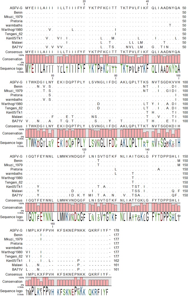
The ASFV-G open reading frame (ORF) I177L encodes a 177-amino-acid protein and is positioned on the reverse strand between nucleotide positions 174471 and 175004 of the ASFV-G genome (Fig. 1). The degree of I177L conservation among ASFV isolates was examined by multiple-sequence alignment using the CLC Genomics Workbench software (CLC bio, Aarhus, Denmark). The ASFV I177L protein sequences were derived from all sequenced isolates of ASFV representing African, European, and Caribbean isolates from domestic pig, wild pig, and tick sources. I177L has a predicted protein length of 161 to 177 amino acid residues depending on the isolate. Most of the isolates contain a protein with a length of 177 amino acids, with a few isolates showing a truncated C terminus that yield a protein of 161 to 162 amino acids. It is predicted that I177L contains a possible N-terminal transmembrane helix (data not shown). I177L is sometimes annotated in recent isolates using a different start codon that occurs at position 112; however, this has recently been shown to be a sequencing mistake in the annotation of these genomes (16). Il177L at the amino acid level revealed a very high degree of amino acid identity among isolates compared to isolates containing the same or different forms of I177L (Fig. 1). It should be mentioned that the ASFV-G I177L gene is identical to those recently reported for virus isolates from Belgium and Estonia (GenBank accession numbers LR536725.1 and LS478113.1). In addition, the I177L gene sequence in an ASFV Benin isolate was identical to the gene sequence of isolates from Italy, OURT, NHV, L60, and E75, and the gene in BA71V was identical to isolates from Kenya and Uganda (GenBank accession numbers AM712240.1, KM262846, KM262844, FN557520.1, NC_001659.2, AY261360, and MH025917.1).
FIG 1.
Multiple-sequence alignment of the indicated ASFV isolates of viral protein I177L, with matching residues represented as periods and gaps in the sequence represented by dashes. The degree of conservation between the sequences is represented below the sequences.
I177L gene is transcribed as a late gene during the virus replication cycle.
The time of transcription of the I177L gene was determined by microarray evaluating total RNA extracted from primary swine macrophage cell cultures infected with ASFV-G at 3, 6, 9, 12, 15, and 18 hours postinfection (hpi) (representing approximately one life cycle of ASFV replication).
Figure 2 shows the microarray signal intensities of three ASFV open reading frames at the six time points sampled. The CP204L gene (encoding ASFV protein p30) was expressed at approximate 41,000 photons per pixel at 3 hpi, which agrees with its known early gene expression after ASFV infection. The expression gradually decreased at 6 and 9 hpi and then significantly increased by more than 9-fold, reaching a plateau at 12 hpi. In contrast, B646L, a p72 virus capsid protein gene known for its late expression, was practically not expressed at 3 hpi, being at a level of <20 photons per pixel. p72 expression significantly increased to >44,000 at 6 hpi and reached a plateau at 12 hpi. I177L appears to be a late-expressed gene, much like B646L. The I177L gene was transcribed at less than 50 photons per pixel at 3 hpi. Unlike the p72 gene, I177L expression increased linearly at a much lower rate, and its expression remained low at 18 hpi. No expression was observed for any of the valuated genes in mock control samples taken at the indicated time points.
FIG 2.
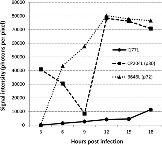
Time course of I177L gene transcriptional activity. Shown are averaged microarray signal intensities (photons per pixel) of ASFV I177L, CP204L, and B646L open reading frame RNA prepared from ex vivo pig macrophages infected with ASFV at 3, 6, 9, 12, 15, and 18 h postinfection.
Development of the I177L gene deletion mutant of the ASFV-Georgia isolate.
To determine the role of I177L during ASFV infection in cell cultures and virulence in swine, a recombinant virus lacking the I177L gene was designed (ASFV-G-ΔI177L). ASFV-G-ΔI177L was constructed from the highly pathogenic ASFV Georgia 2010 (ASFV-G) isolate by homologous recombination procedures as described in Materials and Methods. The I177L gene was replaced by a cassette containing the fluorescent gene mCherry under the ASFV p72 promoter (Fig. 3). Recombinant virus was selected after 10 rounds of limiting dilution purification based on the fluorescent activity. The virus population obtained from the last round of purification was amplified in primary swine macrophage cell cultures to obtain a virus stock.
FIG 3.
Diagram indicating the position of the I177L open reading frame in the ASFV-G genome (top). A donor plasmid with arms homologous to ASFV-G and the mCherry under the control of the p72 promoter was used to introduce the final genomic changes (bottom) to create ASFV-Georgia-ΔI177L, where the sequence of the donor plasmid mCherry reporter (red) is introduced to replace the ORF of I177L, as indicated.
To evaluate the accuracy of the genetic modification, the integrity of the genome, and the purity of the recombinant virus stock, full-genome sequences of ASFV-G-ΔI177L and parental ASFV-G were obtained using next-generation sequencing (NGS) for comparison. The DNA sequence assemblies of ASFV-G-ΔI177L and ASFV-G revealed a deletion of 112 nucleotides (between nucleotide positions 174530 and 174671) from the I177L gene corresponding with the introduced modification. The consensus sequence of the ASFV-G-ΔI177L genome showed an insertion of 3,944 nucleotides corresponding to the p72mCherryΔI177L cassette sequence introduced within the 112-nucleotide deletion in the I177L gene. Besides the designed changes, no unwanted additional mutations were detected in the rest of the ASFV-G-ΔI177L genome.
Replication of ASFV-G-ΔI177L in primary swine macrophages.
In vitro growth characteristics of ASFV-G-ΔI177L were evaluated in primary swine macrophage cell cultures, the primary cell targeted by ASFV during infection in swine, and compared to those of parental ASFV-G in multistep growth curves (Fig. 4). Cell cultures were infected at a multiplicity of infection (MOI) of 0.01, and samples were collected at 2, 24, 48, 72, and 96 h postinfection (hpi). The results demonstrated that ASFV-G-ΔI177L displayed a decreased growth kinetic compared to that of parental ASFV-G. ASFV-G-ΔI177L yields are approximately 100- to 1,000-fold lower than those of ASFV-G depending on the time point considered. Therefore, deletion of the I177L gene decreased the ability of ASFV-G-ΔI177L, relative to the parental ASFV-G isolate, to replicate in vitro in primary swine macrophage cell cultures.
FIG 4.
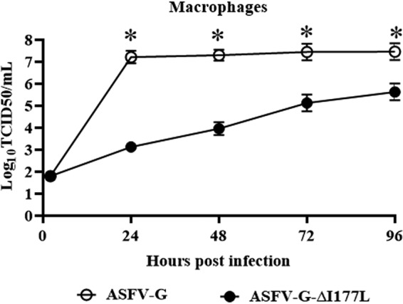
In vitro growth characteristics of ASFV-Georgia-ΔI177L and parental ASFV-G. Primary swine macrophage cell cultures were infected (MOI, 0.01) with each of the viruses and the virus yield titrated at the indicated times postinfection. Data represent the means of the results from three independent experiments. The sensitivity of virus detection is ≥1.8 log10 HAD50/ml. Significant differences (*) in viral yields between the two viruses at specific times points were determined using the Holm-Sidak method (α = 0.05), without assuming a consistent standard deviation. All calculations were conducted on the software GraphPad Prism version 8. TCID50, 50% tissue culture infective dose.
Assessment of ASFV-G-ΔI177L virulence in swine.
To evaluate the degree of attenuation reached by ASFV-G-ΔI177L, a preliminary experiment was performed using low virus load. A group of 80- to 90-pound pigs were inoculated via intramuscularly (i.m.) with 102 50% hemadsorbing doses (HAD50) ASFV-G-ΔI177L and compared with animals inoculated with 102 HAD of parental ASFV-G. All five animals inoculated with ASFV-G had increased body temperature (>40°C) by day 5 postinfection, presenting with clinical signs associated with the disease, including anorexia, depression, purple skin discoloration, staggering gait, and diarrhea (Table 1). Signs of the disease aggravated progressively over time, and animals either died or were euthanized in extremis by 7 days postinfection (pi). Conversely, the five animals inoculated i.m. with ASFV-G-ΔI177L did not present with any ASF-related signs, remaining clinically normal during the entire 28-day observation period.
TABLE 1.
Swine survival and fever response following infection with 102 HAD50 doses of ASFV-G-ΔI177L or parental ASFV-G
| Virus | No. of survivors (n = 5) | Time to death (mean [SD]) (days) | Data for fever (mean [SD]) |
||
|---|---|---|---|---|---|
| No. of days to onset | Duration (days) | Maximum daily temp (°C) | |||
| ASFV-G | 0 | 7 (0)a | 5 (0) | 1 (0) | 41.2 (0.77) |
| ASFV-G-ΔI177L | 5 | 39.2 (0.31) | |||
All animals were euthanized due to humanitarian reasons, following the corresponding IACUC protocol.
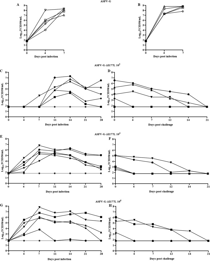
Animals infected with ASFV-G presented with expected high homogenous titers (107.5 to 108.5 HAD50/ml) on day 4 pi, increasing (around 108.5 HAD50/ml) by day 7 pi, when all animals were euthanized. Conversely, ASFV-G-ΔI177L revealed a different pattern, with low viremia (101.8 to 102.3 HAD50/ml) at day 4 pi, reaching peak values (103.8 to 107.5 HAD50/ml) by day 11 pi and then having decreasing titers (102.3 to 104 HAD50/ml) until day 28 pi (Fig. 5). It should be noted that one of the five animals inoculated with ASFV-G-ΔI177L showed a remarkably lower level of viremia (1,000- to 10,000-fold lower depending on the time point considered) than the average viremia values of the other animals in the group. Therefore, deletion of the I177L gene produced complete attenuation of the parental highly virulent ASFV-G virus when inoculated at a low dose, with the infected animals presenting long viremias with relatively low values.
FIG 5.
Viremia titers detected in pigs i.m. inoculated with either 102 HAD50 of ASFV-Georgia-ΔI177L or 102 HAD50 of ASFV-G. Each curve represents values from individual animals in each group. The sensitivity of virus detection was ≥1.8 log10 HAD50/ml.
To investigate the potential presence of residual virulence in ASFV-G-ΔI177L, a second experiment was performed where different groups of five pigs were infected i.m. with either 102, 104, or 106 HAD50 of ASFV-G-ΔI177L and their behavior compared with that of naive animals inoculated with 102 HAD50 of parental ASFV-G. In addition, a mock-infected animal cohabitated in each of the groups, acting as a sentinel to detect potential virus shedding from the infected animals.
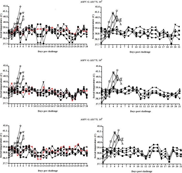
As in the first experiment, animals inoculated with ASFV-G exhibited all typical clinical signs of the disease and were euthanized in extremis by day 7 pi (Table 2). Interestingly, all animals inoculated with ASFV-G-ΔI177L, including those receiving 106 HAD50, did not present any ASF-related signs, remaining clinically normal during the entire observation period (28 days). Similarly, all sentinel animals remained clinically normal (Fig. 6).
TABLE 2.
Swine survival and fever response following infection with different doses of ASFV-G-ΔI177L or parental ASFV-G
| Virus | No. of survivors (n = 5) | Time to death (mean [SD]) (days) | Data for fever (mean [SD]) |
||
|---|---|---|---|---|---|
| No. of days to onset | Duration (days) | Maximum daily temp (°C) | |||
| ASFV-G | 0 | 4.8 (0.84)a | 4.8 (0.84) | 0.8 (0.84) | 40.7 (0.33) |
| ASFV-G-ΔI177L at HAD50 of: | |||||
| 102 | 5 | 39.4 (0.32) | |||
| 104 | 5 | 39.3 (0.32) | |||
| 106 | 5 | 39.3 (0.27) | |||
All animals were euthanized due to humanitarian reasons, following the corresponding IACUC protocol.
FIG 6.
Kinetics of body temperature values in pigs i.m. inoculated with either 102, 104, or 106 HAD50 (top of each panel) of ASFV-Georgia-ΔI177L (filled symbols), mock inoculated (sentinels, shown in red), or 102 HAD50 of ASFV-G (open symbols) (left) and after the challenge with 102 HAD50 of ASFV-G (right). Each curve represents an individual animal’s values in each group. Data from sentinel animals are depicted in red.
Viremia kinetics in ASFV-G-infected animals presented high titers (103.5 to 108 HAD50/ml) on day 4 pi increasing (around 107.5 HAD50/ml) by day 7 pi, when all animals were euthanized (Fig. 7). Animals infected with 102 HAD50/ml of ASFV-G-ΔI177L showed results similar to those seen in the previous experiment, although this time, viremias were not detected until day 11 pi (with the exception of one animal), and two out of the five animals presented evidently lower titers than did the other three animals in the group. In the groups of animals infected with 104 or 106 HAD50/ml of ASFV-G-ΔI177L, viremias were clearly detectable at 4 days pi, with average values remarkably higher (1,000- to 10,000-fold) than those of the group inoculated with 102 HAD50/ml, particularly at 4 and 7 days pi. Heterogeneity in the viremia measurements is also seen in the groups inoculated with the higher doses of ASFV-G-ΔI177L, particularly at 21 and 28 days pi, when 3 animals in each group presented remarkably lower titers than did the other three animals in the group. Therefore, deletion of the I177L gene produced a complete attenuation of the parental highly virulent ASFV-G virus even when used at high dosage, with the infected animals presenting low-titer viremias that persisted throughout the duration of the 28-day observational period. Interestingly, no virus was detected in any of the samples (all sampled blood time points as well as tonsil and spleen samples obtained at 28 days pi) obtained from sentinel animals (data not shown), indicating that ASFV-G-ΔI177L-infected animals did not shed enough virus to infect naive pigs during the 28 days of cohabitation.
FIG 7.
Viremia titers detected in pigs i.m. inoculated with either ASFV-Georgia-ΔI177L or ASFV-G. (A to H) Animals were inoculated with either 102 HAD50 of ASFV-G (A and B) or ASFV-Georgia-ΔI177L at doses of 102 (C and D), 104 (E and F), or 106 (G and H) HAD50. (C to H) Viremias in ASFV-Georgia-ΔI177L-inoculated animals before (C, E, and G) or after (D, F, and H) the challenge with 102 HAD50 of ASFV-G. Each curve represents values from individual animals in each group. The sensitivity of virus detection was ≥1.8 log10 HAD50/ml50/ml. Data from sentinel animals are depicted in red.
Protective efficacy of ASFV-G-ΔI177L against challenge with parental ASFV-G.
To assess the ability of ASFV-G-ΔI177L infection to induce protection against challenge with highly virulent parental virus ASFV-G, all animals infected with ASFV-G-ΔI177L were challenged 28 days later with 102 HAD50 of ASFV-G by the i.m. route. Five naive animals were challenged as a mock-inoculated control group.
All mock animals started showing disease-related signs by 3 to 4 days postchallenge (dpc), with rapidly increasing disease severity in the following hours; they were euthanized around 5 dpc (Table 3). On the other hand, the three groups of animals infected with ASFV-G-ΔI177L remained clinically healthy, not showing any significant signs of disease during the 21-day observational period. Therefore, ASFV-G-ΔI177L-treated animals are protected against clinical disease when challenged with the highly virulent parental virus.
TABLE 3.
Swine survival and fever response in ASFV-G-ΔI177L-infected animals challenged with ASFV-G virus 28 days later
| Virus | No. of survivors/total no. of animals | Time to death (mean [SD]) (days) | Data for fever (mean [SD]) |
||
|---|---|---|---|---|---|
| No. of days to onset | Duration (days) | Maximum daily temp (°C) | |||
| Mock | 0/5 | 5.6 (0.55)a | 4.2 (0.84) | 1.4 (0.88) | 40.9 (0.43) |
| ASFV-G-ΔI177L at HAD50 of: | |||||
| 102 | 10/10 | 39.3 (0.38) | |||
| 104 | 5/5 | 39.4 (0.21) | |||
| 106 | 5/5 | 39.4 (0.24) | |||
All animals were euthanized due to humanitarian reasons, following the corresponding IACUC protocol.
Analysis of viremia in animals infected with ASFV-G presented with expected high titers (107.3 to 108.3 HAD50/ml) on day 4 pi, increasing (averaging 108.5 HAD50/ml) by day 7 pi, when all animals were euthanized. After challenge, none of the ASFV-G-ΔI177L-infected animals had viremias with values higher than those present at challenge, and viremia values decreased progressively until the end of the experimental period (21 days after challenge) when, importantly, no circulating virus could be detected in blood from any of these animals (Fig. 7). Interestingly, postchallenge viremia titers, calculated by hemadsorption (HA), exactly coincide with those calculated by fluorescence, suggesting a lack (or at least a very low rate) of replication by the challenge virus. To assess the potential replication of the challenge virus, the presence of ASFV-G was tested in blood samples taken at day 4 postchallenge, when the highest viremia titers occur after challenge (Fig. 7). Using an I177L-specific real-time PCR to detect only challenge virus (with a demonstrated sensitivity of approximately 10 HAD50), all blood samples tested negative except one from an animal infected with 102 HAD50 of ASFV-G-ΔI177L (data not shown). Furthermore, tonsil and spleen samples were obtained from all ASFV-G-ΔI177L-infected animals at the end of the observational period (21 days postchallenge) and tested for the presence of virus (detected by hemadsorption) using swine macrophage cultures. Most of the animals in each group had infectious virus either in the tonsils or spleen (data not shown). All positive samples were then assessed using the I177L-specific real-time PCR, detecting the presence of the challenge virus in only one spleen belonging to the same animal initially infected with 102 HAD50/ml of ASFV-G-ΔI177L, which also had challenge virus in the blood (data not shown). These results suggest that replication of challenge virus was absent in all infected animals receiving 104 HAD50/ml or higher and most of the animals receiving 102 HAD50/ml of ASFV-G-ΔI177L.
Host antibody response in animals infected with ASFV-G-ΔI177L.
Host immune mechanisms mediating protection against virulent strains of ASFV in animals infected with attenuated strains of virus are not well identified (17–19). Our previous experience indicated that the only parameter consistently associated with protection against challenge is the level of circulating antibodies (20). In order to gain additional understanding of immune mechanisms in ASFV-G-ΔI177L-infected animals, we attempted to correlate the presence of anti-ASFV circulating antibodies with protection. ASFV-specific antibody response was detected in the sera of these animals using two in-house-developed direct enzyme-linked immunosorbent assays (ELISAs) (20). All animals infected with ASFV-G-ΔI177L, regardless of the dose of virus received, possessed similar high titers of circulating anti-ASFV antibodies (Fig. 8). An antibody response mediated by IgM and IgG isotypes was detected in all three groups by day 12 pi. By day 14 pi, the response mediated by both antibody isotypes reached maximum levels in all groups. The IgM-mediated antibody response disappeared in all animals by day 21 pi, while the IgG-mediated response remained high with minimal fluctuation until day 28 pi without significant differences between animals in the three groups inoculated with ASFV-G-ΔI177L. Therefore, as described in our previous reports (14, 20), there is a close correlation between the presence of anti-ASFV antibodies at the moment of challenge and protection. It should be mentioned that no antibodies were detected in any serum sample obtained from the sentinel animals, corroborating the virological data indicating that sentinel animals were not infected from ASFV-G-ΔI177L-infected animals in any of the three groups (Fig. 8).
FIG 8.
(A to F) Anti-ASFV antibody (IgM mediated [A, C, and E] and IgG mediated [B, D, and F]) titers detected by ELISA in pigs i.m. inoculated with either 102, 104, or 106 HAD50 of ASFV-Georgia-ΔI177L. Each curve represents values from individual animals in each group.
DISCUSSION
The use of attenuated strains is currently the most plausible approach to develop an effective ASF vaccine. Rational development of attenuated strains by genetic manipulation is a valid alternative, and perhaps safer methodology, compared to the use of naturally attenuated isolates. Several attenuated strains, obtained by genetic manipulation consisting of deletions of single genes or a group of genes, have been shown to induce protection against the virulent parental virus (10–13, 15, 21, 22). Here, the identification of a previously uncharacterized ASFV gene, I177L, as a viral genetic determinant of virulence is described. The deletion of I177L completely attenuates ASFV-G in swine, even when used at doses as high as 106 HAD50. Only two other genetic modifications have been shown to completely abolish virulence in the highly virulent ASFV Georgia isolate, those being deletion of the 9GL gene (particularly potentiated by the additional deletion of the UK gene) and deletion of a group of six genes from the MGF360 and 530 (12, 13, 22). The attenuation observed by deleting the I177L gene is a remarkable discovery since ASFV-G has not been efficiently attenuated by the deletion of any other genes that have been associated with attenuation in other ASFV isolates (13, 23). Based on the cumulative efforts supported by several studies, it is apparent that the genetic background where the deletion is operated plays a critical role in the effect of a particular gene in virus virulence, supporting the concept that AFV virulence is the result of the interactive effect of several virus genes.
As initially described (3), in the genome of ASFV-G 2007, the I177L gene was annotated from nucleotide positions 174471 to 17467, encoding a 66-amino-acid protein, while an adjacent ORF, ASFV-G-ACD_0176, was annotated at positions 174920 to 175003. Recent corrections, using new technology (16), identified a single A deletion at position 174954 in the initial version of the genome, and the corrected genome annotation now joins the ASFV-G-CD_0176 gene to the I177L gene, resulting in a single open reading frame (this open reading frame is similar to that in other virus genomes, as shown in Fig. 1). The recombination plasmid p72mCherryΔI177L was designed according to the original sequence of ASFV Georgia/2007 (3). To further add complexity to the I177L gene, a recent preprint (29) analyzing the transcriptome of ASFV, only the shortened version of I177L, as originally annotated (3), was detected as a late protein transcript, while the full-length transcript of I177L was not detected. We suggest reannotation of the I177L gene to the shortened ORF. In either case, as designed, our recombinant virus ASFV-G-ΔI177L would only result in a theoretical 25-amino-acid product matching the first 18 residues in the I177L ORF before a frameshift occurs plus an additional 7 additional amino acid residues being produced before a stop codon. The former ASFV-G-ACD_0176 ORF would only produce the predicted N-terminal transmembrane domain of I177L.
Although ASFV-G-ΔI177L-infected animals remained clinically normal, all of them presented with viremia by 28 days pi, in some cases with relatively high titers. It is interesting to note these relatively high levels of viremias, while ASFV-G-ΔI177L appears to grow at a decreased rate compared to the parental ASFV-G in swine macrophage cultures. In our experience, the presence of long viremias or virus persistence is not a rare event in animals infected with attenuated ASFV strains (12, 13, 21). As shown in those reports, the presence of the attenuated virus was associated with protection against the challenge with the virulent parental ASFV-G. Interestingly, no infectious virus could be detected in any of the three sentinels, indicating that transmission of ASFV-G-ΔI177L from infected to naive animals is not a frequent event, a desirable characteristic for a potential candidate live attenuated vaccine. The absence of infectious ASFV-G nor its genome, along with the lack of ASFV serum-specific antibody in the sentinels, strongly supports the suggestion that no virus is shed from the ASFV-G-ΔI177L-inoculated animals to the sentinels.
Importantly, animals infected with ASFV-G-ΔI177L were effectively protected when challenged at 28 dpi. Protection was achieved with doses as low as 102 HAD50 of ASFV-G-ΔI177L, while even the administration of 106 HAD50 ASFV-G-ΔI177L did not produce any disease-associated signs (not even a transient rise in body temperature), emphasizing the safety of ASFV-G-ΔI177L as a potential vaccine candidate. Importantly, it appears that replication of the challenge virus in the ASFV-G-ΔI177L-infected animals is quite restricted since challenge virus was isolated from only one of the animals inoculated with the low dose of ASFV-G-ΔI177L.
Although the host mechanisms mediating protection against ASFV infection remain under discussion (1, 25), in our experience with different live attenuated vaccine candidates, we have been observing a close association between the presence of circulating virus-specific antibodies and protection (12–14, 22, 24). In this report, we were also able to associate the presence of virus-specific antibodies and protection. Interestingly, regardless of the ASFV-G-ΔI177L dose used, all animals had similar antibody titers at the time of challenge, supporting the fact that low doses of ASFV-G-ΔI177L were as effective as the highest dose. Of note is the fact that by day 14 pi, all animals reached maximum antibody titers. Although in this report, challenge was not performed at 14 dpi, these data agree with those of previous published reports demonstrating that animals inoculated with vaccine candidate ASFV-G-Δ9GL/ΔUK presenting with circulating antibodies were protected against challenge at 2 weeks postinfection (21).
We believe that the results presented here demonstrate that ASFV-G-ΔI177L can be considered a strong vaccine candidate to protect animals against the ASFV Georgia isolate and its derivatives currently causing outbreaks in a wide geographical area from Central Europe to China and Southeast Asia. The complete lack of residual virulence, even when administered at high doses, apparent low levels of transmissibility to naive animals, and its high efficacy in inducing protection even at low doses make ASFV-G-ΔI177L a promising novel vaccine candidate.
MATERIALS AND METHODS
Cell culture and viruses.
Primary swine macrophage cell cultures were prepared from defibrinated swine blood, as previously described (15). Briefly, heparin-treated swine blood was incubated at 37°C for 1 h to allow sedimentation of the erythrocyte fraction. Mononuclear leukocytes were separated by flotation over a Ficoll-Paque density gradient (Pharmacia, Piscataway, NJ; specific gravity, 1.079). The monocyte/macrophage cell fraction was cultured in plastic Primaria tissue culture flasks (Falcon; Becton, Dickinson Labware, Franklin Lakes, NJ) containing macrophage medium composed of RPMI 1640 medium (Life Technologies, Grand Island, NY) with 30% L929 supernatant and 20% heat-inactivated fetal bovine serum (HI-FBS; Thermo Scientific, Waltham, MA) for 48 h at 37°C under 5% CO2. Adherent cells were detached from the plastic by using 10 mM EDTA in phosphate-buffered saline (PBS) and were then reseeded into Primaria T25 flasks and in 6- or 96-well dishes at a density of 5 × 10⁶ cells per ml for use in assays conducted 24 h later.
Comparative growth curves between ASFV-G and ASFV-G-ΔI177L viruses were performed in primary swine macrophage cell cultures. Preformed monolayers were prepared in 24-well plates and infected at an MOI of 0.01 (based on the HAD50 previously determined in primary swine macrophage cell cultures). After 1 h of adsorption at 37°C under 5% CO2, the inoculum was removed, and the cells were rinsed two times with PBS. The monolayers were then rinsed with macrophage medium and incubated for 2, 24, 48, 72, and 96 h at 37°C under 5% CO2. At appropriate times postinfection, the cells were frozen at <−70°C, and the thawed lysates were used to determine titers by HAD50/ml in primary swine macrophage cell cultures. All samples were run simultaneously to avoid interassay variability.
Virus titration was performed on primary swine macrophage cell cultures in 96-well plates. Virus dilutions and cultures were performed using macrophage medium. The presence of virus was assessed by hemadsorption (HA), and virus titers were calculated by the Reed and Muench method (26).
ASFV Georgia (ASFV-G) was a field isolate kindly provided by Nino Vepkhvadze from the Laboratory of the Ministry of Agriculture (LMA) in Tbilisi, Republic of Georgia.
Microarray analysis.
The microarray data of ASFV open reading frames were obtained from the data set deposited in NCBI databases from our previous study (27). In brief, total RNA was extracted from primary swine macrophage cell cultures infected with ASFV Georgia strain or mock infected at 3, 6, 9, 12, 15, and 18 h postinfection (hpi). A custom-designed porcine microarray manufactured by Agilent Technologies (Chicopee, MA) was used for this study. Both infected and mock-infected RNA samples were labeled with Cy3 and Cy5 using a low-input RNA labeling kit (Agilent Technologies). Cy5-labeled infected or mock-infected samples were cohybridized with Cy3-labeled mock-infected or infected samples in one array, respectively, for each time point using a dye-swap design. The entire procedure of microarray analysis was conducted according to protocols, reagents, and equipment provided or recommended by Agilent Technologies. Array slides were scanned using a GenePix 4000B scanner (Molecular Devices, San Jose, CA) with the GenePix Pro 6.0 software at 5-μm resolution. Background signal correction and data normalization of the microarray signals and statistical analysis were performed using the LIMMA package. The signal intensities of ASFV open reading frame RNA were averaged from both the Cy3 and Cy5 channels.
Construction of the recombinant ASFV-G-ΔI177L.
Recombinant ASFVs were generated by homologous recombination between the parental ASFV-G genome and recombination transfer vector by infection and transfection procedures using swine macrophage cell cultures (15). Recombinant transfer vector (p72mCherryΔI177L) containing flanking genomic regions to amino acids 112 through 150 of the I177L gene, mapping approximately 1 kbp to the left and right of these amino acids, and a reporter gene cassette containing the mCherry gene with the ASFV p72 late gene promoter, p72mCherry, were used. This construction created a 112-bp deletion in the I177L ORF (Fig. 1). Recombinant transfer vector p72mCherryΔI177L was obtained by DNA synthesis (Epoch Life Sciences, Missouri City, TX, USA).
Next-generation sequencing of ASFV genomes.
ASFV DNA was extracted from infected cells and quantified as described earlier. The full-length sequence of the virus genome was determined as described previously (28) using an Illumina NextSeq 500 sequencer.
Animal experiments.
Animal experiments were performed under biosafety level 3-Agriculture (3-AG) conditions in the animal facilities at Plum Island Animal Disease Center (PIADC) following a protocol approved by the PIADC Institutional Animal Care and Use Committee of the U.S. Department of Agriculture and U.S. Department of Homeland Security (protocol number 225.04-16-R, 09-07-16).
ASFV-G-ΔI177L was assessed for its virulence phenotype relative to the virulent parental ASFV-G virus using 80- to 90-pound commercial breed swine. Groups of pigs (n = 5) were inoculated intramuscularly (i.m.) either with 102 to 106 HAD50 of ASFV-G ΔI177L or 102 HAD50 of parental ASFV-G virus. Clinical signs (anorexia, depression, fever, purple skin discoloration, staggering gait, diarrhea, and cough) and changes in body temperature were recorded daily throughout the experiment. In protection experiments, animals inoculated with ASFV-G ΔI177L were 28 days later i.m. challenged with 102 HAD50 of the parental virulent ASFV-G strain. The presence of clinical signs associated with the disease was recorded as described earlier (13).
Detection of anti-ASFV antibodies.
ASFV antibody detection used an in-house indirect ELISA, developed as described previously (20). Briefly, ELISA antigen was prepared from ASFV-infected Vero cells. MaxiSorp ELISA plates (Nunc, St. Louis, MO, USA) were coated with 1 μg per well of infected or uninfected cell extract. The plates were blocked with phosphate-buffered saline containing 10% skim milk (Merck, Kenilworth, NJ, USA) and 5% normal goat serum (Sigma, St. Louis, MO). Each swine serum was tested at multiple dilutions against both infected and uninfected cell antigen. ASFV-specific antibodies in the swine sera were detected by an anti-swine IgM- or IgG-horseradish peroxidase conjugate (KPL, Gaithersburg, MD, USA) and SureBlue Reserve peroxidase substrate (KPL). Plates were read at an optical density at 630 nm (OD630) in an ELx808 plate reader (BioTek, Shoreline, WA, USA). Serum titers were expressed as the log10 of the highest dilution, where the OD630 reading of the tested sera at least duplicates the reading of the mock-infected sera.
ACKNOWLEDGMENTS
We thank the Plum Island Animal Disease Center Animal Care Unit staff for excellent technical assistance. We particularly thank Melanie V. Prarat for editing the manuscript.
This research was supported in part by an appointment to the Plum Island Animal Disease Center (PIADC) Research Participation Program administered by the Oak Ridge Institute for Science and Education (ORISE) through an interagency agreement between the U.S. Department of Energy (DOE) and the U.S. Department of Agriculture (USDA). ORISE is managed by ORAU under DOE contract DE-SC0014664. This project was partially funded through an interagency agreement with the Science and Technology Directorate of the U.S. Department of Homeland Security under awards 70RSAT19KPM000056 and 70RSAT18KPM000134.
All opinions expressed in this paper are those of the authors and do not necessarily reflect the policies and views of the USDA, ARS, APHIS, DHS, DOE, or ORAU/ORISE.
Douglas P. Gladue and Manuel V. Borca have a patent application filed by the U.S. Department of Agriculture for ASFV-G-ΔI177L as a live attenuated vaccine for African swine fever.
REFERENCES
- 1.Tulman ER, Delhon GA, Ku BK, Rock DL. 2009. African swine fever virus, p 43–87. In Van Etten JL. (ed), Lesser known large dsDNA viruses, vol 328 Springer-Verlag, Berlin, Germany. [Google Scholar]
- 2.Costard S, Wieland B, de Glanville W, Jori F, Rowlands R, Vosloo W, Roger F, Pfeiffer DU, Dixon LK. 2009. African swine fever: how can global spread be prevented? Philos Trans R Soc Lond B Biol Sci 364:2683–2696. doi: 10.1098/rstb.2009.0098. [DOI] [PMC free article] [PubMed] [Google Scholar]
- 3.Chapman DA, Darby AC, Da Silva M, Upton C, Radford AD, Dixon LK. 2011. Genomic analysis of highly virulent Georgia 2007/1 isolate of African swine fever virus. Emerg Infect Dis 17:599–605. doi: 10.3201/eid1704.101283. [DOI] [PMC free article] [PubMed] [Google Scholar]
- 4.Coggins L. 1974. African swine fever virus. Pathogenesis. Prog Med Virol 18:48–63. [PubMed] [Google Scholar]
- 5.Forman AJ, Wardley RC, Wilkinson PJ. 1982. The immunological response of pigs and guinea pigs to antigens of African swine fever virus. Arch Virol 74:91–100. doi: 10.1007/bf01314703. [DOI] [PubMed] [Google Scholar]
- 6.Kihm UAM, Mueller H, Pool R. 1987. Approaches to vaccination African swine fever. Martinus Nijhoff Publishing, Boston, MA. [Google Scholar]
- 7.Mebus CA. 1988. African swine fever. Adv Virus Res 35:251–269. doi: 10.1016/s0065-3527(08)60714-9. [DOI] [PubMed] [Google Scholar]
- 8.Hamdy FM, Dardiri AH. 1984. Clinical and immunologic responses of pigs to African swine fever virus isolated from the Western Hemisphere. Am J Vet Res 45:711–714. [PubMed] [Google Scholar]
- 9.Ruiz-Gonzalvo FC, Bruyel V. 1981. Immunological responses of pigs to partially attenuated ASF and their resistance to virulent homologous and heterologous viruses. Food and Agriculture Organization of the United Nations, Rome, Italy. [Google Scholar]
- 10.Lewis T, Zsak L, Burrage TG, Lu Z, Kutish GF, Neilan JG, Rock DL. 2000. An African swine fever virus ERV1-ALR homologue, 9GL, affects virion maturation and viral growth in macrophages and viral virulence in swine. J Virol 74:1275–1285. doi: 10.1128/jvi.74.3.1275-1285.2000. [DOI] [PMC free article] [PubMed] [Google Scholar]
- 11.Moore DM, Zsak L, Neilan JG, Lu Z, Rock DL. 1998. The African swine fever virus thymidine kinase gene is required for efficient replication in swine macrophages and for virulence in swine. J Virol 72:10310–10315. doi: 10.1128/JVI.72.12.10310-10315.1998. [DOI] [PMC free article] [PubMed] [Google Scholar]
- 12.O'Donnell V, Holinka LG, Gladue DP, Sanford B, Krug PW, Lu X, Arzt J, Reese B, Carrillo C, Risatti GR, Borca MV. 2015. African swine fever virus Georgia isolate harboring deletions of MGF360 and MGF505 genes is attenuated in swine and confers protection against challenge with virulent parental virus. J Virol 89:6048–6056. doi: 10.1128/JVI.00554-15. [DOI] [PMC free article] [PubMed] [Google Scholar]
- 13.O'Donnell V, Holinka LG, Krug PW, Gladue DP, Carlson J, Sanford B, Alfano M, Kramer E, Lu Z, Arzt J, Reese B, Carrillo C, Risatti GR, Borca MV. 2015. African swine fever virus Georgia 2007 with a deletion of virulence-associated gene 9GL (B119L), when administered at low doses, leads to virus attenuation in swine and induces an effective protection against homologous challenge. J Virol 89:8556–8566. doi: 10.1128/JVI.00969-15. [DOI] [PMC free article] [PubMed] [Google Scholar]
- 14.O'Donnell V, Holinka LG, Sanford B, Krug PW, Carlson J, Pacheco JM, Reese B, Risatti GR, Gladue DP, Borca MV. 2016. African swine fever virus Georgia isolate harboring deletions of 9GL and MGF360/505 genes is highly attenuated in swine but does not confer protection against parental virus challenge. Virus Res 221:8–14. doi: 10.1016/j.virusres.2016.05.014. [DOI] [PubMed] [Google Scholar]
- 15.Zsak L, Lu Z, Kutish GF, Neilan JG, Rock DL. 1996. An African swine fever virus virulence-associated gene NL-S with similarity to the herpes simplex virus ICP34.5 gene. J Virol 70:8865–8871. doi: 10.1128/JVI.70.12.8865-8871.1996. [DOI] [PMC free article] [PubMed] [Google Scholar]
- 16.Forth JH, Forth LF, King J, Groza O, Hubner A, Olesen AS, Hoper D, Dixon LK, Netherton CL, Rasmussen TB, Blome S, Pohlmann A, Beer M. 2019. A deep-sequencing workflow for the fast and efficient generation of high-quality African swine fever virus whole-genome sequences. Viruses 11:846. doi: 10.3390/v11090846. [DOI] [PMC free article] [PubMed] [Google Scholar]
- 17.Onisk DV, Borca MV, Kutish G, Kramer E, Irusta P, Rock DL. 1994. Passively transferred African swine fever virus antibodies protect swine against lethal infection. Virology 198:350–354. doi: 10.1006/viro.1994.1040. [DOI] [PubMed] [Google Scholar]
- 18.Ruiz Gonzalvo F, Carnero ME, Caballero C, Martínez J. 1986. Inhibition of African swine fever infection in the presence of immune sera in vivo and in vitro. Am J Vet Res 47:1249–1252. [PubMed] [Google Scholar]
- 19.Oura CA, Denyer MS, Takamatsu H, Parkhouse RM. 2005. In vivo depletion of CD8+ T lymphocytes abrogates protective immunity to African swine fever virus. J Gen Virol 86:2445–2450. doi: 10.1099/vir.0.81038-0. [DOI] [PubMed] [Google Scholar]
- 20.Carlson J, O’Donnell V, Alfano M, Velazquez Salinas L, Holinka LG, Krug PW, Gladue DP, Higgs S, Borca MV. 2016. Association of the host immune response with protection using a live attenuated African swine fever virus model. Viruses 8:291. doi: 10.3390/v8100291. [DOI] [PMC free article] [PubMed] [Google Scholar]
- 21.Zsak L, Caler E, Lu Z, Kutish GF, Neilan JG, Rock DL. 1998. A nonessential African swine fever virus gene UK is a significant virulence determinant in domestic swine. J Virol 72:1028–1035. doi: 10.1128/JVI.72.2.1028-1035.1998. [DOI] [PMC free article] [PubMed] [Google Scholar]
- 22.O’Donnell V, Risatti GR, Holinka LG, Krug PW, Carlson J, Velazquez-Salinas L, Azzinaro PA, Gladue DP, Borca MV. 2017. Simultaneous deletion of the 9GL and UK genes from the African swine fever virus Georgia 2007 isolate offers increased safety and protection against homologous challenge. J Virol 91:e01760-16. doi: 10.1128/JVI.01760-16. [DOI] [PMC free article] [PubMed] [Google Scholar]
- 23.Ramirez-Medina E, Vuono E, O’Donnell V, Holinka LG, Silva E, Rai A, Pruitt S, Carrillo C, Gladue DP, Borca MV. 2019. Differential effect of the deletion of African swine fever virus virulence-associated genes in the induction of attenuation of the highly virulent Georgia strain. Viruses 11:599. doi: 10.3390/v11070599. [DOI] [PMC free article] [PubMed] [Google Scholar]
- 24.Sanford B, Holinka LG, O'Donnell V, Krug PW, Carlson J, Alfano M, Carrillo C, Wu P, Lowe A, Risatti GR, Gladue DP, Borca MV. 2016. Deletion of the thymidine kinase gene induces complete attenuation of the Georgia isolate of African swine fever virus. Virus Res 213:165–171. doi: 10.1016/j.virusres.2015.12.002. [DOI] [PubMed] [Google Scholar]
- 25.Costard S, Porphyre V, Messad S, Rakotondrahanta S, Vidon H, Roger F, Pfeiffer DU. Exploratory multivariate analysis for differentiating husbandry practices relevant to disease risk for pig farmers in Madagascar. Food and Agriculture Organization of the United Nations, Rome, Italy. [Google Scholar]
- 26.Reed LJ, Muench H. 1938. A simple method of estimating fifty percent endpoints. Am J Hyg 27:493–497. doi: 10.1093/oxfordjournals.aje.a118408. [DOI] [Google Scholar]
- 27.Zhu JJ, Ramanathan P, Bishop EA, O'Donnell V, Gladue DP, Borca MV. 2019. Mechanisms of African swine fever virus pathogenesis and immune evasion inferred from gene expression changes in infected swine macrophages. PLoS One 14:e0223955. doi: 10.1371/journal.pone.0223955. [DOI] [PMC free article] [PubMed] [Google Scholar]
- 28.Borca MV, Holinka LG, Berggren KA, Gladue DP. 2018. CRISPR-Cas9, a tool to efficiently increase the development of recombinant African swine fever viruses. Sci Rep 8:3154. doi: 10.1038/s41598-018-21575-8. [DOI] [PMC free article] [PubMed] [Google Scholar]
- 29.Cackett G, Matelska D, Sýkora M, Portugal R, Malecki M, Bähler J, Dixon L, Werner F. 2019. Temporal transcriptome and promoter architecture of the African swine fever virus. bioRxiv doi: 10.1101/847343. [DOI]


