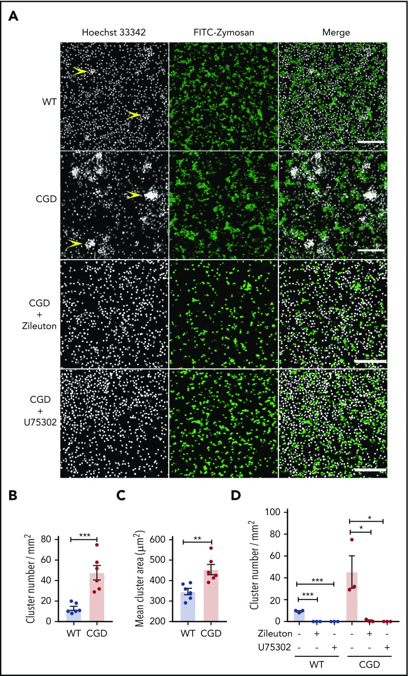
Figure 3.
CGD neutrophils spontaneously formed larger and increased amounts of clusters in the presence of zymosan in vitro. Mouse BM neutrophils (4 × 10⁶/mL) stained with 5 µg/mL Hoechst 33342 were stimulated for 1 hour with FITC-zymosan (MOI = 2). Images were acquired with LASX software (Leica Microsystems). (A) Representative images showing the neutrophil nuclei (white) and FITC-zymosan (green) and merged images of the 2 channels acquired by confocal microscopy. Examples of clusters are indicated by yellow arrowheads. Scale bars, 100 µm. (B) Quantification of the clusters and (C) cluster area. n = 6 in each group from 3 separate experiments. (D) CGD mouse BM neutrophils (4 × 10⁶/mL) stained with 5 µg/mL Hoechst 33342 were preincubated with 30 µM zileuton or 10 µM U75302 for 10 minutes and then stimulated for 1 hour with FITC-zymosan (MOI = 2) in the presence of 30 µM zileuton or 10 µM U75302. Images were acquired by confocal microscope. (E) Quantification of the cluster number. n = 3 in each group from 2 separate experiments. Data are means ± standard error of the mean. *P < .05, **P < .01; ***P < .001, by Student t test. MOI, multiplicity of infection.

