Emotional disorders are common comorbid conditions that further exacerbate the severity and chronicity of chronic pain. However, individuals show considerable vulnerability to the development of chronic pain under similar pain conditions.
Keywords: CaMKIIα, central amygdala, chronic pain, pain vulnerability, SIRT1
Abstract
Emotional disorders are common comorbid conditions that further exacerbate the severity and chronicity of chronic pain. However, individuals show considerable vulnerability to the development of chronic pain under similar pain conditions. In this study on male rat and mouse models of chronic neuropathic pain, we identify the histone deacetylase Sirtuin 1 (SIRT1) in central amygdala as a key epigenetic regulator that controls the development of comorbid emotional disorders underlying the individual vulnerability to chronic pain. We found that animals that were vulnerable to developing behaviors of anxiety and depression under the pain condition displayed reduced SIRT1 protein levels in central amygdala, but not those animals resistant to the emotional disorders. Viral overexpression of local SIRT1 reversed this vulnerability, but viral knockdown of local SIRT1 mimicked the pain effect, eliciting the pain vulnerability in pain-free animals. The SIRT1 action was associated with CaMKIIα downregulation and deacetylation of histone H3 lysine 9 at the CaMKIIα promoter. These results suggest that, by transcriptional repression of CaMKIIα in central amygdala, SIRT1 functions to guard against the emotional pain vulnerability under chronic pain conditions. This study indicates that SIRT1 may serve as a potential therapeutic molecule for individualized treatment of chronic pain with vulnerable emotional disorders.
SIGNIFICANCE STATEMENT Chronic pain is a prevalent neurological disease with no effective treatment at present. Pain patients display considerably variable vulnerability to developing chronic pain, indicating individual-based molecular mechanisms underlying the pain vulnerability, which is hardly addressed in current preclinical research. In this study, we have identified the histone deacetylase Sirtuin 1 (SIRT1) as a key regulator that controls this pain vulnerability. This study reveals that the SIRT1–CaMKIIaα pathway in central amygdala acts as an epigenetic mechanism that guards against the development of comorbid emotional disorders under chronic pain, and that its dysfunction causes increased vulnerability to the development of chronic pain. These findings suggest that SIRT1 activators may be used in a novel therapeutic approach for individual-based treatment of chronic pain.
Introduction
Chronic pain is a prevalent neurological disease, for which an effective treatment is currently lacking. Chronic pain consists of a sensory-discriminative component and an affective-emotional component with exacerbating interactions between the two components (Price, 2000; Liu and Chen, 2014). However, for the last few decades, basic research on pain mechanisms has mainly focused on the sensory component of chronic pain. The affective-emotional component of chronic pain, whose importance is increasingly recognized recently, is mainly manifested by chronic sensory pain-induced emotional disorders, such as anxiety and depression (Liu and Chen, 2014; Usdin and Dimitrov, 2016), which further exacerbate the severity and chronicity of the pain condition, leading to a vicious cycle that exerts a devastating effect on the quality of life in patients with chronic pain (Wiech and Tracey, 2009; Failde et al., 2018; Kosson et al., 2018).
Another important feature of chronic pain is individual vulnerability, as clinical data clearly show that some patients are more vulnerable, while others are relatively resilient, to the development of chronic pain under similar pain conditions (Denk et al., 2014). Nevertheless, the underlying mechanisms for this individual vulnerability are unknown and hardly addressed in current preclinic pain studies on animal models of chronic pain. Recently, epigenetic mechanisms have emerged with increasing prominence in individual susceptibility to various environmental factors, mediating the development of multiple diseases including neuropsychiatric disorders, drug addiction, and cardiometabolic disorders (Sillivan et al., 2017; Egervari et al., 2018; Palumbo et al., 2018; Wang et al., 2018).
Sirtuins (SIRTs) 1–7, NAD+-dependent histone deacetylases (HDACs), regulate diverse cellular processes including aging, inflammation, apoptosis, autophagy, and cell metabolism by modifying the acetylation status of histone and nonhistone proteins (Herranz and Serrano, 2010; Herskovits and Guarente, 2014; Chalkiadaki and Guarente, 2015). Recent studies have suggested that SIRTs have important roles in the pathophysiology of emotional disorders. Decreased SIRT1 and SIRT2 levels and increased SIRT6 level in hippocampus have been shown to contribute to the chronic stress-elicited depression-like behavior (Liu et al., 2015; Abe-Higuchi et al., 2016). Increased SIRT1 levels in the nucleus accumbens (NAc) plays an essential role in regulating mood-related behavioral abnormalities induced by chronic social defeat stress (Kim et al., 2016). SIRT2 knockout blocks the development of social defeat stress-induced depression-like behavior (Zhang et al., 2018). Clinical studies also suggest that SIRT1 and SIRT2 gene polymorphisms are associated with depressive disorder (Porcelli et al., 2013; Nivoli et al., 2016), and decreased expression of SIRT1, SIRT2, and SIRT6 genes has been found in patients with a mood disorder (Abe et al., 2011; Luo and Zhang, 2016). These data suggest that SIRT1, SIRT2, and SIRT6 in different brain regions play important roles in the emotional disorders. However, how SIRTs are involved in chronic pain-induced emotional disorders or pain vulnerability remains unexplored.
In this study, we investigated the roles of SIRT1, SIRT2, and SIRT6 in the central nucleus of the amygdala (CeA), a key brain structure that regulates emotion-related behaviors (Gilpin et al., 2015; Janak and Tye, 2015; Neugebauer, 2015; Thompson and Neugebauer, 2017; Babaev et al., 2018), in the development of chronic pain-induced emotional disorders of affective pain component, focusing on individual variances in the epigenetic changes and behavioral responses for individual vulnerability to chronic pain.
Materials and Methods
Animals.
Adult male Wistar rats (weight, 200–300 g) and 8-week-old male mice were used in this study. B6;129-Sirt1tm1Ygu/J (SIRT1flox/wt) mice were purchased from The Jackson Laboratory. SIRT1flox/wt mice were intercrossed to generate SIRT1w/w and SIRT1flox/flox mice. Genotypes of mice were identified by PCR using the following primer sequences: 5′-GGT TGA CTT AGG TCT TGT CTG-3′ (forward) and 5′-CGT CCC TTG TAA TGT TTC CC-3′ (reverse). All animals were on a 12 h light/dark cycle and received food and water ad libitum. Animal care and experimental procedures were approved by and performed in accordance with the Institutional Animal Care and Use Committee.
Model of chronic neuropathic pain.
The model of chronic trigeminal neuropathic pain was established by chronic constriction injury (CCI) of the unilateral infraorbital nerve (ION) according to the method described in previous studies (Kim et al., 2014). Under anesthesia, an animal was restrained on an operation table in a supine position. An 8- to 10-mm-long incision was made along the gingiva-buccal margin in the buccal mucosa, beginning distal to the first molar. The ION was freed from the surrounding connective tissue and tied loosely with two chromic gut ligatures (4.0) under a surgical microscope. The incision was sutured with 4.0 silk sutures. Animals in sham groups underwent the same surgical procedure but without the ION ligation.
Measurement of mechanical pain.
Mechanical allodynia was evaluated as described in previous reports (Wei et al., 2008; Kim et al., 2014). After a 30 min accommodation period, animals were held loosely and a series of von Frey filaments (North Coast Medical) of varying force was applied to the vibrissa pad region. A brisk head withdrawal, escape or attack reactions, or short-lasting facial grooming behaviors were defined as positive responses. Each von Frey filament was applied five times at an interval of 10 s. The EF50 value, defined as the von Frey filament force (g) that produced a 50% positive response, was calculated to evaluate mechanical pain sensitivity.
Open field test.
Open field test was performed as described previously (Fernando and Robbins, 2011; Haller and Alicki, 2012). Twenty-eight days after CCI-ION or sham surgery, an animal was placed in the corner of an open arena (72 × 72 × 30 cm for rats; 42 × 42 × 30 cm for mice) and allowed to move freely for 15 min. Locomotor activity of the animal was video recorded and analyzed by an automated video-tracking system (EthoVision XT, Noldus; or ANY-maze, Stoelting). In an analysis for individual variance, the average time the sham control animals spent in the central zone (central time) during the test was used to divide the CCI-ION rats into a vulnerable group (central time below the sham average) and a resistant group (central time above the sham average).
Forced swim test.
A forced swim test was performed as previously described (Fernando and Robbins, 2011; Slattery and Cryan, 2012). The next day after the open field test (29 d after CCI-ION or sham surgery), a 15 min pretest was performed. An animal was placed in a clear cylinder (height × diameter: rats, 45 × 30 cm; mice, 25 × 12 cm) filled with water (depth: rats, 30 cm; mice, 12 cm) at 25 ± 1°C. Twenty-four hours after the pretest, the animal was allowed to swim in the cylinder for 6 min and the immobility time during the last 5 min period was recorded. Immobility was defined as the cessation of all active swimming and escaping activities. Similarly, based on the averaged immobility time of sham animals, CCI-ION animals were divided into a vulnerable group (immobility time above the sham average) and a resistant group (immobility time below the sham average) in the analysis.
Adeno-associated virus vectors and viral microinjections.
An adeno-associated virus (AAV)2/9-CaMKIIα-SIRT1–3*flag-GFP (AAV-SIRT1) vector and a control vector AAV2/9-CaMKIIα-GFP (AAV-GFP) were obtained from Hanbio and microinjected bilaterally (1 μl each side) into the CeA of rats (−2.1 mm anteroposterior, ±4.5 mm mediolateral, −8.6 mm dorsoventral from the bregma) at a rate of 0.1 μl/min. An AAV9-CaMKIIα-Cre-EGFP (AAV-Cre) vector and a control vector AAV9-CaMKIIα-EGFP-3flag (AAV-EGFP) were obtained from Shanghai Genechem and microinjected bilaterally (0.5 μl each side) into the CeA of mice (−1.2 mm anteroposterior, ±2.75 mm mediolateral, −4.8 mm dorsoventral from the bregma) at a rate of 0.1 μl/min. Stereotaxic surgeries were performed to microinject the viral vectors into the CeA. After the injections, animals were allowed to recover for 2 weeks. Fifteen days after the vector injection, CeA tissues were collected for analysis. Viral injection sites were histologically verified by confirming the GFP or EGFP signal in the CeA of brain slices with a fluorescence microscope.
Immunofluorescence staining.
Animals were deeply anesthetized with chloral hydrate and transcardially perfused with 4% paraformaldehyde in PBS. The brain tissues were removed and postfixed in 4% paraformaldehyde for 4–6 h at 4°C, followed by dehydration with 30% sucrose solution. Coronal slices (30 μm) containing the amygdala (−1.8 to −3.3 mm from bregma) were obtained with a cryostat (Leica) at −20°C. After reaction blockade with 10% normal goat serum in PBS and 0.3% Triton X-100, the sections were incubated with the following primary antibodies: mouse anti-SIRT1 (1:200; Abcam); rabbit anti-CaMKIIα (1:200; Abcam); rabbit anti-GAD67 (1:100; Cell Signaling Technology); and rabbit anti-vGluT2 (1:50; Cell Signaling Technology). After washes, sections were then incubated with suitable secondary antibodies, as follows: Alexa Fluor 594 goat anti-mouse IgG or Alexa Fluor 488 goat anti-rabbit IgG (Thermo Fisher Scientific) in dark for 2 h. The stained sections were examined with an LSM880 confocal laser-scanning microscope (Zeiss). Quantitative analyses were performed by using tissue sections from three rats (three sections per rat) and examining an area of 213 × 213 μm in CeA by an experimenter blinded to the treatment groups.
Western blotting analysis.
The frozen CeA tissues were homogenized with N-PER Neuronal Protein Extraction Reagent (Thermo Fisher Scientific) containing Phosphatase Inhibitor Cocktails 2, 3, and a Protease Inhibitor Cocktail (Sigma-Aldrich). Supernatants were collected after centrifugation at 13,000 × g for 10 min at 4°C, and protein concentrations were determined with DC Protein Assay Kits according to the manufacturer instruction (Bio-Rad). Equal amounts of protein samples were separated by SDS-PAGE and transferred onto a nitrocellulose membrane. The membrane was incubated with the primary antibodies including anti-SIRT1 (Cell Signaling Technology), anti-CaMKIIα (Cell Signaling Technology), and anti-β-actin (BioWorld), followed by incubation with the appropriate secondary antibodies (Li-Cor). An Infrared Imaging System (Gene Company Limited) was used to detect immune-reactive bands.
Chromatin immunoprecipitation assay.
Chromatin immunoprecipitation (ChIP) assays were performed with an EpiQuik ChIP Kit (Epigentek). After chromatin cross-linking with 1% formaldehyde and DNA shearing by sonication, chromatin–protein complexes were immunoprecipitated with antibodies against acetylated H3K9 (H3K9Ac; Abcam) and acetylated H4K16 (H4K16Ac; Cell Signaling Technology). An antibody against normal IgG (anti-mouse) was used as a negative control. Immunoprecipitated DNA was subjected to quantitative real-time PCR with the following primers: CaMKIIα (rat): forward, 5′-ACAGGGCGTTTCGG-3′, CaMKIIα (rat): reverse, 5′-TCGGGACTAGGACTGG-3′; CaMKIIα (mouse): forward, 5′-ACGGACTCAGGAAGAGGGATA-3′; and CaMKIIα (mouse): reverse, 5′-CTTGCTCCTCAGAATCCATTG-3′. The cumulative fluorescence for each amplification was normalized to input amplification.
Experimental designs and statistical analyses.
Most experiments used a between-subject design for comparisons between the treatment group and corresponding control group, such as comparisons for a nerve injury-induced pain group versus a sham-operated group, an AAV-SIRT1-overexpressing group versus an AAV-GFP group, and SIRT1-knock-down mice versus corresponding wild-type (WT) mice. The pain group was further divided into vulnerable and resistant groups based on behavioral parameters of individual data, and each was compared with the control group. Behavioral responses of sensory pain were monitored daily by repeated measures on the same animals. The experimenter performing the behavioral tests was blind to the treatment groups of animals. Each emotional test was conducted only once on each animal. All individual data from each animal were included and analyzed. Number of animals for each group was determined by power analysis and based on our previous results of similar experiments and on individual data distribution. Two-group comparisons were made by the Student's t test, and three-group comparisons were performed with one-way ANOVA followed by LSD post hoc test. Behavioral data of sensory pain thresholds with multiple measurements were analyzed by two-way ANOVA for repeated measures with LSD post hoc analysis. A χ2 test was performed to analyze the percentage of animals in vulnerable and resistant groups, and a Pearson correlation test was performed to analyze cross-vulnerability of each animal to anxiety and depression. p < 0.05 was considered statistically significant. All statistical analyses were performed with Prism software version 7. Data are expressed as the mean ± SEM.
Results
Chronic sensory pain induces anxiety- and depression-like behaviors with significant individual variance
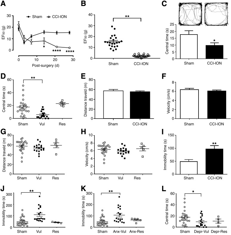
We used CCI of the ION to induce chronic trigeminal neuropathic pain, and an EF50 value was determined to evaluate mechanical pain sensitivity. As shown in Figure 1A, rats with CCI-ION, compared with sham-operated rats, displayed significant mechanical allodynia with reduced EF50 values in the ipsilateral ION territory on days 14 (p < 0.05), 21 (p < 0.01), and 28 (p < 0.01) after the CCI-ION surgery, demonstrating chronic sensitization to mechanical pain. A transient decrease in EF50 values was observed in sham rats on day 3 after surgery, which might be caused by surgical incision and local inflammation. To examine individual variance in this pain sensitization, we analyzed the EF50 values of individual rats on day 28 after surgery. As shown in Figure 1B, there was nearly no overlap in EF50 values between sham rats and CCI-ION rats, suggesting that all rats developed a similar degree of mechanical allodynia or that there is little individual variance in the nerve injury-induced sensory pain sensitization.
Figure 1.
Chronic sensory pain induces anxiety- and depression-like behaviors with significant individual variance. A, Mechanical pain thresholds in sham-operated rats and in rats with CCI of the ION up to 28 d after the surgery. B, Distribution of mechanical pain thresholds (EF50) in individual rats 28 d after CCI-ION surgery. C, Traces of locomotor activity (top) and the time rats spent in the central zone (central time, bottom) in an open field test in sham rats and CCI-ION rats. D, Individual data of central time in the same rats as in C, with the CCI-ION rats divided into a vulnerable group (Vul; defined by the central time of each CCI-ION rat below the averaged central time in sham rats, n = 17) and a resistant group (Res; with central time above the sham average, n = 5). E, F, Average group data of distance traveled (E) and travel velocity (F) in the sham rats and CCI-ION rats. G, H, Individual data of distance traveled (G) and travel velocity (H) in the same rats as in E and F. I, J, Group averages (I) and individual data (J) of immobility time in the forced swim test in the sham rats and CCI-ION rats. CCI-ION rats were divided into a vulnerable group (immobility time above sham average, n = 18) and a resistant group (immobility time below sham average, n = 4). K, Immobility time of depressive behavior in sham rats, in anxiety-vulnerable CCI-ION rats (Anx-Vul; n = 17), and in anxiety-resistant CCI-ION rats (Anx-Res; n = 5). L, Central time of anxiety behavior in sham rats, in depression-vulnerable CCI-ION rats (Depr-Vul; n = 18), and in depression-resistant CCI-ION rats (Depr-Res; n = 4). N = 22 both in the sham group and the CCI-ION group. Errors bars are SEM. *p < 0.05, **p < 0.01, ****p < 0.0001.
We then determined anxiety- and depression-like behaviors in sham rats and CCI-ION rats with chronic pain, using the open field test and the forced swim test, respectively. We found that, compared with sham rats, CCI-ION rats showed significant anxiety behavior with decreased time spent in the central zone (central time, p < 0.05) in open field test 28 d after the surgery (Fig. 1C). To determine individual variance and vulnerability to anxiety, we analyzed the distribution of central times for the variance of individual rats. Rats in the sham group displayed a large variance in central times, suggesting that individual animals are largely variant in response to environmental stimulus of anxiety in normal conditions. For the purpose of analyzing individual vulnerability to anxiety under the pain condition, we used the averaged central time in sham rats as an arbitrary cutoff line and a reference index of anxiety vulnerability to approximately divide the CCI-ION rats into a vulnerable group (central time below the sham average) and a resistant group (central time above the sham average). We found that the averaged central time in the vulnerable group of CCI-ION rats was significantly less than that in sham rats (p < 0.01), but the resistant group of CCI-ION rats was not significantly different from sham rats in the averaged central time (Fig. 1D). The difference in the variability of anxiety (distribution of central times) between the sham group and the CCI-ION group was not significant (coefficient of variance: sham, 73.88%; CCI-ION, 91.89%; F = 0.09). There was no significant difference in total distance traveled and travel velocity between sham rats and CCI-ION rats (Fig. 1E,F), or among sham rats, vulnerable CCI-ION rats, and resistant CCI-ION rats (Fig. 1G,H), indicating that the locomotor activity of the rats was not affected by CCI-ION 28 d after the surgery. These results suggest that the chronic sensory pain induces significant anxiety-like behavior of negative emotion in CCI-ION group. In reference to the average of control rats, the pain rats have large individual variance in the pain-induced anxiety behavior, with some pain rats significantly more vulnerable to anxiety, but other pain rats resistant to anxiety and similar to the control average.
In the forced swim test on the same rats, we found similar individual variance in data distribution and individual vulnerability to depression-like behavior. The CCI-ION rats as a group showed significant depression-like behavior with increased immobility time when compared with the sham group (Fig. 1I). Individually, those CCI-ION rats assigned as vulnerable similarly with immobility time above the average of the sham rats displayed depression-like behavior with significantly increased immobility time than sham controls (p < 0.01), but those resistant CCI-ION rats (immobility time below the average of sham rats) were compatible to sham rats in immobility time (Fig. 1J). These results indicate that chronic sensory pain also induces depression-like behavior of negative emotion with large individual variance, and, similarly, those pain rats below the average depression reference of control rats are significantly more vulnerable to depression under the pain condition.
We further analyzed the data to determine whether individual rats were consistent in their vulnerability to developing the anxiety and depression behaviors. Our results showed that those anxiety-vulnerable CCI-ION rats as a group also displayed significant depression-like behavior, with significantly increased immobility time when compared with sham rats (p < 0.01), whereas those anxiety-resistant CCI-ION rats did not show significant depression-like behavior (Fig. 1K). Conversely, depression-vulnerable CCI-ION rats also displayed significant anxiety-like behavior (p < 0.05; Fig. 1L). However, depression-resistant CCI-ION rats also showed, to a lesser degree, anxiety-like behavior (Fig. 1L). For each individual animal, however, there was no significant correlation in its cross-vulnerability to anxiety and depression in the CCI-ION group (anxiety-vulnerable vs depression vulnerability: r = 0.04, p = 0.89; depression-vulnerable vs anxiety vulnerability: r = −0.17, p = 0.50), and in the sham group (anxiety-vulnerable vs depression vulnerability: r = 0.17, p = 0.59; depression-vulnerable vs anxiety vulnerability: r = 0.16, p = 0.70). These results suggest that, under chronic pain conditions, rats that are vulnerable to developing anxiety also tend to be more vulnerable to developing depression, and vice versa. Individually, however, an anxiety-vulnerable animal is not necessarily also vulnerable to depression, and vice versa, either in normal condition or with chronic pain. Thus, it appears that the anxiety test can be used to approximately measure and simulate individual vulnerability to chronic sensory pain-induced behaviors of negative emotion or emotional pain vulnerability, which may largely reflect the characteristic attribute of the individual rather than the types of emotional behaviors.
SIRT1 in GABAergic CeA neurons is involved in emotional pain vulnerability
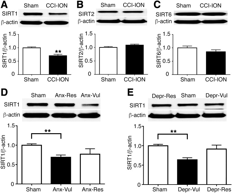
To investigate the underlying molecular mechanism for the individual pain vulnerability of negative emotion, we determined the role of sirtuin family members SIRT1, SIRT2, and SIRT6 in the CeA of the same groups of sham and CCI-ION rats as above. We found that the protein level of SIRT1 in CeA of CCI-ION rats was significantly decreased (p < 0.01), but not that of SIRT2 or SIRT6, when compared with sham rats (Fig. 2A–C). We then determined whether SIRT1 was associated with the emotional pain vulnerability. Interestingly, we found that both anxiety-vulnerable rats and depression-vulnerable rats displayed a significantly reduced level of SIRT1 in CeA, but no change was found in anxiety-resistant rats or depression-resistant rats (Fig. 2D,E). These data indicate that SIRT1 may act as a key epigenetic regulator in the pain vulnerability to negative emotion.
Figure 2.
SIRT1 in CeA neurons is involved in emotional pain vulnerability. A–C, Western blots (top) and group data (bottom) of SIRT1, SIRT2, SIRT6, and β-actin proteins in the CeA in sham rats and CCI-ION rats 28 d after the sham or CCI-ION surgery. D, Western blots (top) and group data (bottom) of SIRT1 and β-actin proteins in the CeA of sham rats, anxiety-vulnerable CCI-ION rats (n = 17), and anxiety-resistant CCI-ION rats (n = 5). E, Similar data of CeA SIRT1 and β-actin proteins in sham rats, in depression-vulnerable CCI-ION rats (n = 18), and in depression-resistant CCI-ION rats (n = 4). N = 22 both in the sham group and in the CCI-ION group. **p < 0.01.
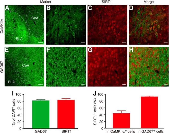
To determine the cell types for SIRT1 location in CeA neurons, we used immunohistochemistry to examine the colocalization of SIRT1 and a cell marker in CeA cells that expressed CaMKIIα or the GABAergic cell marker GAD-67. We found that, in DAPI-labeled CeA cells, ∼84.5 ± 3.6% were stained for SIRT1 and 82.6 ± 2.6% expressed GAD-67. In CaMKIIα-positive cells, ∼44.5 ± 7.1% were also labeled for SIRT1, and in GAD67-positive cells, 93.4 ± 1.4% also expressed SIRT1 (Fig. 3). These immunohistochemical data suggest that the majority of CeA cells are GABAergic cells, consistent with previous reports (Tye et al., 2011), and SIRT1 is widely expressed in CeA cells and predominantly in GABAergic cells.
Figure 3.
Cellular localization of SIRT1 in CeA cells. A–H, Representative images with immunohistochemical staining for CaMKIIα (green; A, B), GAD67 (green; E, F), and for SIRT1 (red; C, G), and merged images of B and C for colocalization of CaMKIIα and SIRT1 (D) and merged images of F and G for colocalization of GAD67 and SIRT1 (H). Scale bars: A, E [showing CeA and basolateral amygdala (BLA)], 100 μm; B–D and F–H (showing a randomly selected area in CeA), 20 μm. I, J, Graphs showing percentage of GAD67-positive (+) and SIRT1+ cells in DAPI-labeled CeA cells (I), and the percentage of SIRT1+ cells in CaMKIIα-labeled and in GAD67-labeled CeA cells (J).
SIRT1 overexpression in CeA decreases emotional pain vulnerability
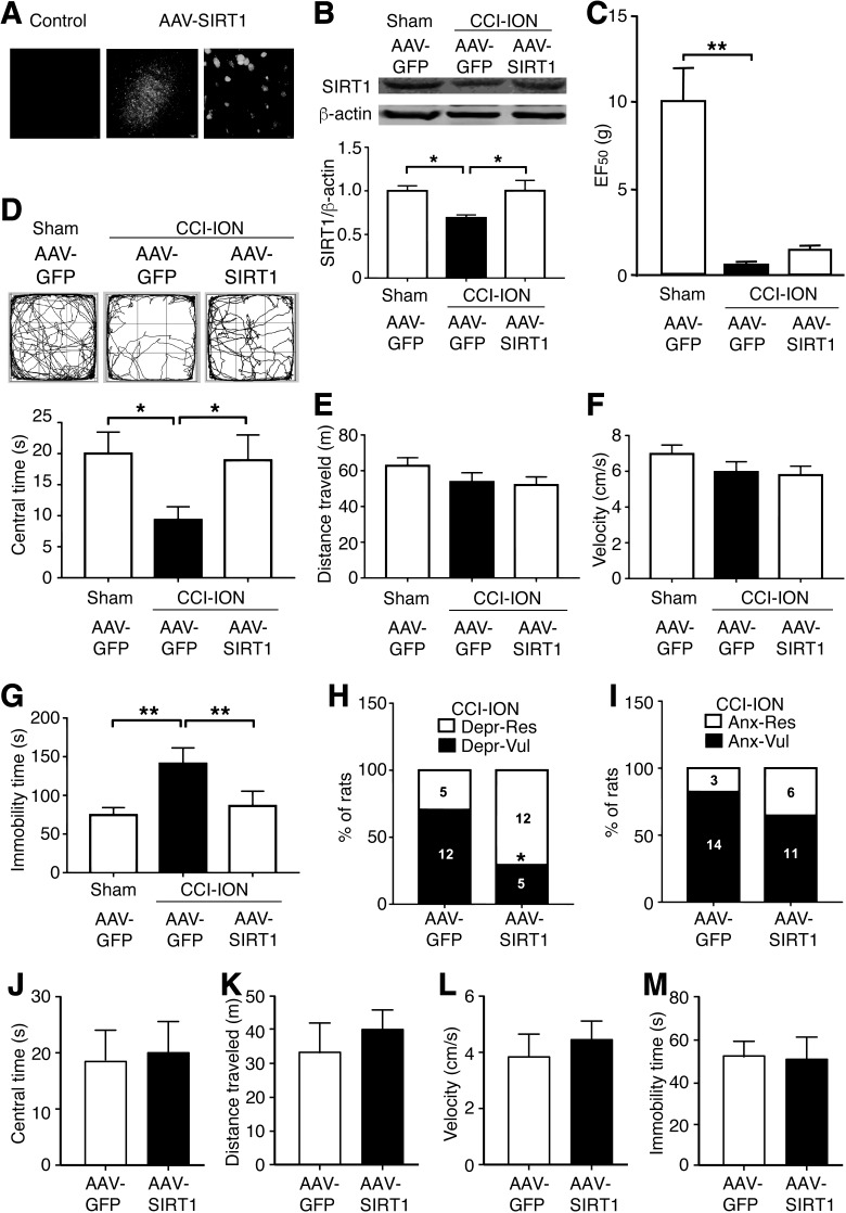
We then investigated whether overcoming the pain-induced SIRT1 reduction, by SIRT1 overexpression in CeA neurons, would alter anxiety- and depression-like behaviors in separate groups of sham and CCI-ION rats. An AAV vector expressing SIRT1 and GFP under the control of CaMKIIα promoter (AAV-SIRT1) was microinjected into the CeA of CCI-ION rats. Control rats were injected with an AAV vector that expresses GFP only. Sensory pain and emotion behaviors were evaluated 15 d after the AAV microinjection and 28 d after the CCI-ION surgery. GFP fluorescence images demonstrated that CeA cells were successfully transfected virally in CCI-ION rats (Fig. 4A). We found that intra-CeA infusion of AAV-SIRT1 significantly upregulated the SIRT1 protein in CCI-ION rats, reversing the pain-induced SIRT1 reduction (p < 0.05; Fig. 4B). Interestingly, the SIRT1 overexpression did not alter the mechanical threshold of sensory pain (Fig. 4C). However, the SIRT1 overexpression reversed the effect of chronic sensory pain on anxiety behavior, with significantly increased central time in open field test in the CCI-ION rats after SIRT1 overexpression when compared with the AAV-GFP-infused CCI-ION rats (p < 0.05; Fig. 4D). There was no change in total distance traveled or travel velocity among the three groups of sham and AAV-GFP-infused or AAV-SIRT1-infused CCI-ION rats (Fig. 4E,F). In the forced swim test, SIRT1 overexpression in CeA also inhibited the pain effect with significantly decreased immobility time in CCI-ION rats (p < 0.05; Fig. 4G). Furthermore, SIRT1 overexpression significantly increased the number of depression-resistant CCI-ION rats compared with total CCI-ION rats from 5 of 17 (29%) to 12 of 17 (71%; p < 0.05; Fig. 4H). The SIRT1 overexpression also increased the number of anxiety-resistant rats from 3 of 17 (18%) to 6 of 17 (35%), although it did not reach statistical significance (Fig. 4I). These results suggest that the overexpression of SIRT1 in CeA neurons is able to counteract and inhibit chronic sensory pain-induced behaviors of negative emotion, reducing the vulnerability to emotional pain.
Figure 4.
SIRT1 overexpression in CeA decreases emotional pain vulnerability. A, GFP fluorescence in CeA after CeA infusion of AAV-GFP (control) or AAV-SIRT1 under the control of CaMKIIα promoter in low- and high-magnification images. B, Western blots (top) and group data (bottom) of SIRT1 and β-actin proteins 28 d after the surgery in sham and CCI-ION rats with CeA infusion of AAV-GFP, and in CCI-ION rats with CeA infusion of AAV-SIRT1. N = 6 each group. C, Mechanical pain thresholds in the same three groups of rats as in B (n = 17 each group). D–F, Locomotor traces (top) and central time (bottom; D), distance traveled (E), and travel velocity (F) in the open field test in the same three groups of rats as above. G, Immobility time of depression-like behavior in forced swim test in the same three groups of rats. H, Percentage of depression-vulnerable and depression-resistant rats (numbers in columns refer to the number of rats) in CCI-ION rats with CeA infusion of AAV-GFP or AAV-SIRT1 (n = 17 each group). I, The percentage of anxiety-vulnerable and anxiety-resistant rats in AAV-GFP and AAV-SIRT1-infused CCI-ION rats (n = 17 each group). J–L, Central time (J), distance traveled (K), and travel velocity (L) in open field test in sham rats with CeA infusion of AAV-GFP or AAV-SIRT1 28 d after the surgery. M, Immobility time of depression-like behavior in forced swim test in sham rats with CeA infusion of AAV-GFP or AAV-SIRT1 30 d after the surgery. N = 8 each group. *p < 0.05, **p < 0.01.
To determine whether SIRT1 overexpression in CeA would alter the emotional behaviors regardless of the pain condition, we examined the effects of SIRT1 overexpression in sham animals. We found that similar SIRT1 overexpression in the CeA of sham rats had no significant effect on the anxiety-like or depression-like behavior (Fig. 4J–M). This suggests that increasing the CeA level of SIRT1 may not affect emotional behaviors in general under normal conditions.
SIRT1 knockdown in CeA increases emotional pain vulnerability
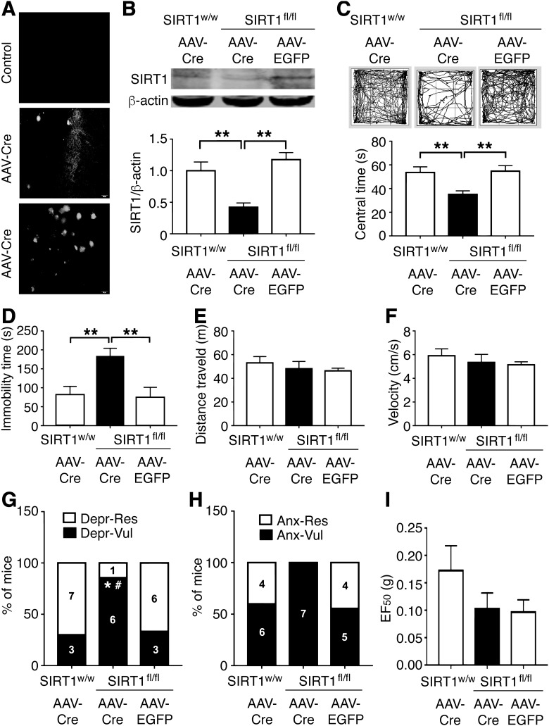
Next, to further confirm the causal role of SIRT1 in the emotional pain vulnerability, we infused an AAV vector expressing Cre recombinase and EGFP under the control of CaMKIIα promoter into the CeA of SIRT1flox/flox mice to knock down CeA SIRT1. WT mice (SIRT1w/w) with CeA infusion of Cre and SIRT1flox/flox mice with CeA infusion of AAV-EGFP were used as two control groups. An EGFP fluorescence image showed that CeA cells were transfected virally in Cre-infused SIRT1flox/flox mice (Fig. 5A). The AAV-Cre infusion significantly knocked down the protein level of SIRT1 in the CeA of SIRT1flox/flox mice when compared with both of the two control groups (Fig. 5B). Behaviorally, we found that the SIRT1 knockdown induced significant anxiety- and depression-like behaviors in AAV-Cre-infused SIRT1flox/flox mice without chronic sensory pain, manifested by reduced central time in the open field test (Fig. 5C) and increased immobility time in the forced swim test (Fig. 5D), respectively. The total distance traveled or travel velocity in the open field test was not changed by the SIRT1 knockdown (Fig. 5E,F). Interestingly, after the knockdown of CeA SIRT1, the number of depression-vulnerable mice significantly increased from 3 of 10 (30%) and 3 of 9 (33%) in the two control groups to 6 of 7 (86%) in the SIRT1 knock-down group (Fig. 5G). For anxiety behavior, the knockdown of CeA SIRT1 also increased the number of anxiety-vulnerable mice, although it did not reach statistical significance when compared with the two control groups of mice (Fig. 5H). On sensory pain, the SIRT1flox/flox mice showed a small, nonsignificant decrease in baseline pain threshold when compared with WT mice for unclear reasons; however, the SIRT1 knockdown did not alter this baseline threshold of mechanical pain (Fig. 5I). Thus, it appears that the decreased function of SIRT1 in CeA neurons is sufficient to cause behaviors of negative emotion with increased vulnerability to negative emotion in the absence of chronic pain.
Figure 5.
SIRT1 knockdown in CeA increases emotional pain vulnerability. A, Staining images of EGFP in CeA after CeA infusion of AAV-EGFP (control) or AAV-Cre under control of CaMKIIα promoter in low- and high-magnification images. B, Western blots (top) and group data (bottom) of SIRT1 and β-actin proteins 15 d after the intra-CeA infusions in WT type (SIRT1w/w) and SIRT1-floxed (SIRT1fl/fl) mice (n = 6 each group). C, Locomotor traces (top) and central time (bottom) in an open field test 15 d after intra-CeA infusion of AAV-Cre in SIRT1w/w mice (n = 10) and in SIRT1fl/fl mice (n = 7), and AAV-EGFP in SIRT1f/f mice (n = 9). D, Immobility time in a forced swim test 15 d after similar CeA infusions of AAV-Cre in SIRT1w/w mice (n = 10) and in SIRT1fl/fl mice (n = 7), and AAV-EGFP in SIRT1fl/fl mice (n = 9). E, F, Distance traveled (E) and travel velocity (F) in an open field test in the same three groups as in C. G, H, Percentage of depression-vulnerable rats (G) and anxiety-vulnerable rats (H) after similar intra-CeA infusions of the AAV vectors in the three groups of mice. I, Thresholds of mechanical pain in the three groups of mice (SIRT1w/w+AAV-Cre, n = 10; SIRT1fl/fl+AAV-Cre, n = 7; SIRT1fl/fl+AAV-EGFP, n = 9). *p < 0.05 (SIRT1fl/fl+AAV-Cre vs SIRT1w/w+AAV-Cre); #p < 0.05 (SIRT1fl/fl+AAV-Cre vs SIRT1fl/fl+AAV-EGFP). **p < 0.01.
SIRT1-mediated decrease in emotional pain vulnerability is associated with downregulation of CaMKIIaα and deacetylation of histone H3K9
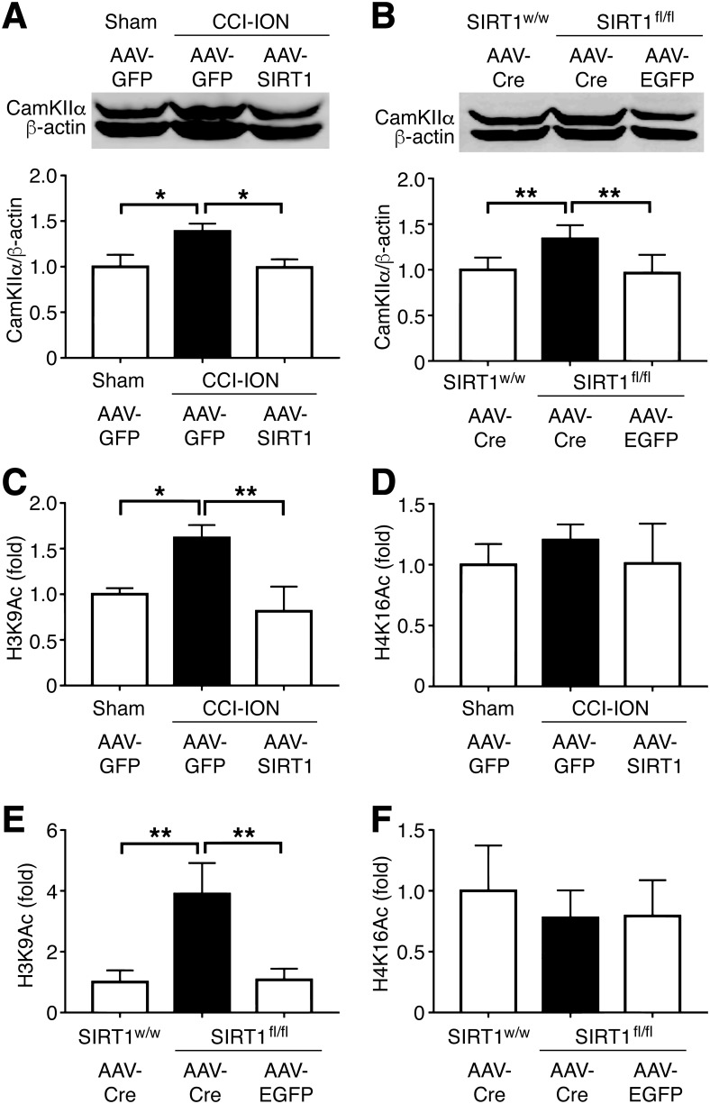
Finally, we determined whether SIRT1 directly regulated CaMKIIα as a regulation target in CeA for its effect on the emotional pain vulnerability. We found that CaMKIIα protein in CeA was significantly increased in CCI-ION rats with chronic pain 28 d after surgery (p < 0.05), and, importantly, this increase was completely reversed by AAV-SIRT1-mediated SIRT1 overexpression in CeA (p < 0.05; Fig. 6A). Moreover, in AAV-Cre-infused SIRT1flox/flox mice with no chronic sensory pain, the knockdown of CeA SIRT1 that induced behaviors of negative emotion (Fig. 5C,D) also significantly increased the CeA level of CaMKIIα protein (Fig. 6B), as in CCI-ION rats with chronic sensory pain. These findings suggest a repressive relationship between SIRT1 and CaMKIIα in CeA and indicate that SIRT1 may decrease the emotional pain vulnerability by downregulating CaMKIIα in CeA.
Figure 6.
A SIRT1-mediated decrease in emotional pain vulnerability is associated with the downregulation of CaMKIIα and the deacetylation of histone H3K9. A, Western blots (top) and group data (bottom) of CaMKIIα and β-actin proteins in the CeA of AAV-GFP-infused sham rats and CCI-ION rats, and of AAV-SIRT1-infused CCI-ION rats 28 d after the surgery (n = 7 each group). B, Similar data of CaMKIIα and β-actin proteins in AAV-Cre-infused SIRT1w/w mice and SIRT1fl/fl mice. C, D, Levels of H3K9Ac (C) and H4K16Ac (D) on the CaMKIIα promoter in the same three groups of rats as in A, 28 d after the surgery. E, F, Levels of H3K9Ac (E) and H4K16Ac (F) on the CaMKIIα promoter in the same three groups of mice as in B, 15 d after the vector infusion. N = 6 each group. *p < 0.05, **p < 0.01.
As an HDAC, SIRT1 has been reported to directly regulate many genes by deacetylating histone H3 at lysine 9 (H3K9) and histone H4 at lysine 16 (H4K16) at the promoters of these genes, leading to their transcriptional repression (Imai et al., 2000; Dang et al., 2009; Bagul et al., 2015; Jing and Lin, 2015). To determine whether SIRT1 decreased the CaMKIIα level in CeA by direct deacetylation, we used ChIP assays to examine the levels of H3K9Ac and H4K16Ac at the promoter of CaMKIIα. Our results showed that the H3K9Ac level on the CaMKIIα promoter was significantly increased in CCI-ION rats with chronic pain (p < 0.05), and this increase was reversed by SIRT1 overexpression (p < 0.01; Fig. 6C). In contrast, compared with controls, the H4K16Ac level was not altered by CCI-ION-induced chronic pain or by SIRT1 overexpression, with no significant difference in the H4K16Ac levels among the three groups (Fig. 6D). Furthermore, the knockdown of CeA SIRT1 also significantly increased, as chronic pain in rats, the level of H3K9Ac (Fig. 6E), but not that of H4K16Ac (Fig. 6F), on the CaMKIIα promoter in AAV-Cre-infused SIRT1flox/flox mice without chronic sensory pain. Together, these results indicate that, under chronic pain, decreased SIRT1 in CeA may cause reduced H3K9 deacetylation, or increased H3K9 acetylation, at the CaMKIIα promoter region, which promotes the transcription of CaMKIIα in CeA neurons, contributing to the emotional pain vulnerability.
Discussion
Our present results have demonstrated that rats with chronic sensory pain display considerable individual variability and distinct vulnerability to emotional disorders (anxiety and depression), which is likely mediated partly by reduced function of SIRT1 in CeA, causing reduced deacetylation, or increased acetylation, of histone H3K9 at the CaMKIIα promoter in CeA neurons. This may represent an epigenetic mechanism underlying the individual pain vulnerability to emotional disorders under chronic pain conditions.
Clinical studies have shown that emotional disorders such as anxiety and depression are common comorbidities of chronic pain that further exacerbate a pain condition (Poole et al., 2009; Liu and Chen, 2014; Yalcin et al., 2014; Usdin and Dimitrov, 2016). Thus, anti-depression and anxiolytic drugs have been commonly used to treat patients with chronic pain, although with limited success (Fernando and Robbins, 2011; Haller and Alicki, 2012; Denk et al., 2014). However, in preclinical research using animal models of chronic pain, the affective/emotional component of chronic pain has been much less studied, possibly due to the extended time (∼4 weeks) required to induce behaviors of negative emotion (vs minutes to induce sensory pain) in animal models and, importantly, to the large individual variance that often results in nonsignificant changes in comparisons of group averages. Recently, a growing number of preclinical studies have demonstrated that chronic sensory pain can induce anxiety- and depression-like behaviors in rodents (Narita et al., 2006; Caspani et al., 2014; Barthas et al., 2015; Wang et al., 2015, 2017; Gambeta et al., 2018). Nevertheless, in contrast to well documented clinical reports of individual vulnerability to the development of chronic pain in patients (Denk et al., 2014), pain vulnerability remains severely understudied in pain research on animal models. The present study provides original evidence suggesting that individual pain vulnerability lies predominantly in brain processing of emotional responses, rather than in body responses to sensory pain, and that this individual emotional pain vulnerability is largely consistent in response to different types of stressful factors (anxiety and depression) under chronic pain.
SIRTs have been shown to play important roles in cerebral ischemia (Zhang et al., 2011; Yang et al., 2013; Khoury et al., 2018), drug addiction (Renthal et al., 2009; Ferguson et al., 2015; Engel et al., 2016), synaptic plasticity (Herskovits and Guarente, 2014; Kim et al., 2019; Zhang et al., 2019), and cognition and aging (Kim et al., 2007; Gao et al., 2010; Zhang et al., 2011). Recent studies have suggested that SIRT1, SIRT2, and SIRT6 are associated with anxious and depressive disorders (Porcelli et al., 2013; Abe-Higuchi et al., 2016; Kim et al., 2016; Nivoli et al., 2016). CeA is known for its role in the regulation of emotional behaviors and modulation of pain (Veinante et al., 2013; Thompson and Neugebauer, 2017). CeA receives nociceptive information from the spinal cord through the spino–parabrachio–amygdaloid pain pathway (Bernard et al., 1993; Gauriau and Bernard, 2002) and processed polymodal information from the cortex through basolateral amygdala (Neugebauer, 2015). Thus, CeA functions as a pivotal structure to integrate peripheral nociceptive information and process affective information from cortical structures for concerted emotional responses. The present study provides three lines of evidence supporting a causal role of SIRT1 in CeA regulation of emotional pain vulnerability, as follows: (1) CeA SIRT1 level is reduced only in vulnerable animals with anxiodepressive behavior under chronic pain; (2) gain of CeA SIRT1 function inhibited pain-induced negative emotion in vulnerable animals; and (3) loss of CeA SIRT1 function mimics the chronic pain effect, increasing emotional pain vulnerability in normal animals. These SIRT1 manipulations did not change animal responses to sensory pain, indicating that CeA SIRT1 may not be involved in the modulation of sensory pain. This is in striking contrast to previous studies showing that SIRT1 in the spinal cord alleviates sensory neuropathic pain induced by various pain conditions (Yin et al., 2013; Shao et al., 2014; Zhou et al., 2017; Chen et al., 2018; Zhang et al., 2019). Our results highlight the distinct SIRT1 functions in the brain and in the spinal cord in the regulation of different components of chronic pain. Given the known roles of CeA in the regulation of sensory pain, the present results also suggest segregated CeA circuits for processing responses to emotional factors and to nociceptive stimulation, a notion supported by a recent study on CeA circuits (Cai et al., 2018).
A series of recent studies has suggested that CaMKIIα dysfunction may underlie multiple neuropsychiatric disorders including depression and anxiety (Robison, 2014). CaMKIIα, a member of Ca2+/calmodulin-dependent protein kinase family, plays an important role in synaptic transmission, learning, and memory (Lisman et al., 2002, 2012). It has been shown that the upregulation of CaMKIIα in the forebrain leads to increased anxiety-like behavior (Hasegawa et al., 2009), while CaMKIIα knockdown in basolateral amygdala decreases 5-HT-induced anxiety-like behavior and reverses repeated stress-induced depression (Kim et al., 2015; Tran and Keele, 2016). In addition, the suppression of CaMKIIα expression in NAc is beneficial to the antidepressant effect of fluoxetine (Robison et al., 2014). These studies suggest a promoting effect of CaMKIIα on these behaviors of negative emotion in these brain areas. However, it has also been reported that antidepressant drugs desmethylimipramine and SKF83959 promote CaMKIIα activation in hippocampus, frontal cortex, and striatum (Consogno et al., 2001; Hasbi et al., 2009; Jiang et al., 2014). The present results are consistent with a negative emotion-promoting role of CaMKIIα, showing its positive correlation with chronic pain-induced behaviors of negative emotion in CeA, a prominent brain site for emotion regulation.
SIRT1, the most studied sirtuin in recent mammalian research, mediates protein deacetylation in a wide range of target proteins and generally functions as a potent protector against neurodegenerative diseases and neuropathological conditions, including inflammation (Herranz and Serrano, 2010; Paraíso et al., 2013; Herskovits and Guarente, 2014). The current study presents molecular evidence showing that SIRT1 can repress CaMKIIα expression by deacetylating histone H3K9 at the CaMKIIα promoter in CeA neurons. This suggests that the SIRT1–CaMKIIα pathway in CeA may act as an epigenetic mechanism to guard against the development of negative emotion under chronic pain, and its dysfunction increases this pain vulnerability to such development of emotional disorders. This is consistent with the general protective roles of brain SIRT1 in other pathological and disease conditions. It is currently unknown whether this protection mechanism of SIRT1 identified here in CeA is also involved in other SIRT1-related pathological conditions beyond pain. Given the widely recognized roles of SIRT1 in the neuropathological conditions, similar mechanisms of SIRT1 protection in the amygdala are likely involved in the altered emotional behaviors induced by other nonpain triggers.
For the cellular localization of SIRT1, our immunohistochemical results suggest that SIRT1 is predominantly expressed in GAD-67-positive GABAergic cells in CeA. As >90% of CeA cells are GABAergic (Tye et al., 2011), it indicates that GABAergic CeA cells play a major role in the SIRT1 regulation of pain-related emotional behaviors. CaMKIIα is commonly localized in excitatory central neurons (Hudmon and Schulman, 2002). However, recent studies have shown that many inhibitory GABAergic cells, including those in CeA, also express CaMKIIα (Jennings et al., 2013; Dedic et al., 2018). Moreover, in line with the effects of the AAV vectors under the control of the CaMKIIα promoter for manipulating SIRT1 expression in this study, optical activation of neurons in the bed nucleus of the stria terminalis through CaMKIIα promoter-controlled channelrhodopsin produces GABAergic synaptic currents (Jennings et al., 2013). Thus, CaMKIIα is not an exclusive marker for excitatory cells.
It is worth noting that, in our analysis, using the sham average to divide the animals with chronic pain into vulnerable and resistant groups is arbitrary, as the distribution of the emotional parameters (central times and immobility times) in the pain animals is continuous with no clear separation between the two groups. In fact, this continuous distribution is also likely expected in clinical cases where the patients' pain vulnerability could vary from highly vulnerable to highly resistant and anywhere in between. Nevertheless, this simplified analysis allowed simple comparisons for mechanistic studies, and the inclusion of these in between individuals would only underestimate the difference between the vulnerable and resistant animals. Another point worth discussing is the marginal insignificance between the depression-resistant group and sham control in the anxiety test (Fig. 1L). Only 4 of 22 pain animals (18%) were resistant to depression (Fig. 1J), so that the small group size of depression-resistant animals might have contributed to the insignificance. Another possible reason is that this analysis of vulnerability to both anxiety and depression in the same animals introduced yet an additional factor (anxiety) with large individual variance to the analysis of largely variable depression behavior.
In summary, the present study demonstrates that chronic neuropathic pain decreases the function of SIRT1 and results in transcriptional activation of CaMKIIα in emotion-regulating CeA, which likely mediates individual vulnerability to development of affective disorders of negative emotion under chronic pain conditions. These results suggest that SIRT1 activators may be used in a novel therapeutic approach for individual-based treatment of chronic pain.
Footnotes
This work was supported by National Institutes of Health, National Institute of Dental and Craniofacial Research Grant DE-025943; National Institute of Neurological Disorders and Stroke Grant NS-113256; National Natural Science Foundation of China Grant 81573481; and the Key Subject of Colleges and Universities Natural Science Foundation of Jiangsu Province Grant 18KJA320008.
The authors declare no competing financial interests.
References
- Abe N, Uchida S, Otsuki K, Hobara T, Yamagata H, Higuchi F, Shibata T, Watanabe Y (2011) Altered sirtuin deacetylase gene expression in patients with a mood disorder. J Psychiatr Res 45:1106–1112. 10.1016/j.jpsychires.2011.01.016 [DOI] [PubMed] [Google Scholar]
- Abe-Higuchi N, Uchida S, Yamagata H, Higuchi F, Hobara T, Hara K, Kobayashi A, Watanabe Y (2016) Hippocampal sirtuin 1 signaling mediates depression-like behavior. Biol Psychiatry 80:815–826. 10.1016/j.biopsych.2016.01.009 [DOI] [PubMed] [Google Scholar]
- Babaev O, Piletti Chatain C, Krueger-Burg D (2018) Inhibition in the amygdala anxiety circuitry. Exp Mol Med 50:18. 10.1038/s12276-018-0063-8 [DOI] [PMC free article] [PubMed] [Google Scholar]
- Bagul PK, Deepthi N, Sultana R, Banerjee SK (2015) Resveratrol ameliorates cardiac oxidative stress in diabetes through deacetylation of NFkB-p65 and histone 3. J Nutr Biochem 26:1298–1307. 10.1016/j.jnutbio.2015.06.006 [DOI] [PubMed] [Google Scholar]
- Barthas F, Sellmeijer J, Hugel S, Waltisperger E, Barrot M, Yalcin I (2015) The anterior cingulate cortex is a critical hub for pain-induced depression. Biol Psychiatry 77:236–245. 10.1016/j.biopsych.2014.08.004 [DOI] [PubMed] [Google Scholar]
- Bernard JF, Alden M, Besson JM (1993) The organization of the efferent projections from the pontine parabrachial area to the amygdaloid complex: a phaseolus vulgaris leucoagglutinin (PHA-L) study in the rat. J Comp Neurol 329:201–229. 10.1002/cne.903290205 [DOI] [PubMed] [Google Scholar]
- Cai YQ, Wang W, Paulucci-Holthauzen A, Pan ZZ (2018) Brain circuits mediating opposing effects on emotion and pain. J Neurosci 38:6340–6349. 10.1523/JNEUROSCI.2780-17.2018 [DOI] [PMC free article] [PubMed] [Google Scholar]
- Caspani O, Reitz MC, Ceci A, Kremer A, Treede RD (2014) Tramadol reduces anxiety-related and depression-associated behaviors presumably induced by pain in the chronic constriction injury model of neuropathic pain in rats. Pharmacol Biochem Behav 124:290–296. 10.1016/j.pbb.2014.06.018 [DOI] [PubMed] [Google Scholar]
- Chalkiadaki A, Guarente L (2015) The multifaceted functions of sirtuins in cancer. Nat Rev Cancer 15:608–624. 10.1038/nrc3985 [DOI] [PubMed] [Google Scholar]
- Chen K, Fan J, Luo ZF, Yang Y, Xin WJ, Liu CC (2018) Reduction of SIRT1 epigenetically upregulates NALP1 expression and contributes to neuropathic pain induced by chemotherapeutic drug bortezomib. J Neuroinflammation 15:292. 10.1186/s12974-018-1327-x [DOI] [PMC free article] [PubMed] [Google Scholar]
- Consogno E, Racagni G, Popoli M (2001) Modifications in brain CaM kinase II after long-term treatment with desmethylimipramine. Neuropsychopharmacology 24:21–30. 10.1016/S0893-133X(00)00176-7 [DOI] [PubMed] [Google Scholar]
- Dang W, Steffen KK, Perry R, Dorsey JA, Johnson FB, Shilatifard A, Kaeberlein M, Kennedy BK, Berger SL (2009) Histone H4 lysine 16 acetylation regulates cellular lifespan. Nature 459:802–807. 10.1038/nature08085 [DOI] [PMC free article] [PubMed] [Google Scholar]
- Dedic N, Kühne C, Jakovcevski M, Hartmann J, Genewsky AJ, Gomes KS, Anderzhanova E, Pöhlmann ML, Chang S, Kolarz A, Vogl AM, Dine J, Metzger MW, Schmid B, Almada RC, Ressler KJ, Wotjak CT, Grinevich V, Chen A, Schmidt MV, et al. (2018) Chronic CRH depletion from GABAergic, long-range projection neurons in the extended amygdala reduces dopamine release and increases anxiety. Nat Neurosci 21:803–807. 10.1038/s41593-018-0151-z [DOI] [PMC free article] [PubMed] [Google Scholar]
- Denk F, McMahon SB, Tracey I (2014) Pain vulnerability: a neurobiological perspective. Nat Neurosci 17:192–200. 10.1038/nn.3628 [DOI] [PubMed] [Google Scholar]
- Egervari G, Ciccocioppo R, Jentsch JD, Hurd YL (2018) Shaping vulnerability to addiction- the contribution of behavior, neural circuits and molecular mechanisms. Neurosci Biobehav Rev 85:117–125. 10.1016/j.neubiorev.2017.05.019 [DOI] [PMC free article] [PubMed] [Google Scholar]
- Engel GL, Marella S, Kaun KR, Wu J, Adhikari P, Kong EC, Wolf FW (2016) Sir2/Sirt1 links acute inebriation to presynaptic changes and the development of alcohol tolerance, preference, and reward. J Neurosci 36:5241–5251. 10.1523/JNEUROSCI.0499-16.2016 [DOI] [PMC free article] [PubMed] [Google Scholar]
- Failde I, Dueñas M, Ribera MV, Gálvez R, Mico JA, Salazar A, de Sola H, Pérez C (2018) Prevalence of central and peripheral neuropathic pain in patients attending pain clinics in spain: factors related to intensity of pain and quality of life. J Pain Res 11:1835–1847. 10.2147/JPR.S159729 [DOI] [PMC free article] [PubMed] [Google Scholar]
- Ferguson D, Shao N, Heller E, Feng J, Neve R, Kim HD, Call T, Magazu S, Shen L, Nestler EJ (2015) SIRT1-FOXO3a regulate cocaine actions in the nucleus accumbens. J Neurosci 35:3100–3111. 10.1523/JNEUROSCI.4012-14.2015 [DOI] [PMC free article] [PubMed] [Google Scholar]
- Fernando AB, Robbins TW (2011) Animal models of neuropsychiatric disorders. Annu Rev Clin Psychol 7:39–61. 10.1146/annurev-clinpsy-032210-104454 [DOI] [PubMed] [Google Scholar]
- Gambeta E, Batista MA, Maschio GP, Turnes JM, Araya EI, Chichorro JG (2018) Anxiety- but not depressive-like behaviors are related to facial hyperalgesia in a model of trigeminal neuropathic pain in rats. Physiol Behav 191:131–137. 10.1016/j.physbeh.2018.04.025 [DOI] [PubMed] [Google Scholar]
- Gao J, Wang WY, Mao YW, Gräff J, Guan JS, Pan L, Mak G, Kim D, Su SC, Tsai LH (2010) A novel pathway regulates memory and plasticity via SIRT1 and miR-134. Nature 466:1105–1109. 10.1038/nature09271 [DOI] [PMC free article] [PubMed] [Google Scholar]
- Gauriau C, Bernard JF (2002) Pain pathways and parabrachial circuits in the rat. Exp Physiol 87:251–258. 10.1113/eph8702357 [DOI] [PubMed] [Google Scholar]
- Gilpin NW, Herman MA, Roberto M (2015) The central amygdala as an integrative hub for anxiety and alcohol use disorders. Biol Psychiatry 77:859–869. 10.1016/j.biopsych.2014.09.008 [DOI] [PMC free article] [PubMed] [Google Scholar]
- Haller J, Alicki M (2012) Current animal models of anxiety, anxiety disorders, and anxiolytic drugs. Curr Opin Psychiatry 25:59–64. 10.1097/YCO.0b013e32834de34f [DOI] [PubMed] [Google Scholar]
- Hasbi A, Fan T, Alijaniaram M, Nguyen T, Perreault ML, O'Dowd BF, George SR (2009) Calcium signaling cascade links dopamine D1–D2 receptor heteromer to striatal BDNF production and neuronal growth. Proc Natl Acad Sci U S A 106:21377–21382. 10.1073/pnas.0903676106 [DOI] [PMC free article] [PubMed] [Google Scholar]
- Hasegawa S, Furuichi T, Yoshida T, Endoh K, Kato K, Sado M, Maeda R, Kitamoto A, Miyao T, Suzuki R, Homma S, Masushige S, Kajii Y, Kida S (2009) Transgenic up-regulation of alpha-CaMKII in forebrain leads to increased anxiety-like behaviors and aggression. Mol Brain 2:6. 10.1186/1756-6606-2-6 [DOI] [PMC free article] [PubMed] [Google Scholar]
- Herranz D, Serrano M (2010) SIRT1: recent lessons from mouse models. Nat Rev Cancer 10:819–823. 10.1038/nrc2962 [DOI] [PMC free article] [PubMed] [Google Scholar]
- Herskovits AZ, Guarente L (2014) SIRT1 in neurodevelopment and brain senescence. Neuron 81:471–483. 10.1016/j.neuron.2014.01.028 [DOI] [PMC free article] [PubMed] [Google Scholar]
- Hudmon A, Schulman H (2002) Neuronal CA2+/calmodulin-dependent protein kinase II: the role of structure and autoregulation in cellular function. Annu Rev Biochem 71:473–510. 10.1146/annurev.biochem.71.110601.135410 [DOI] [PubMed] [Google Scholar]
- Imai S, Armstrong CM, Kaeberlein M, Guarente L (2000) Transcriptional silencing and longevity protein Sir2 is an NAD-dependent histone deacetylase. Nature 403:795–800. 10.1038/35001622 [DOI] [PubMed] [Google Scholar]
- Janak PH, Tye KM (2015) From circuits to behaviour in the amygdala. Nature 517:284–292. 10.1038/nature14188 [DOI] [PMC free article] [PubMed] [Google Scholar]
- Jennings JH, Sparta DR, Stamatakis AM, Ung RL, Pleil KE, Kash TL, Stuber GD (2013) Distinct extended amygdala circuits for divergent motivational states. Nature 496:224–228. 10.1038/nature12041 [DOI] [PMC free article] [PubMed] [Google Scholar]
- Jiang B, Wang F, Yang S, Fang P, Deng ZF, Xiao JL, Hu ZL, Chen JG (2014) SKF83959 produces antidepressant effects in a chronic social defeat stress model of depression through BDNF-TrkB pathway. Int J Neuropsychopharmacol 18:pyu096. 10.1093/ijnp/pyu096 [DOI] [PMC free article] [PubMed] [Google Scholar]
- Jing H, Lin H (2015) Sirtuins in epigenetic regulation. Chem Rev 115:2350–2375. 10.1021/cr500457h [DOI] [PMC free article] [PubMed] [Google Scholar]
- Khoury N, Koronowski KB, Young JI, Perez-Pinzon MA (2018) The NAD(+)-dependent family of sirtuins in cerebral ischemia and preconditioning. Antioxid Redox Signal 28:691–710. 10.1089/ars.2017.7258 [DOI] [PMC free article] [PubMed] [Google Scholar]
- Kim D, Nguyen MD, Dobbin MM, Fischer A, Sananbenesi F, Rodgers JT, Delalle I, Baur JA, Sui G, Armour SM, Puigserver P, Sinclair DA, Tsai LH (2007) SIRT1 deacetylase protects against neurodegeneration in models for Alzheimer's disease and amyotrophic lateral sclerosis. EMBO J 26:3169–3179. 10.1038/sj.emboj.7601758 [DOI] [PMC free article] [PubMed] [Google Scholar]
- Kim H, Kim S, Choi JE, Han D, Koh SM, Kim HS, Kaang BK (2019) Decreased neuron number and synaptic plasticity in SIRT3-knockout mice with poor remote memory. Neurochem Res 44:676–682. 10.1007/s11064-017-2417-3 [DOI] [PubMed] [Google Scholar]
- Kim HD, Hesterman J, Call T, Magazu S, Keeley E, Armenta K, Kronman H, Neve RL, Nestler EJ, Ferguson D (2016) SIRT1 mediates depression-like behaviors in the nucleus accumbens. J Neurosci 36:8441–8452. 10.1523/JNEUROSCI.0212-16.2016 [DOI] [PMC free article] [PubMed] [Google Scholar]
- Kim TK, Kim JE, Park JY, Lee JE, Choi J, Kim H, Lee EH, Kim SW, Lee JK, Kang HS, Han PL (2015) Antidepressant effects of exercise are produced via suppression of hypocretin/orexin and melanin-concentrating hormone in the basolateral amygdala. Neurobiol Dis 79:59–69. 10.1016/j.nbd.2015.04.004 [DOI] [PubMed] [Google Scholar]
- Kim YS, Chu Y, Han L, Li M, Li Z, LaVinka PC, Sun S, Tang Z, Park K, Caterina MJ, Ren K, Dubner R, Wei F, Dong X (2014) Central terminal sensitization of TRPV1 by descending serotonergic facilitation modulates chronic pain. Neuron 81:873–887. 10.1016/j.neuron.2013.12.011 [DOI] [PMC free article] [PubMed] [Google Scholar]
- Kosson D, Malec-Milewska M, Galazkowski R, Rzonca P (2018) Analysis of anxiety, depression and aggression in patients attending pain clinics. Int J Environ Res Public Health 15:1. 10.3390/ijerph15010001 [DOI] [PMC free article] [PubMed] [Google Scholar]
- Lisman J, Schulman H, Cline H (2002) The molecular basis of CaMKII function in synaptic and behavioural memory. Nat Rev Neurosci 3:175–190. 10.1038/nrn753 [DOI] [PubMed] [Google Scholar]
- Lisman J, Yasuda R, Raghavachari S (2012) Mechanisms of CaMKII action in long-term potentiation. Nat Rev Neurosci 13:169–182. 10.1038/nrn3192 [DOI] [PMC free article] [PubMed] [Google Scholar]
- Liu MG, Chen J (2014) Preclinical research on pain comorbidity with affective disorders and cognitive deficits: challenges and perspectives. Prog Neurobiol 116:13–32. 10.1016/j.pneurobio.2014.01.003 [DOI] [PubMed] [Google Scholar]
- Liu R, Dang W, Du Y, Zhou Q, Jiao K, Liu Z (2015) SIRT2 is involved in the modulation of depressive behaviors. Sci Rep 5:8415. 10.1038/srep08415 [DOI] [PMC free article] [PubMed] [Google Scholar]
- Luo XJ, Zhang C (2016) Down-regulation of SIRT1 gene expression in major depressive disorder. Am J Psychiatry 173:1046. 10.1176/appi.ajp.2016.16040394 [DOI] [PubMed] [Google Scholar]
- Narita M, Kaneko C, Miyoshi K, Nagumo Y, Kuzumaki N, Nakajima M, Nanjo K, Matsuzawa K, Yamazaki M, Suzuki T (2006) Chronic pain induces anxiety with concomitant changes in opioidergic function in the amygdala. Neuropsychopharmacology 31:739–750. 10.1038/sj.npp.1300858 [DOI] [PubMed] [Google Scholar]
- Neugebauer V. (2015) Amygdala pain mechanisms. Handb Exp Pharmacol 227:261–284. 10.1007/978-3-662-46450-2_13 [DOI] [PMC free article] [PubMed] [Google Scholar]
- Nivoli A, Porcelli S, Albani D, Forloni G, Fusco F, Colom F, Vieta E, Serretti A (2016) Association between sirtuin 1 gene rs10997870 polymorphism and suicide behaviors in bipolar disorder. Neuropsychobiology 74:1–7. 10.1159/000446921 [DOI] [PubMed] [Google Scholar]
- Palumbo S, Mariotti V, Iofrida C, Pellegrini S (2018) Genes and aggressive behavior: epigenetic mechanisms underlying individual susceptibility to aversive environments. Front Behav Neurosci 12:117. 10.3389/fnbeh.2018.00117 [DOI] [PMC free article] [PubMed] [Google Scholar]
- Paraíso AF, Mendes KL, Santos SH (2013) Brain activation of SIRT1: role in neuropathology. Mol Neurobiol 48:681–689. 10.1007/s12035-013-8459-x [DOI] [PubMed] [Google Scholar]
- Poole H, White S, Blake C, Murphy P, Bramwell R (2009) Depression in chronic pain patients: prevalence and measurement. Pain Pract 9:173–180. 10.1111/j.1533-2500.2009.00274.x [DOI] [PubMed] [Google Scholar]
- Porcelli S, Salfi R, Politis A, Atti AR, Albani D, Chierchia A, Polito L, Zisaki A, Piperi C, Liappas I, Alberti S, Balestri M, Marsano A, Stamouli E, Mailis A, Biella G, Forloni G, Bernabei V, Ferrari B, Lia L, et al. (2013) Association between sirtuin 2 gene rs10410544 polymorphism and depression in Alzheimer's disease in two independent european samples. J Neural Transm 120:1709–1715. 10.1007/s00702-013-1045-6 [DOI] [PubMed] [Google Scholar]
- Price DD. (2000) Psychological and neural mechanisms of the affective dimension of pain. Science 288:1769–1772. 10.1126/science.288.5472.1769 [DOI] [PubMed] [Google Scholar]
- Renthal W, Kumar A, Xiao G, Wilkinson M, Covington HE 3rd, Maze I, Sikder D, Robison AJ, LaPlant Q, Dietz DM, Russo SJ, Vialou V, Chakravarty S, Kodadek TJ, Stack A, Kabbaj M, Nestler EJ (2009) Genome-wide analysis of chromatin regulation by cocaine reveals a role for sirtuins. Neuron 62:335–348. 10.1016/j.neuron.2009.03.026 [DOI] [PMC free article] [PubMed] [Google Scholar]
- Robison AJ. (2014) Emerging role of CaMKII in neuropsychiatric disease. Trends Neurosci 37:653–662. 10.1016/j.tins.2014.07.001 [DOI] [PubMed] [Google Scholar]
- Robison AJ, Vialou V, Sun HS, Labonte B, Golden SA, Dias C, Turecki G, Tamminga C, Russo S, Mazei-Robison M, Nestler EJ (2014) Fluoxetine epigenetically alters the CaMKIIalpha promoter in nucleus accumbens to regulate DeltaFosB binding and antidepressant effects. Neuropsychopharmacology 39:1178–1186. 10.1038/npp.2013.319 [DOI] [PMC free article] [PubMed] [Google Scholar]
- Shao H, Xue Q, Zhang F, Luo Y, Zhu H, Zhang X, Zhang H, Ding W, Yu B (2014) Spinal SIRT1 activation attenuates neuropathic pain in mice. PLoS One 9:e100938. 10.1371/journal.pone.0100938 [DOI] [PMC free article] [PubMed] [Google Scholar]
- Sillivan SE, Joseph NF, Jamieson S, King ML, Chévere-Torres I, Fuentes I, Shumyatsky GP, Brantley AF, Rumbaugh G, Miller CA (2017) Susceptibility and resilience to posttraumatic stress disorder-like behaviors in inbred mice. Biol Psychiatry 82:924–933. 10.1016/j.biopsych.2017.06.030 [DOI] [PMC free article] [PubMed] [Google Scholar]
- Slattery DA, Cryan JF (2012) Using the rat forced swim test to assess antidepressant-like activity in rodents. Nat Protoc 7:1009–1014. 10.1038/nprot.2012.044 [DOI] [PubMed] [Google Scholar]
- Thompson JM, Neugebauer V (2017) Amygdala plasticity and pain. Pain Res Manag 2017:8296501. 10.1155/2017/8296501 [DOI] [PMC free article] [PubMed] [Google Scholar]
- Tran L, Keele NB (2016) CaMKIIalpha knockdown decreases anxiety in the open field and low serotonin-induced upregulation of GluA1 in the basolateral amygdala. Behav Brain Res 303:152–159. 10.1016/j.bbr.2016.01.053 [DOI] [PMC free article] [PubMed] [Google Scholar]
- Tye KM, Prakash R, Kim SY, Fenno LE, Grosenick L, Zarabi H, Thompson KR, Gradinaru V, Ramakrishnan C, Deisseroth K (2011) Amygdala circuitry mediating reversible and bidirectional control of anxiety. Nature 471:358–362. 10.1038/nature09820 [DOI] [PMC free article] [PubMed] [Google Scholar]
- Usdin TB, Dimitrov EL (2016) The effects of extended pain on behavior: recent progress. Neuroscientist 22:521–533. 10.1177/1073858416633104 [DOI] [PubMed] [Google Scholar]
- Veinante P, Yalcin I, Barrot M (2013) The amygdala between sensation and affect: a role in pain. J Mol Psychiatry 1:9. 10.1186/2049-9256-1-9 [DOI] [PMC free article] [PubMed] [Google Scholar]
- Wang J, Hodes GE, Zhang H, Zhang S, Zhao W, Golden SA, Bi W, Menard C, Kana V, Leboeuf M, Xie M, Bregman D, Pfau ML, Flanigan ME, Esteban-Fernández A, Yemul S, Sharma A, Ho L, Dixon R, Merad M, et al. (2018) Epigenetic modulation of inflammation and synaptic plasticity promotes resilience against stress in mice. Nat Commun 9:477. 10.1038/s41467-017-02794-5 [DOI] [PMC free article] [PubMed] [Google Scholar]
- Wang W, Li C, Cai Y, Pan ZZ (2017) Pain vulnerability and DNA methyltransferase 3a involved in the affective dimension of chronic pain. Mol Pain 13:1–12. 10.1177/1744806917726713 [DOI] [PMC free article] [PubMed] [Google Scholar]
- Wang XQ, Zhong XL, Li ZB, Wang HT, Zhang J, Li F, Zhang JY, Dai RP, Xin-Fu Z, Li CQ, Li ZY, Bi FF (2015) Differential roles of hippocampal glutamatergic receptors in neuropathic anxiety-like behavior after partial sciatic nerve ligation in rats. BMC Neurosci 16:14. 10.1186/s12868-015-0150-x [DOI] [PMC free article] [PubMed] [Google Scholar]
- Wei F, Guo W, Zou S, Ren K, Dubner R (2008) Supraspinal glial-neuronal interactions contribute to descending pain facilitation. J Neurosci 28:10482–10495. 10.1523/JNEUROSCI.3593-08.2008 [DOI] [PMC free article] [PubMed] [Google Scholar]
- Wiech K, Tracey I (2009) The influence of negative emotions on pain: behavioral effects and neural mechanisms. Neuroimage 47:987–994. 10.1016/j.neuroimage.2009.05.059 [DOI] [PubMed] [Google Scholar]
- Yalcin I, Barthas F, Barrot M (2014) Emotional consequences of neuropathic pain: insight from preclinical studies. Neurosci Biobehav Rev 47:154–164. 10.1016/j.neubiorev.2014.08.002 [DOI] [PubMed] [Google Scholar]
- Yang Y, Duan W, Li Y, Yan J, Yi W, Liang Z, Wang N, Yi D, Jin Z (2013) New role of silent information regulator 1 in cerebral ischemia. Neurobiol Aging 34:2879–2888. 10.1016/j.neurobiolaging.2013.06.008 [DOI] [PubMed] [Google Scholar]
- Yin Q, Lu FF, Zhao Y, Cheng MY, Fan Q, Cui J, Liu L, Cheng W, Yan CD (2013) Resveratrol facilitates pain attenuation in a rat model of neuropathic pain through the activation of spinal Sirt1. Reg Anesth Pain Med 38:93–99. 10.1097/AAP.0b013e3182795b23 [DOI] [PubMed] [Google Scholar]
- Zhang F, Wang S, Gan L, Vosler PS, Gao Y, Zigmond MJ, Chen J (2011) Protective effects and mechanisms of sirtuins in the nervous system. Prog Neurobiol 95:373–395. 10.1016/j.pneurobio.2011.09.001 [DOI] [PMC free article] [PubMed] [Google Scholar]
- Zhang Z, Zhang P, Qi GJ, Jiao FJ, Wang QZ, Yan JG, He F, Zhang Q, Lv ZX, Peng X, Cai HW, Chen X, Sun N, Tian B (2018) CDK5-mediated phosphorylation of Sirt2 contributes to depressive-like behavior induced by social defeat stress. Biochim Biophys Acta Mol Basis Dis 1864:533–541. 10.1016/j.bbadis.2017.11.012 [DOI] [PubMed] [Google Scholar]
- Zhang Z, Ding X, Zhou Z, Qiu Z, Shi N, Zhou S, Du L, Zhu X, Wu Y, Yin X, Zhou C (2019) SIRT1 alleviates diabetic neuropathic pain by regulating synaptic plasticity of spinal dorsal horn neurons. Pain 160:1082–1092. 10.1097/j.pain.0000000000001489 [DOI] [PubMed] [Google Scholar]
- Zhou CH, Zhang MX, Zhou SS, Li H, Gao J, Du L, Yin XX (2017) SIRT1 attenuates neuropathic pain by epigenetic regulation of mGluR1/5 expressions in type 2 diabetic rats. Pain 158:130–139. 10.1097/j.pain.0000000000000739 [DOI] [PubMed] [Google Scholar]


