. 2020 Feb 5;9:e51247. doi: 10.7554/eLife.51247

Figure 3. The LtgA helix 30 mutant shows morphological abnormalities.

(a) Morphological differences between the wild type and strains expressing the mutant lytic transglycosylases were determined by fluorescence microscopy (left panel) and scanning electron microscopy (SEM) (rightpanel). White arrows in the images of ΔltgAltgAΔ30 strain (left panel) point to cells defective in division and separation, as well as extracellular material. White arrows in the right panel points to, irregular cell surfaces, high-molecular-weight blebs (not observed in other strains), asymmetrical diplococci, and ghost cells (see Figure 3—figure supplement 1 for other images detailing additional morphological abnormalities). (b) Quantification of the confocal microscopy data. The different fields were manually counted to evaluate the number of cells per unit. Each unit is defined as an isolated cluster of cells that it is not in contact with other cells. Whenever in contact two cells were defined as apart of the same unit.

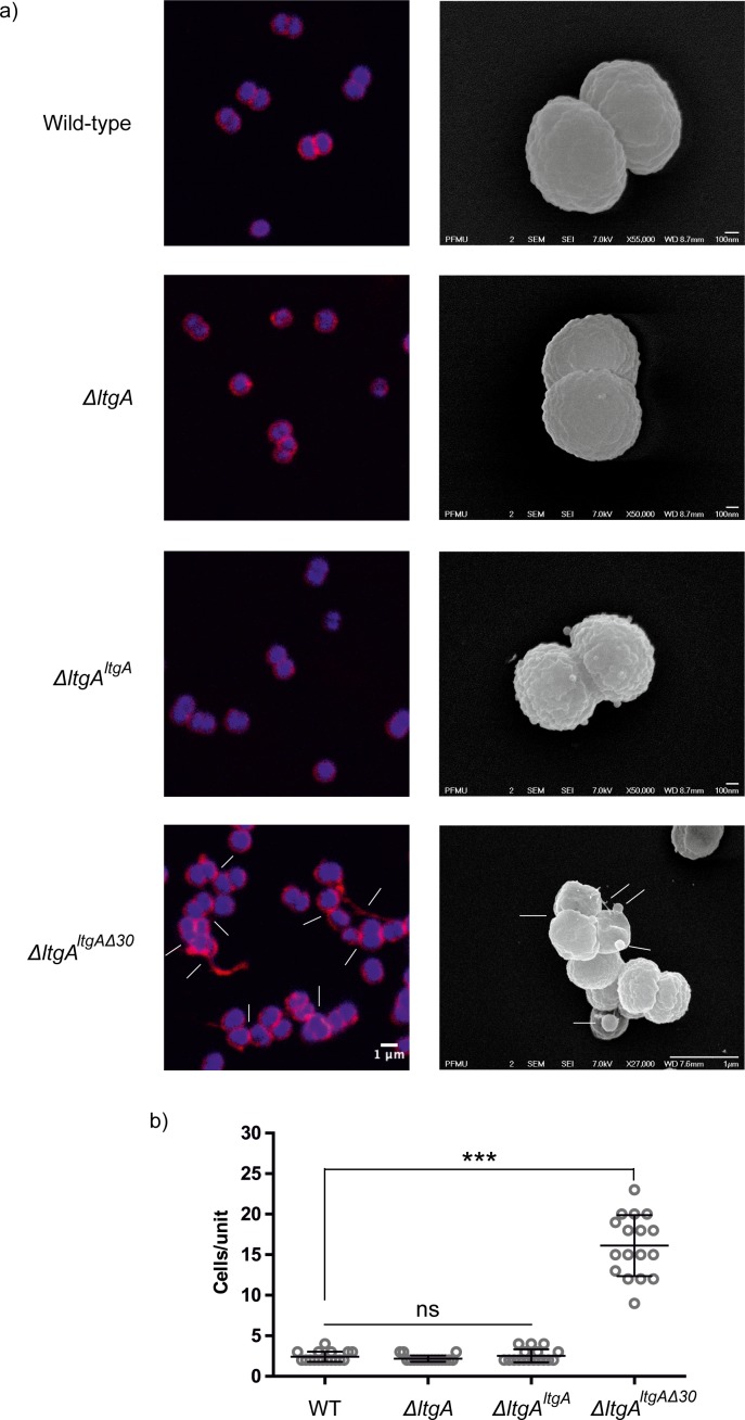
Figure 3.

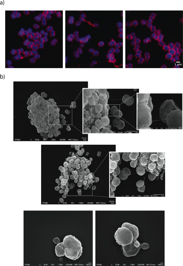
Figure 3—figure supplement 1. Morphological abnormalities of LtgA alpha helix 30 mutant.

Figure 3—figure supplement 1.

(a) Fluorescent microscopy of ΔltgAltgAΔ30 strain highlighting aggregated cells that are defective in division and separation. (b) Scanning electron microscopy of ΔltgAltgAΔ30 strain detailing additional morphological abnormalities such as, aggregation of extracellular material that resembles type IV pilin protein structures that stretches between diplococcic bacteria.
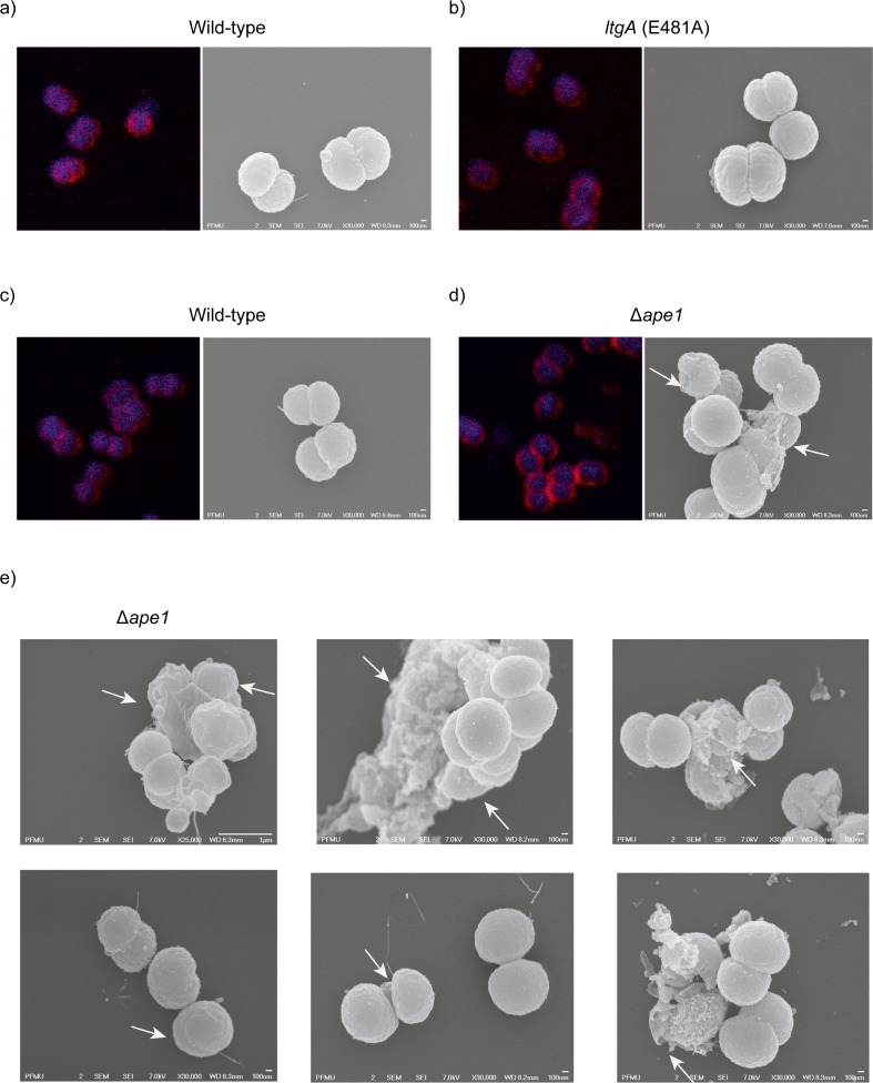
Figure 3—figure supplement 2. Morphological abnormalities of the Δape1 strain.

Figure 3—figure supplement 2.

(a) Strains (8013) expressing wild-type lytic transglycosylase were imaged using fluorescent microscopy (right panel) and scanning electron microscopy (SEM) (left panel). (b) Strains (8013) expressing mutant lytic transglycosylases (E481A) were imaged by fluorescent microscopy (right panel) and scanning electron microscopy (SEM) (left panel). (c) Strains (MC58) expressing wild-type lytic transglycosylase were imaged using fluorescent microscopy (right panel) and scanning electron microscopy (SEM) (left panel), (d–e) The Δape1 strains were imaged using fluorescent microscopy (right panel) and scanning electron microscopy (SEM) (left panel). White arrows in the images points to, irregular cell surfaces, high-molecular-weight blebs (not observed in other strains), asymmetrical diplococci, and lysed bacteria.