Abstract
BACKGROUND:
Anatomic variations in palatal canal morphology in maxillary first molars (MFMs) are relatively rare occurrences. Therefore, omission is common unless clinicians recognize their presence.
CASE REPORT:
The aim of this report is to point out new signs that can be viewed as indicators of the existence of additional canals in the palatal root (PR) in this upper first molar endodontic retreatment case. Moreover, the role of preoperative cone-beam computed tomography (CBCT) in both discovering and determining the location of those additional canals will also be discussed.
CONCLUSION:
Besides formerly discussed signs that indicate the existence of this canal, clinicians should also pay attention to other signals on periapical radiograph, including the aberrant divergence of a palatal canal at apical third and an unusual lesion occurring laterally in the periapical area of palatal root.
Keywords: Cone-beam computed tomography, Upper first molar, Endodontic retreatment, Bifurcated palatal canal, Second mesiobuccal canal, S-shaped canal
Introduction
For successful endodontic treatment, clinicians should pay attention to anatomic variations in root canal systems besides canal instrumentation and obturation [1]. The canal configurations of maxillary first molars (MFMs) is complex as a result of the presence of additional canals with an extremely high prevalence of the second mesiobuccal (MB2) canal, at over 60% [2], [3], [4], [5]. In contrast, only one single canal, which has one orifice and one foramen, is found in the palatal root (PR) [2], [4]. Nevertheless, there have been reports [6], [7], [8] about cases in which canals split at the last third of the PR are classified type V (1-2 anatomy) according to Vertucci’s classification [9]. This means one orifice for the canal and two distinct apical foramina. Ali Nosrat et al., [10] also presented 5 clinical cases involving the treatment of upper first molar whose canal morphology was far from 1-1 anatomy, four out of which, were classified type V.
The omission of root canals with such internal complexities is one of the principal causes of the failure in treating MFM [11]. To uncover the morphology and variations of root canal systems, lots of researches have been carried out with many different approaches; and a combination of techniques such as dental loupes and microscopes, periapical radiographs or CBCT have been employed [2], [12]. Among all of these, CBCT scanning is considered as an ideal tool to easily visualize root canal systems thanks to its ability to produce high-resolution three-dimensional images [8]. However, compliance with the ALARA rule – “the radiation should be as low as reasonably achievable” – is essential upon the use of CBCT.
This report presents a clinical case that involved the application of preoperative CBCT in retreatment of upper first molar whose previous therapeutic attempt had been a failure due to missed S-shaped MB2 canal and bifurcated palatal canal. This work will also review earlier reports on the same issue and point out new signs on two-dimensional images that suggest the existence of additional palatal canals.
Case Report
A 23-year-old male medical student visited our endodontic department due to the dull and persistent pain on his right maxillary first molar (tooth #16) while eating, especially hard food. The dental history showed that the tooth had received previous endodontic treatment and got a porcelain-fused-to-metal (PFM) crown because of asymptomatic irreversible pulpitis 10 months ago. There was also no medical history of systemic diseases or allergy.
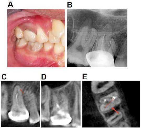
The clinical examination revealed: (1) a marginal deficiency at the buccal surface of the crown (Figure 1A); (2) no percussion sensitivity; and (3) a negative result of pulp test. In addition, there were no signs of swelling, sinus tract, abnormal mobility, and pathological periodontal pocket.
Figure 1.

The preoperative examination; A) A marginal deficiency at the buccal surface of the crown; B) The abnormal lesions were indicated in the periapical radiograph; C) A bifurcated palatal canal at the apical third on the coronal slices of CBCT; D) Remaining obturating material in the mesiodistal root; E) Axial slices showed an untreated MB2 orifice which is located along the MB groove, heading 2 mm palatally from the main MB orifice
The periapical radiograph (Figure 1B) of tooth #16 showed abnormal signs: (1) an incomplete endodontic therapy with underfilled obturating materials in the palatal canal; (2) atypically enlarged periodontal ligament that surrounds the J-formed mesiobuccal (MB) root, suggesting the appearance of curved canals; and (3) and a periapical radiolucent lesion towards the distal side in PR and a vague distal divergence of the palatal canal.
To clarify the dubious signs in the periapical film, a CBCT evaluation of high-resolution scanning (Planmeca ProMax 3D Max; Planmeca Oy, Helsinki, Finland) was performed. CBCT scanning were obtained following the parameters: 96 KV (tube voltage), 5 mA (tube current), a field of view (FOV) 80 X 80 mm, and 150 μm voxel size. The result of CBCT images illustrated: - Firstly, there was a branched canal diverging to the mesial side at the last third of the PR (Figure 1C). The precise location and dimension of the apical lesion around this root were determined; Secondly, remaining root-filling material, though unclear on the periapical radiograph at a coronal third of the mesiodistal root, was observed on the sagittal slices (Figure 1D); and Lastly, a second mesiobuccal (MB2) canal was detected on the axial slices. It was located along the MB groove, heading 2mm palatally, from the main MB orifice (Figure 1E).
Based on clinical and radiographical evaluations, the final diagnosis of tooth #16 was symptomatic apical periodontitis after root canal therapy and non-surgical endodontic retreatment was indicated. The retreatment was performed after written consent had been obtained.
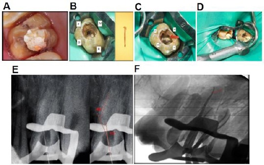
First, the PFM crown was removed by Transmetal bur (Dentsply Sirona, Victoria, Australia). The abutment tooth #16 was exposed, displaying temporary filling material and a protruded portion of gutta-percha at the occlusal surface (Figure 2A) which could be the root canal filling material of distobuccal canal poorly obturated with single cone technique. The tooth was then isolated by applying a rubber dam. The Endo Z bur (Dentsply Maillefer, Ballaiges, Switzerland) allowed us to regain direct access to the pulp chamber. During the procedure, a dental loupe with three-time magnification was used to assist in the inspection of the location of orifices. As all intrapulpal materials were removed, it was easier to visualize the pulpal chamber comprising palatal, distobuccal and mesiobuccal orifices. In particular, a conspicuous pink of gutta-percha inside the palatal canal was exposed.
Figure 2.

During the procedure; A) The abutment tooth #16 was exposed with temporary filling material and Gutta-percha at the occlusal surface; B) The elimination of filling materials from palatal canal using both mechanical and chemical methods; C) The locating of the MB2 root canal based on CBCT; D) The Glide-Path preparation was carried out with PathFile rotary files; E) S-shaped MB2 canal on the working-length radiograph; (F) The master cone radiograph
Therefore, the next plan was eliminating the root filling materials using both mechanical and chemical methods (Figure 2B): - Firstly, removed a coronal portion of gutta-percha with a Gates-Glidden drill #3 (Mani, Tochigi, Japan); - Next, put Eucalyptol solvent (Cerkamed, Stalowa Wola, Polska) into the palatal canal within 2 minutes to dissolve the remaining gutta-percha; and Finally, used a C+ file #15 (Dentsply Maillefer) to penetrate into the remaining gutta-percha and remove it.
In the following treatment, the detection of only 3 orifices and the existence of MB2 canal on preoperative CBCT also showed that this canal was omitted in the previous dental treatment and had to be located.
MB2 canal was identified based on initial assessments on the axial slices. In sequence, the access cavity was enlarged mesially using the Endo Z bur (Dentsply Maillefer) to enhance the observation of the orifices, making the outline of the access cavity transform into rhombus shape. Then, the dentin layer covering the MB2 orifice was removed following a path that run along the MB groove and headed 2 mm towards the palatal canal from the main MB orifice. This allowed MB2 to be located using DG 16 explorer (Hu-Friedy, Chicago, United States). The MB2 canal at the coronal third was then flared with Gate-Glidden drill #3 (Mani) (Figure 2C). Following the determination of four orifices, the #10 K-files (Dentsply Maillefer), which were initially curved following the canal direction on CBCT images, were applied to negotiate the root canals.
The working-length (WL) of each canal was measured by using Propex II apex locator (Dentsply Maillefer) and then verified through the working-length radiographs. On the confirmation film of MB2’s working-length, an S-shaped curvature was revealed and then measured by Schneider’s method [13]. The angle of the first curvature portion was 13O while the second one was 42O. (Figure 2E). In the glide-path preparation, the PathFile rotary files (Dentsply Maillefer) were inserted into the root canals corresponding to the ascertained working-length in the following order: Pathfile 13/.02, 16/.02 and 19/.02 (Figure 2D).
At the second appointment, the complete canal preparation was performed with the Protaper Next X1 (17/.04) and X2 (25/.06) rotary file (Dentsply Maillefer). The branched canal in the palatal root is shaped with precurved hand files from size #10/.04 to #30/.04. Throughout the instrumentation, The Intracanal lubricant with 15% EDTA was applied and the canals were then irrigated with sonic-activated sodium hypochlorite 3% between file insertions. Finally, the root canals were flushed with chlorhexidine 2% and rinsed with saline. Utilized intracanal medicament was calcium hydroxide and CavitTM (3MTM ESPE, United States) was applied as a temporary sealing material.
On the next visit, the root canals were filled with the pre-curved ProTaper Next gutta-percha (GP) X2 and evaluated by means of radiographs (Figure 2F). After drying canals, the GP cones were coated with the AH Plus sealer (Dentsply Maillefer) and inserted into the canals to working-length. The obturating procedure was achieved using continuous wave technique with System B-Cordless Obturation (SybronEndo, Orange, CA). The completed obturation process was evaluated through an immediate postoperative radiograph. Finally, the access cavity was directly restored with composite (Surefil SDR and Ceram X universal; Dentsply Sirona). The male patient, eventually, was introduced to the prosthetic department for full crown restoration.
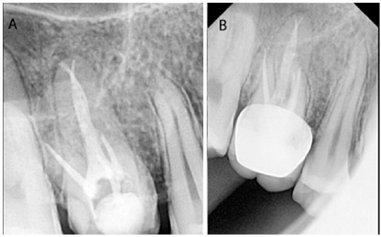
After 6 months of following up, the tooth #16 was asymptomatic as well as complete healing was shown on the radiograph (Figure 3B).
Figure 3.

After the procedure; A) Immediate-postoperative image; B) 6-months-follow-up radiograph
Discussion
The case in question represents the root canal retreatment of a right MFM missing MB2 canal with the bifurcated palatal canal. The canal bifurcation is classified as type V (1-2) according to Vertucci’s classification [9]. Previous studies have reported rare cases of PR containing more than one root canals in MFM, especially that of the Vertucci type V. In one research conducted by Tian et al., [8], CBCT scanning method was applied to study the canal morphology of 1558 MFMs. The findings then revealed statistics on the presence of additional canals, at only 0.7% in the palatal root and an extremely low figure for type V canal anatomy, at 0.05%. As stated in the study done with the same method by Neelakantan et al., [7], the corresponding figures for additional canals and 1-2 canal morphology canal were 9.6% and 1.3% respectively while Vertucci type I (1-1) palatal canal was prevalent in these studies. This indicates that clinicians should carefully examine the existence of bifurcated canals in palatal root through initial radiograph images or clinical inspection besides their typical anatomy (Vertucci type I).
Earlier studies provided two clues about the bifurcation of the palatal canal on periapical radiograph: 1) the unusual diverging gutta-percha on the master cone radiograph or the weird divergence of a master apical hand-file on the working-length film [10], 2) the abnormal divergence of root canal filling material which was obturated previously in palatal canal [10]. In this case, the upper first molar undergoing incomplete endodontic treatment with underfilled obturating material in the palatal canal posed an obstacle to discovering the canal morphology. However, observations of preoperative radiograph showed that the discreet periapical lesion was located on the distal portion of the apical root, suggesting an inflamed canal on that side. Moreover, a distal deviation of the palatal canal at apical third was also disclosed at the film. We supposed that detecting this deviation without gutta-percha point or working-length file was possible because of the canal shaping and cleaning phase in previous treatment. This procedure facilitated the removal of debris, calcified tissues as well as apparently shaped the pathway of the palatal canal. As a consequence, the canal became more radiopaque on the two-dimensional film. This leads to the conclusion that endodontic clinicians should identify both mentioned clues and these following features when determining the possible presence of a bifurcated canal: (1) The aberrant divergence of a palatal canal at apical third on the preoperative radiograph, and (2) An unusual lesion occurring laterally in the periapical area of palatal root.
Once there are signs of the bifurcated palatal canal, clinicians need to re-examine and determine its presence with certainty. On the one hand, according to earlier reports on the case, the canal was only detected after instrumentation with the support of a microscope [6], [10]. More seriously, clinical dentists made a mistake when assuming the palatal root had one canal and one foramen (1-1 anatomy) on the initial periapical film, resulting in failure in the first treatment [10]. On the other hand, the branched canal in the case, which was located at the last third of the PR and barely visible on the two-dimensional radiograph, demonstrated complicated root canal morphology and the need for further examination on the CBCT scan [14]. Therefore, applying preoperative high-resolution CBCT scanning was needed and this method made it possible to confirm the existence and exact position of the bifurcated canal. The use of CBCT not only helped clinically locate additional canals but also avoid complications during canal preparation such as root canal perforation and ledging [15]. In this case, high-resolution CBCT images were taken with the protocol: a FOV 80 x 80 mm, voxel size 150 mm, anode parameters (96 KV and 5 mA). High-resolution scanning mode with voxel size 0.125 mm has been reported to reveal bifurcated canal at apical third of roots as opposed to modes with larger voxel size ( 0.20 mm and 0.25 mm) [16]. The branched canal located at apical third is likely to be identified under CBCT scanning with a voxel size below 0.20 mm.
The process of canal instrumentation for the type V palatal canal is also worth considering. Ultrasonic irrigation combined with irrigants should be applied in the branched canal at the last third of the root due to the capability of removing smear layer efficiently [17]. Once the blockage at the apical third has disappeared, precurved files can slide down the entire working length easily and the risk of separating instruments will be minimized.
Managing the S-shaped curvature MB2 canal is also a major challenge for clinicians. Besides the application of double flare technique [18], the Ni-Ti rotary files by Dentsply, Pathfiles which prove to be flexible, should be applied to create a smooth glide-path in curved canals before the shaping phase with rotary files. Compared to previous manual stainless steel files, conducting canal preparation with Pathfiles considerably minimize canal defects at curvature portion [19] as well as periapical infections of the root [20]. Thus, glide path preparation with these files enables rotary shaping files to not only eliminate infected tissues more effectively but also contribute to obturating the curved canal more tightly in three-dimensions.
Last but not least, the removal of previous filling material in the retreatment should also be considered. When the palatal canal is poorly obturated with underfilled gutta-percha (GP) using single cone technique, manual removal of GP with rigid C+ file and solvent is the chosen method. By adopting this approach, the risk of excessive enlargement at the coronal third of the canal can be reduced. However, the solvent should be injected into the canal through a small-sized needle to avoid being pushed down to the periapical area.
In conclusion, figures have indicated that the existence of type V palatal canals in MFM is just roughly 1%, which is extremely low. Besides formerly discussed signs that indicate the existence of this canal, clinicians should also pay attention to other signals on periapical radiograph, including the aberrant divergence of a palatal canal at apical third and an unusual lesion occurring laterally in the periapical area of palatal root. In addition, further research should be conducted to determine appropriate parameters on CBCT for identifying the presence of branched canals, which can help lower the risks of treatment failure.
Ethics statement
All study protocols were approved by a local ethics committee of the School of Odonto Stomatology, Hanoi Medical University, and have therefore been performed in accordance with the ethical standards laid down in the 1964 Declaration of Helsinki and its later amendments.
Footnotes
Funding: This research did not receive any financial support
Competing Interests: The authors have declared that no competing interests exist
References
- 1.Weine FS, et al. Canal configuration in the mesiobuccal root of the maxillary first molar and its endodontic significance. Oral Surg Oral Med Oral Pathol. 1969;28(3):419–25. doi: 10.1016/0030-4220(69)90237-0. https://doi.org/10.1016/0030-4220(69)90237-0. [DOI] [PubMed] [Google Scholar]
- 2.Kim Y, Lee SJ, Woo J. Morphology of maxillary first and second molars analyzed by cone-beam computed tomography in a korean population:variations in the number of roots and canals and the incidence of fusion. J Endod. 2012;38(8):1063–8. doi: 10.1016/j.joen.2012.04.025. https://doi.org/10.1016/j.joen.2012.04.025 PMid:22794206. [DOI] [PubMed] [Google Scholar]
- 3.Betancourt P, et al. Cone-beam computed tomography study of prevalence and location of MB2 canal in the mesiobuccal root of the maxillary second molar. Int J Clin Exp Med. 2015;8(6):9128–34. [PMC free article] [PubMed] [Google Scholar]
- 4.Alavi AM, et al. Root and canal morphology of Thai maxillary molars. Int Endod J. 2002;35(5):478–85. doi: 10.1046/j.1365-2591.2002.00511.x. https://doi.org/10.1046/j.1365-2591.2002.00511.x PMid:12059921. [DOI] [PubMed] [Google Scholar]
- 5.Shetty H, et al. A Cone Beam Computed Tomography (CBCT) evaluation of MB2 canals in endodontically treated permanent maxillary molars. A retrospective study in Indian population. J Clin Exp Dent. 2017;9(1):e51–e55. doi: 10.4317/jced.52716. https://doi.org/10.4317/jced.52716 PMid:28149463 PMCid:PMC5268108. [DOI] [PMC free article] [PubMed] [Google Scholar]
- 6.Holderrieth S, Gernhardt CR. Maxillary molars with morphologic variations of the palatal root canals:a report of four cases. J Endod. 2009;35(7):1060–5. doi: 10.1016/j.joen.2009.04.029. https://doi.org/10.1016/j.joen.2009.04.029 PMid:19567335. [DOI] [PubMed] [Google Scholar]
- 7.Neelakantan P, et al. Cone-beam computed tomography study of root and canal morphology of maxillary first and second molars in an Indian population. J Endod. 2010;36(10):1622–7. doi: 10.1016/j.joen.2010.07.006. https://doi.org/10.1016/j.joen.2010.07.006 PMid:20850665. [DOI] [PubMed] [Google Scholar]
- 8.Tian XM, et al. Analysis of the Root and Canal Morphologies in Maxillary First and Second Molars in a Chinese Population Using Cone-beam Computed Tomography. J Endod. 2016;42(5):696–701. doi: 10.1016/j.joen.2016.01.017. https://doi.org/10.1016/j.joen.2016.01.017 PMid:26994598. [DOI] [PubMed] [Google Scholar]
- 9.Vertucci FJ. Root canal anatomy of the human permanent teeth. Oral Surg Oral Med Oral Pathol. 1984;58(5):589–99. doi: 10.1016/0030-4220(84)90085-9. https://doi.org/10.1016/0030-4220(84)90085-9. [DOI] [PubMed] [Google Scholar]
- 10.Nosrat A, et al. Variations of Palatal Canal Morphology in Maxillary Molars:A Case Series and Literature Review. J Endod. 2017;43(11):1888–1896. doi: 10.1016/j.joen.2017.04.006. https://doi.org/10.1016/j.joen.2017.04.006 PMid:28673493. [DOI] [PubMed] [Google Scholar]
- 11.Nair PN. On the causes of persistent apical periodontitis:a review. Int Endod J. 2006;39(4):249–81. doi: 10.1111/j.1365-2591.2006.01099.x. https://doi.org/10.1111/j.1365-2591.2006.01099.x PMid:16584489. [DOI] [PubMed] [Google Scholar]
- 12.Alacam T, et al. Second mesiobuccal canal detection in maxillary first molars using microscopy and ultrasonics. Aust Endod J. 2008;34(3):106–9. doi: 10.1111/j.1747-4477.2007.00090.x. https://doi.org/10.1111/j.1747-4477.2007.00090.x PMid:19032644. [DOI] [PubMed] [Google Scholar]
- 13.Schneider SW. A comparison of canal preparations in straight and curved root canals. Oral Surgery, Oral Medicine, Oral Pathology and Oral Radiology. 1971;32(2):271–275. doi: 10.1016/0030-4220(71)90230-1. https://doi.org/10.1016/0030-4220(71)90230-1. [DOI] [PubMed] [Google Scholar]
- 14.Patel S, et al. European Society of Endodontology position statement:the use of CBCT in endodontics. Int Endod J. 2014;47(6):502–4. doi: 10.1111/iej.12267. https://doi.org/10.1111/iej.12267 PMid:24815882. [DOI] [PubMed] [Google Scholar]
- 15.Durack C, Patel S. Cone beam computed tomography in endodontics. Braz Dent J. 2012;23(3):179–91. doi: 10.1590/s0103-64402012000300001. https://doi.org/10.1590/S0103-64402012000300001 PMid:22814684. [DOI] [PubMed] [Google Scholar]
- 16.Ji Y, et al. Could cone-beam computed tomography demonstrate the lateral accessory canals? BMC Oral Health. 2017;17(1):142. doi: 10.1186/s12903-017-0430-1. https://doi.org/10.1186/s12903-017-0430-1 PMid:29187181 PMCid:PMC5708093. [DOI] [PMC free article] [PubMed] [Google Scholar]
- 17.Mozo S, et al. Effectiveness of passive ultrasonic irrigation in improving elimination of smear layer and opening dentinal tubules. J Clin Exp Dent. 2014;6(1):e47–52. doi: 10.4317/jced.51297. https://doi.org/10.4317/jced.51297 PMid:24596635 PMCid:PMC3935905. [DOI] [PMC free article] [PubMed] [Google Scholar]
- 18.Reuben J, et al. Endodontic management of a maxillary second premolar with an S-shaped root canal. J Conserv Dent. 2008;11(4):168–70. doi: 10.4103/0972-0707.48842. https://doi.org/10.4103/0972-0707.48842 PMid:20351976 PMCid:PMC2843539. [DOI] [PMC free article] [PubMed] [Google Scholar]
- 19.Kim Y, Love R, George R. Surface Changes of PathFile after Glide Path Preparation:An Ex Vivo and In Vivo Study. J Endod. 2017;43(10):1674–1678. doi: 10.1016/j.joen.2017.04.008. https://doi.org/10.1016/j.joen.2017.04.008 PMid:28689701. [DOI] [PubMed] [Google Scholar]
- 20.Dagna A, et al. Comparison of apical extrusion of intracanal bacteria by various glide-path establishing systems:an in vitro study. Restor Dent Endod. 2017;42(4):316–323. doi: 10.5395/rde.2017.42.4.316. https://doi.org/10.5395/rde.2017.42.4.316 PMid:29142880 PMCid:PMC5682148. [DOI] [PMC free article] [PubMed] [Google Scholar]


