Figure 3.
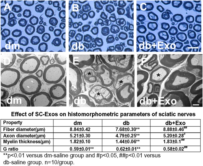
Effect of SC-Exos on histomorphometric changes of sciatic nerves. Representative images of semithin toluidine blue–stained cross sections of sciatic nerves from nondiabetic mice (dm) (A), diabetic mice treated with saline (db) (B), and diabetic mice treated with SC-Exos (db + Exo) (C). TEM images of ultrastructure of sciatic nerve from dm (D), db (E), db + Exo (F) mice. Intact myelinated axons with few mitochondria (arrows) in a dm mouse (D). Demyelinated and damaged axons (asterisks) in a db mouse (E). Axon with thin myelin (asterisk) in a db + Exo mouse, indicating remyelination (F). The table shows the quantitative data of a histomorphometric parameter of toluidine blue–stained sciatic nerve. Scale bars = 25 μm (C) and 1 μm (F).

