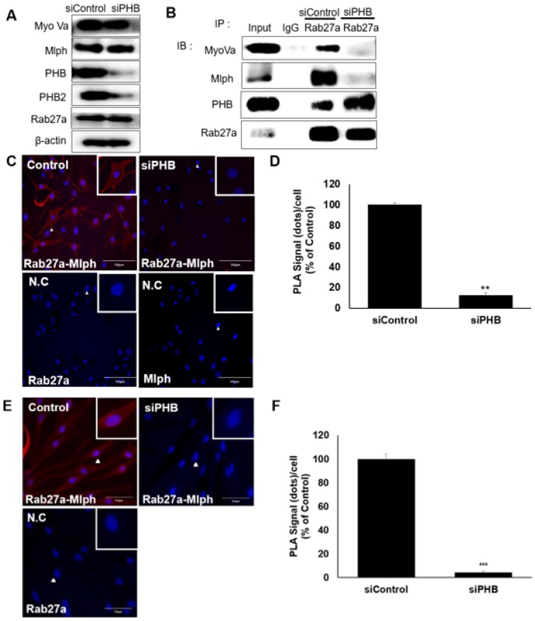
Figure 7.
PHB is necessary for binding of Rab27a and Mlph. A IB analysis for Rab27a, PHB, Mlph, and myosin-Va after transfection of siPHB (20 nM) for 72h. B Anti-Rab27a immunoprecipitated (IP) was performed for O/N at 4 °C followed by western blot analysis to check co-IP proteins (WB). Whole cell lysates were analyzed for total protein levels. C, D PLA assay was performed with control and PHB-KD Melan-a cells (scale bar=100 µm). PLA signals (red) indicated Rab27a-Mlph interaction events. The cells were stained with DAPI to supply evidence for the nuclei. For negative control, the cells were stained with anti-Rab27a or Mlph-IgG antibodies only (C representative picture, D quantification)/ (mean ± standard deviation, n = 3). **p < 0.01. E,F PLA assay was performed with control and PHB-KD NHEM cells (scale bar=100 µm). PLA signals (red) indicated Rab27a-Mlph interaction events. The cells were stained with DAPI to supply evidence for the nuclei. For negative control, the cells were stained with anti-Rab27a or Mlph-IgG antibodies only (E representative picture, F quantification) (mean ± standard deviation, n = 3). ***p < 0.001.

