Abstract
White spot syndrome virus (WSSV) is a major shrimp viral pathogen responsible for large economic losses to shrimp aquaculture all over the world. The RNAi mediated by siRNA contributes a new strategy to control this viral disease. However, the efficient approach to deliver the siRNA into shrimp remains to be addressed. In this investigation, an antiviral vp28-siRNA was encapsulated in β-1,3-d-glucan, and then the β-1,3-d-glucan-encapsulated vp28-siRNA particles (GeRPs) were delivered into Marsupenaeus japonicus shrimp. The results showed that the vp28-siRNA in GeRPs could be released in hemocytes of shrimp. It was found that the GeRPs containing the vp28-siRNA inhibited the replication of WSSV in vivo, which presented a better antiviral activity than the non-encapsulated vp28-siRNA. Further evidence indicated that the mortality of WSSV-infected shrimp was significantly delayed by the GeRPs containing vp28-siRNA. Therefore, our study presented that the glucan-encapsulated siRNA might represent a novel potential therapeutic or preventive approach to control the shrimp disease.
Keywords: WSSV; siRNA; β-1,3-d-Glucan; Marsupenaeus japonicus
Introduction
White spot syndrome, caused by white spot syndrome virus (WSSV), has become the most hazardous and devastating disease in shrimp culture. The virus can lead to 100% shrimp mortality within 7∼10 days in commercial shrimp farms, resulting in large economic losses (Escobedo-Bonilla et al. 2008). WSSV, a rod-shaped enveloped DNA virus, contains a double-stranded DNA genome with 184 potential open reading frames (Yang et al. 2001). Recently, the virus is classified as a new genus Whispovirus of a new family Nimaviridae (Mayo 2002). Due to the importance of viral envelop proteins in virus infection, many WSSV structural proteins have been characterized (Tsai et al. 2004; Wu et al. 2005). Among the WSSV structural proteins, the VP28 protein, located in the viral envelope (Zhang et al. 2002), plays very important roles in the invasion of WSSV into shrimp (Witteveldt et al. 2004a, b; Wu et al. 2005; Xu et al. 2007). It has been reported that the VP28 protein, when being injected intramuscularly or administered orally, provides higher and prolonged survival rates after WSSV challenge (Witteveldt et al. 2004a, b). Interestingly, the silencing of vp28 gene by a specific 21-bp short interfering RNA (vp28-siRNA) in vivo leads to the eradication of WSSV in the virus-infected shrimp (Xu et al. 2007), which sheds a light on the control of WSSV infection in shrimp industry by administration of RNAi.
RNA interference (RNAi) is an evolutionarily conserved mechanism by which double-stranded RNA (dsRNA) initiates posttranscriptional silencing of homologous genes (Fire et al. 1998; Elbashir et al. 2001; Hammond et al. 2001; Hannon 2002). Higher eukaryotes can mount antiviral immune responses induced by dsRNA. RNAi is sequence-specific and can therefore be used to target gene expression (Cullen 2002). In invertebrates, RNAi studies are commonly performed with large dsRNA molecules, which are cleaved into smaller 21–25-bp siRNAs by the host enzymes (Hammond et al. 2000). However, it is reported that the 21-bp siRNAs are the most effective at triggering RNAi in vitro (Kim et al. 2004). In fact, RNAi can be used as an antiviral immune defense strategy in Caenorhabditis elegans during VSV infection (Wilkins et al. 2005). RNAi has been widely applied to suppress the infection or replication of many viruses, including several important human pathogens such as poliovirus (Gitlin et al. 2002), HIV-1 (Jacque et al. 2002), hepatitis B virus (McCaffrey et al. 2003), hepatitis C virus (Kapadia et al. 2003), foot and mouth disease virus (Kahana et al. 2004), influenza virus (Tompkins et al. 2004), and SARS coronavirus (Li et al. 2005). In shrimp, it is reported that the injection of sequence-dependent siRNA, targeting the vp28 gene of WSSV, can eliminate the WSSV in virus-infected cells (Xu et al. 2007). RNAi technology has been recently extended to shrimp as a therapeutic approach and efficient strategy for viral disease prevention in the aquaculture industry (Shekhar and Lu 2009; Westenberg et al. 2005; Wu et al. 2007; Xu et al. 2007). However, the efficient approach for delivery of siRNA into shrimp remains to be addressed.
A new siRNA delivery system has been developed on the basis of the utilization of yeast cell wall particles (YCWP; Aouadi et al. 2009). YCWPs are hollow and porous microspheres with 2–4 μm diameter which are prepared from yeast β-1,3-d-glucan and chitin. The advantages of siRNA delivery system with YCWP include the high RNA capacity, multiplexable RNA, oral bioavailability, β-1,3-d-glucan receptor targeting to macrophages, cell trafficking to the reticuloendothelial system, and sites of pathology. Based on our previous study (Xu et al. 2007), in this investigation, the antiviral vp28-siRNA was encapsulated with β-1,3-d-glucan and delivered into shrimp. The results showed that the β-1,3-d-glucan-encapsulated vp28-siRNA particles (GeRPs) effectively inhibited the replication of WSSV in shrimp. Further evidence indicated that the mortality of WSSV-infected shrimp was significantly decreased by GeRPs. Our data suggested that the glucan-encapsulated siRNA might represent a novel potential therapeutic or preventive approach to control the shrimp diseases.
Materials and Methods
Shrimp Culture and White Spot Syndrome Virus
Cultures of Marsupenaeus japonicus shrimp, approximately 10 g and 10–12 cm each, were performed keeping in groups of 20 individuals in 80 l aquariums at 20°C. Hemolymph and gill tissues from cultured shrimp were subjected at random to PCR detection with WSSV-specific primers to ensure that the shrimp were WSSV-free before experimental infection. The WSSV inoculum was prepared from the WSSV-infected shrimp according to Zhu et al. (2009). Then 0.1 ml of filtrate was injected intramuscularly into healthy shrimp in the lateral area of the fourth abdominal segment using a syringe with a 29-gauge needle.
Synthesis of siRNA
Based on the previous study, the vp28-siRNA (5′-GACCATCGAAACCCACACA-3′) targeting the vp28 gene of WSSV had the capacity to eliminate the virus in WSSV-infected shrimp (Xu et al. 2007). Therefore, this siRNA was used as a target siRNA to control WSSV in this study. As controls, the sequence of vp28-siRNA was rearranged at random and mutated at one nucleotide, respectively, resulting in the corresponding random-siRNA (5′-CAGACCTCACGACACAACA-3′) and mutation-siRNA (5′-GACCAGCGAAACCCACACA-3′). The siRNAs used in this study consisted of 21-nucleotide double-stranded RNAs, each strand of which contained a 19-nucleotide target sequence and a two-uracil (U) overhang at the 3′ end. The siRNAs were synthesized in vitro using in vitro Transcription T7 Kit (TaKaRa, Japan) according to the manufacturer’s instructions. The synthesized siRNAs were dissolved in siRNA buffer (50 mM Tris–HCl, pH 7.5, 100 mM NaCl) and quantified by spectrophotometry.
Preparation of β-1,3-d-Glucan-Encapsulated siRNA Particles
β-1,3-d-Glucan shells were prepared according to Soto and Ostroff (2008). For loading of vp28-siRNA into GeRPs for in vivo experiments, the procedure by Soto and Ostroff (2008) was followed with an adjusted ratio of 40 pmol of siRNA in 1 × 10⁸ empty GeRPs per dose. Forty picomoles of siRNA was incubated with β-1,3-d-glucan shells at 4°C for 2 h. Subsequently, the GeRPs were trapped with polyethylenimine (50 mg; Sigma, USA) for 20 min at 20–25°C. The siRNA-loaded GeRPs were then washed, resuspended in phosphate-buffered saline, and sonicated to ensure homogeneity of the GeRPs preparation. For each experiment, GeRPs batches were aliquoted into tubes for daily dosing, flash-frozen in liquid nitrogen, and stored at −20°C for later use. The final dosage of GeRPs contains 20 μg/g siRNA directed against the WSSV vp28 gene. For confocal microscopy, the empty β-1,3-d-glucan shells were labeled with 5-(4,6-dichlorotriazinyl) aminofluorescein (Invitrogen, USA), and the siRNA was labeled with rhodamine B (Sigma, USA).
Antiviral Experiment in Shrimp by RNAi
WSSV (107 copies/ml) and 6 μM siRNAs (vp28-siRNA, random-siRNA, or mutation-siRNA in GeRPs) were delivered into shrimp by simultaneous injection into the lateral area of the fourth abdominal segment at 0.1 ml/shrimp using a syringe with a 29-gauge needle. At the same time, the siRNA only and the GeRP only, as well as a negative control (0.9% NaCl) and a positive control (WSSV only), were included in the injections. In antiviral assays, 20 shrimp were used for each treatment. Everyday, the shrimp mortality was monitored and three shrimp specimens from each treatment, selected at random, were subjected to quantitative PCR analysis. All assays described above were carried out in triplicate.
Northern Blot Analysis
Small RNAs were extracted from hemocytes of shrimp at 24 h postinjection using mirVANA miRNA isolation kit (Ambion, USA) according to the manufacturer’s instructions. After treatment with RNase-free DNase I (TakaRa, Japan) for 30 min at 37°C, RNAs were separated by electrophoresis on a 15% polyacrylamide gel in 1× TBE buffer (90 mM Tris–boric acid, 2 mM EDTA, pH 8.0) and transferred to a nitrocellulose membrane (Amersham, USA). The blots were probed with DIG-labeled vp28 probe (5′-CTGGTAGCTTTGGGTGTGT-3′) and DIG-labeled shrimp U6 probe (5′-GGGCCATGCTAATCTTCTCTGTATCGTT-3′), respectively. The DIG labeling and detection were performed following the protocol of DIG High Prime DNA Labeling and Detection Starter Kit II (Roche, Germany).
WSSV Detection and Quantitative Analysis by PCR
Twenty milligrams of gills was collected from shrimp and homogenized in 500 μl of guanidine lysis buffer (50 mM Tris–HCl, 25 mM EDTA, 4 M guanidinium thiocyanate, 0.5% N-lauroylsarcosine, pH 8.0) at room temperature. After centrifugation at 15,000×g for 3 min, 20 μl of silica was added to the supernatant for DNA absorption. Subsequently the mixture was rotated for 5 min, followed by centrifugation at 15,000×g for 30 s. The pellet was rinsed twice with 70% ethanol and resuspended in 20 μl distilled water. Then it was centrifuged at 15,000×g for 2 min. The supernatant was used as PCR template. PCR was performed with two WSSV-specific primers (forward primer 5′-TATTGTCTCTCCTGACGTAC-3′ and reverse primer 5′-CACATTCTTCACGAGTCTAC-3′). The conditions for PCR amplification were as follows: 5 min at 94°C, 40 cycles at 94°C for 45 s and 68°C for 1 min, and extension at 68°C for 5 min. For quantitative analysis of viral DNA, the real-time PCR was conducted. The TaqMan probe was 5′-FAM-TGCTGCCGTCTCCAA-TAMRA-3′.
Statistical Analysis
The numerical data were statistically analyzed using the chi-square test at a significance level of 5%. The relative percent survival values were calculated as relative percent survival, calculated as (1 − vaccinated group mortality/control group mortality) × 100 (Amend 1981).
Results and Discussion
Delivery of vp28-siRNA into Shrimp by β-1,3-d-Glucan-Encapsulated siRNA Particles
Our previous study showed that the injection of sequence-dependent vp28-siRNA could eliminate the WSSV in virus-infected shrimp (Xu et al. 2007). In an attempt to obtain an efficient approach for the delivery of the antiviral vp28-siRNA into shrimp, the vp28-siRNA was encapsulated with β-1,3-d-glucan. The flow cytometry analysis indicated that more than 90% vp28-siRNA was encapsulated. Based on dosage analysis, it was found that when 24 μM of vp28-siRNA was encapsulated in glucan, the GeRPs showed a good antiviral activity (data not shown). The GeRPs containing the vp28-siRNA could function in shrimp for 7 days. Therefore, this concentration of GeRPs was used in this study.
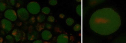
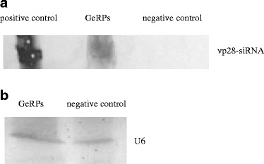
As examined with confocal microscopy, it was shown that the vp28-siRNA was encapsulated into glucan which formed the GeRPs containing the vp28-siRNA (Fig. 1). When the GeRPs were injected into shrimp, the Northern bolts revealed that the vp28-siRNA could be detected in shrimp hemocytes at 24 h after the injection of GeRPs (Fig. 2a). As control, the U6 was also detected in the shrimp hemocytes challenged with GeRPs (Fig. 2b). The results showed that the antiviral vp28-siRNA in GeRPs was delivered into shrimp and further released in hemocytes, suggesting that the encapsulation of siRNA by β-1,3-d-glucan was an efficient strategy for the delivery of siRNA into host. It might be inferred that the GeRPs could efficiently enter the shrimp hemocytes through phagocytosis. The acidic pH in phagosomes could promote the release of siRNA in GeRPs (Aouadi et al. 2009).
Fig. 1.

Confocal microscopy of β-1,3-d-glucan-encapsulated siRNA particles. The vp28-siRNA was labeled with rhodamine B (red). The glucan particles were indicated with green
Fig. 2.

Detection of vp28-siRNA in shrimp hemocytes. The GeRPs containing the vp28-siRNA were injected into shrimp. At 24 h after the injection, the shrimp hemolymph was extracted and subjected to Northern blot analysis using the vp28-siRNA probe (a) or the U6 probe as control (b). The probes were indicated on the right. Positive control the synthesized vp28-siRNA, GeRPs the shrimp hemolymph with GeRPs injection, Negative control the shrimp hemolymph with no GeRPs injection
Inhibition of WSSV Infection in Shrimp by GeRPs Containing vp28-siRNA
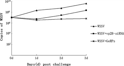
The results of quantitative real-time PCR revealed that the WSSV copies in shrimp were significantly decreased (P < 0.01) when treated with the vp28-siRNA or GeRPs containing the vp28-siRNA by comparison with that of the positive control (WSSV only; Fig. 3), indicating that the virus replication was inhibited by the antiviral vp28-siRNA. It was shown that the inhibitory effect of GeRPs containing the vp28-siRNA against virus was better than that of the non-encapsulated vp28-siRNA at the 2–4 days after WSSV challenge (Fig. 3). The data suggested that the non-encapsulated vp28-siRNA might be degraded before entering the hemocytes and that some siRNAs could not be absorbed by cells, when delivered into shrimp. However, the vp28-siRNA encapsulated in glucan might efficiently enter the shrimp hemocytes through phagocytosis and avoid to be degraded in vivo. Therefore, the GeRPs presented a better antiviral performance than non-encapsulated siRNA in shrimp.
Fig. 3.

Detection of WSSV copies for RNAi. Days post-infection were shown on the abscissa and accumulated virus copies on the ordinate. The solutions used for injection were indicated on the right. The dots represented the means of triplicate assays within ±1% standard deviation
Effects of GeRPs Containing vp28-siRNA on the Mortality of WSSV-Infected Shrimp
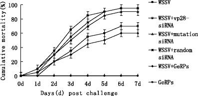
To evaluate the effects of GeRPs containing the vp28-siRNA on the mortality of WSSV-challenged shrimp, WSSV and different siRNAs were co-injected into shrimp. The results showed that the cumulative mortality of virus-infected shrimp was significantly delayed by the vp28-siRNA or GeRPs containing the vp28-siRNA (P < 0.01), while the random-siRNA and mutation-siRNA generated the similar mortality curves as that of the positive control (WSSV only; Fig. 4). The data revealed that RNAi mediated by siRNA was highly specific in shrimp.
Fig. 4.

The cumulative mortality of WSSV-infected shrimp treated with siRNAs. The solutions used for injection were shown on the right. Each point showed the means of triplicate assays within ±1% standard deviation
The results indicated that the shrimp mortality was very low for the control (GeRPs only), whereas the shrimp from the positive control (WSSV only) displayed 95% mortality at 7 days after challenge with WSSV (Fig. 4). The data showed that the GeRPs had no toxicity to shrimp. When the shrimp were co-injected with GeRPs and WSSV, the shrimp mortality was significantly delayed (P < 0.01), indicating that the infection of WSSV could be inhibited by the vp28-siRNA in GeRPs. By comparing the shrimp mortalities between the vp28-siRNA (non-encapsulated) and GeRPs (containing the vp28-siRNA) treatments (Fig. 4), it was found that the GeRPs demonstrated better antiviral activity than that of vp28-siRNA, which was consistent with the detection of WSSV copies, showing a potential use of GeRPs in shrimp against virus infection by using glucan encapsulation.
In our study, the GeRPs containing the vp28-siRNA could inhibit the replication of WSSV and further led to the decrease of virus-infected shrimp mortality. Therefore, our study presented a better strategy to control WSSV in the shrimp industry. It has been reported that the oral administration of β-1,3-glucan extracted from Schizophyllum commune can improve the shrimp immunity against WSSV (Chang et al. 2003). However, β-1,3-glucan, derived from bakers’ yeast Saccharomyces cerevisiae, exhibits no protection of shrimp against WSSV infection (Sukumaran et al. 2010). The documented data show that the improvement of shrimp immunity by β-1,3-glucan is changed with the resource of glucan. In our study, the β-1,3-glucan was derived from S. cerevisiae. Therefore, it could be concluded that the protection of shrimp against WSSV was due to the presence of vp28-siRNA. Our previous study showed that the sustainable presence of vp28-siRNA could lead to the elimination of WSSV in shrimp (Xu et al. 2007). To make the GeRPs containing vp28-siRNA more useful in shrimp industry, the sustainable presence of vp28-siRNA in vivo merited to be further studied.
The glucan particles may be engulfed into cells by phagocytosis which may lead to the enhancement of immunity. The results of our study indicated that the β-1,3-D-glucan-encapsulated vp28-siRNA showed a better antiviral activity than the non-encapsulated vp28-siRNA in shrimp. Based on these data, it could be inferred that the vp28-siRNA in GeRPs entered the shrimp hemocytes through phagocytosis and was released in the hemocytes to function against WSSV. However, the non-encapsulated vp28-siRNA might suffer from degradation before efficiently entering the hemocytes. The problems in the application of siRNA were mainly attributed to inadequate delivery into cells and limited half-life of siRNA in the extracellular environment in shrimp. In this context, the GeRPs shed light on the efficient administration of shrimp diseases. In our future work, the oral administration of glucan-encapsulated siRNA merited to be explored. The glucan encapsulation of antiviral siRNA, as well as other antiviral drugs, represented a novel strategy for the aquaculture disease control.
Acknowledgments
This work was financially supported by National Natural Science Foundation of China (30830084, 31001127), Hi-Tech Research and Development Program of China (863 program of China; 2010AA09Z403), Project of Ministry of Agriculture, China (201103034), and Studies Foundation, Department of Education of Zhejiang Province (Y200908288). We thank Dr. Hongbo Zhang for his useful comments on this investigation.
References
- Amend DF. Potency testing of fish vaccines. In: Anderson DP, Hennessen W, editors. Fish biologics: serodiagnostics and vaccines. Basel: Karger; 1981. pp. 447–454. [Google Scholar]
- Aouadi M, Tesz GJ, Nicoloro SM, Wang MX, Chouinard M, Soto E, Ostroff GR, Czech MP. Orally delivered siRNA targeting macrophage Map4k4 suppresses systemic inflammation. Nature. 2009;458:1180–1184. doi: 10.1038/nature07774. [DOI] [PMC free article] [PubMed] [Google Scholar]
- Chang CF, Su MS, Chen HY, Liao IC. Dietary β-1,3-glucan effectively improves immunity and survival of Penaeus monodon challenged with white spot syndrome virus. Fish Shellfish Immunol. 2003;15:297–310. doi: 10.1016/S1050-4648(02)00167-5. [DOI] [PubMed] [Google Scholar]
- Cullen BR. RNA interference: antiviral defense and genetic tool. Nat Immunol. 2002;3:597–599. doi: 10.1038/ni0702-597. [DOI] [PubMed] [Google Scholar]
- Elbashir SM, Lendeckel W, Tuschl T. RNA interference is mediated by 21- and 22-nucleotide RNAs. Genes Dev. 2001;15:188–200. doi: 10.1101/gad.862301. [DOI] [PMC free article] [PubMed] [Google Scholar]
- Escobedo-Bonilla CM, Alday Sanz V, Wille M, Sorgeloos P, Pensaert MB, Nauwynck HJ. A review on the morphology, molecular characterization, morphogenesis and pathogenesis of white spot syndrome virus. J Fish Dis. 2008;31:1–18. doi: 10.1111/j.1365-2761.2007.00877.x. [DOI] [PubMed] [Google Scholar]
- Fire A, Xu S, Montgomery MK, Kostas SA, Driver SE, Mello CC. Potent and specific genetic interference by double-stranded RNA in Caenorhabditis elegans. Nature. 1998;391:806–811. doi: 10.1038/35888. [DOI] [PubMed] [Google Scholar]
- Gitlin L, Karelsky S, Andino R. Short interfering RNA confers intracellular antiviral immunity in human cells. Nature. 2002;418:430–434. doi: 10.1038/nature00873. [DOI] [PubMed] [Google Scholar]
- Hammond SM, Bernstein E, Beach D, Hannon GJ. An RNA-directed nuclease mediates post-transcriptional gene silencing in Drosophila cells. Nature. 2000;404:293–296. doi: 10.1038/35005107. [DOI] [PubMed] [Google Scholar]
- Hammond SM, Caudy AA, Hannon GJ. Post-transcriptional gene silencing by double-stranded RNA. Nat Rev Genet. 2001;2:110–119. doi: 10.1038/35052556. [DOI] [PubMed] [Google Scholar]
- Hannon GJ. RNA interference. Nature. 2002;418:244–251. doi: 10.1038/418244a. [DOI] [PubMed] [Google Scholar]
- Jacque JM, Triques K, Stevenson M. Modulation of HIV-1 replication by RNA interference. Nature. 2002;418:435–438. doi: 10.1038/nature00896. [DOI] [PMC free article] [PubMed] [Google Scholar]
- Kapadia SB, Brideau-Andersen A, Chisari FV. Interference of hepatitis C virus RNA replication by short interfering RNAs. Proc Natl Acad Sci USA. 2003;100:2014–2018. doi: 10.1073/pnas.252783999. [DOI] [PMC free article] [PubMed] [Google Scholar]
- Kahana R, Kuznetzova L, Rogel A, Shemesh M, Hai D, Yadin H. Inhibition of foot-and-mouth disease virus replication by small interfering RNA. J Gen Virol. 2004;85:3213–3217. doi: 10.1099/vir.0.80133-0. [DOI] [PubMed] [Google Scholar]
- Kim DH, Behlke MA, Rose SD, Chang MS, Choi S, Rossi JJ. Synthetic dsRNA Dicer substrates enhance RNAi potency and efficacy. Nat Biotechnol. 2004;23:222–226. doi: 10.1038/nbt1051. [DOI] [PubMed] [Google Scholar]
- Li BJ, Tang Q, Cheng D, Qin C, Xie FY, Wei Q. Using siRNA in prophylactic and therapeutic regimens against SARS coronavirus in rhesus macaque. Nat Med. 2005;11:944–951. doi: 10.1038/nm1280. [DOI] [PMC free article] [PubMed] [Google Scholar]
- Mayo MA. A summary of taxonomic changes recently approved by ICTV. Arch Virol. 2002;147:1655–1656. doi: 10.1007/s007050200039. [DOI] [PubMed] [Google Scholar]
- McCaffrey AP, Nakai H, Pandey K, Huang Z, Salazar FH, Xu H. Inhibition of hepatitis B virus in mice by RNA interference. Nat Biotechnol. 2003;21:639–644. doi: 10.1038/nbt824. [DOI] [PubMed] [Google Scholar]
- Shekhar MS, Lu YA. Application of nucleic-acid-based therapeutics for viral infections in shrimp aquaculture. Mar Biotechnol. 2009;11:1–9. doi: 10.1007/s10126-008-9155-0. [DOI] [PubMed] [Google Scholar]
- Soto ER, Ostroff GR. Characterization of multilayered nanoparticles encapsulated in yeast cell wall particles for DNA delivery. Bioconjug Chem. 2008;19:840–848. doi: 10.1021/bc700329p. [DOI] [PubMed] [Google Scholar]
- Sukumaran V, Lowman DW, Sajeevan TP, Philip R. Marine yeast glucans confer better protection than that of baker’s yeast in Penaeus monodon against white spot syndrome virus infection. Aqua Res. 2010;41:1799–1805. doi: 10.1111/j.1365-2109.2010.02520.x. [DOI] [Google Scholar]
- Tompkins SM, Lo CY, Tumpey TM, Epstein SL. Protection against lethal influenza virus challenge by RNA interference in vivo. Proc Natl Acad Sci USA. 2004;101:8682–8686. doi: 10.1073/pnas.0402630101. [DOI] [PMC free article] [PubMed] [Google Scholar]
- Tsai JM, Wang HC, Lo CF, Kou GH. Genomic and proteomic analysis of thirty-nine structural proteins of shrimp white spot syndrome virus. J Virol. 2004;78:11360–11370. doi: 10.1128/JVI.78.20.11360-11370.2004. [DOI] [PMC free article] [PubMed] [Google Scholar]
- Westenberg M, Heinhuis B, Zuidema D, Vlak JM. siRNA injection induces sequence-independent protection in Penaeus monodon against white spot syndrome virus. Virus Res. 2005;114:133–139. doi: 10.1016/j.virusres.2005.06.006. [DOI] [PubMed] [Google Scholar]
- Wilkins C, Dishongh R, Moore SC, Whitt MA, Chow M, Machaca K. RNA interference is an antiviral defence mechanism in Caenorhabditis elegans. Nature. 2005;436:1044–1047. doi: 10.1038/nature03957. [DOI] [PubMed] [Google Scholar]
- Witteveldt J, Cifuentes C, Vlak JM, van Hulten MCW. Protection of Penaeus monodon against white spot syndrome virus by oral vaccination. J Virol. 2004;78:2057–2061. doi: 10.1128/JVI.78.4.2057-2061.2004. [DOI] [PMC free article] [PubMed] [Google Scholar]
- Witteveldt J, Vlak JM, van Hulten MCW. Protection of Penaeus monodon against white spot syndrome virus using a WSSV subunit vaccine. Fish Shellfish Immunol. 2004;16:571–579. doi: 10.1016/j.fsi.2003.09.006. [DOI] [PubMed] [Google Scholar]
- Wu WL, Wang L, Zhang XB. Identification of white spot syndrome virus (WSSV) envelope proteins involved in shrimp infection. Virology. 2005;332:578–583. doi: 10.1016/j.virol.2004.12.011. [DOI] [PubMed] [Google Scholar]
- Wu Y, Lü L, Yang LS, Weng SP, Chan SM, He JG. Inhibition of white spot syndrome virus in Litopenaeus vannamei shrimp by sequence-specific siRNA. Aquaculture. 2007;271:21–30. doi: 10.1016/j.aquaculture.2007.06.029. [DOI] [PMC free article] [PubMed] [Google Scholar]
- Xu JY, Han F, Zhang XB. Silencing shrimp white spot syndrome virus (WSSV) genes by siRNA. Antivir Res. 2007;73:126–131. doi: 10.1016/j.antiviral.2006.08.007. [DOI] [PubMed] [Google Scholar]
- Yang F, He J, Lin X, Li Q, Pan D, Zhang X, Xu X. Complete genome sequence of the shrimp white spot bacilliform virus. J Virol. 2001;75:11811–11820. doi: 10.1128/JVI.75.23.11811-11820.2001. [DOI] [PMC free article] [PubMed] [Google Scholar]
- Zhang XB, Huang CH, Xu X, Hew CL. Identification and localization of a prawn white spot syndrome virus gene that encodes an envelope protein. J Gen Virol. 2002;83:1069–1074. doi: 10.1099/0022-1317-83-5-1069. [DOI] [PubMed] [Google Scholar]
- Zhu F, Du HH, Miao ZG, Quan HZ, Xu ZR. Protection of Procambarus clarkii against white spot syndrome virus using inactivated WSSV. Fish Shellfish Immunol. 2009;26:685–690. doi: 10.1016/j.fsi.2009.02.022. [DOI] [PubMed] [Google Scholar]


