Abstract
The present study describes detection of picobirnavirus (PBV) in faecal samples from bovine and buffalo calves employing the polyacrylamide gel electrophoresis (PAGE). A total of 136 faecal samples from buffalo (n = 122) and cow calves (n = 14) exhibiting clinical signs of diarrhoea and from healthy calves were collected during 2007–2010 from subtropical (central India) and tarai area of western temperate Himalayan foothills (Uttarakhand). The dsRNA nature of the virus was confirmed by nuclease treatment (RNase A, RNaseT1 and DNase 1). PAGE results confirmed 3.67% (5/136) positivity for PBV, showing a typical genomic migration pattern with two discrete bands with size of approximately 2.4 and 1.7 kbps for the larger and smaller segments, respectively. Among the five PBV samples identified, three were from buffalo calves and one from cow calf exhibiting clinical signs of acute diarrhoea, while one sample from non-diarrhoeic buffalo calf also showed the presence of PBV. None of the samples showed dual infection of rotavirus and PBV. The preliminary findings indicate sporadic incidences of PBV in bovine calves and emphasize the need for the development of better diagnostics for early detection and genetic characterization of these emerging isolates of farm animals of economic significance.
Keywords: Picobirnavirus, Rotavirus, Gastroenteritis, Bovine calves, RNA-PAGE
Introduction
Viral gastroenteritis is one of the most common diseases affecting young animals throughout the world. The number of viral agents associated with diarrhoeal disease in animals has progressively increased, including rotavirus, astrovirus, calicivirus, coronavirus and picobirnavirus. Picobirnavirus (PBV) classified recently into the family Picobirnaviridae (Fregolente et al. 2009) were first identified in children by Pereira et al. (1988) as bisegmented double-stranded RNA viruses and proposed the name picobirnavirus on the basis of its genomic pattern. PBVs have been identified in humans worldwide (Banyai et al. 2003; Bhattacharya et al. 2007), in a wide variety of farm animals and birds, including pigs (Carruyo et al. 2008), calves (Buzinaro et al. 2003), birds (Tamehiro et al. 2003) and even in wild animals (Masachessi et al. 2007; Wang et al. 2007). It has been identified as an opportunistic pathogen which may cause diarrhoea in immunosuppressed individuals (Giordano et al. 1999). Though these viruses have been identified in both normal and diarrhoeic faecal samples of a broad range of hosts, little information is available regarding their distribution or association with bovine diarrhoea from India, except a report on PBV (strain RUBV-P) from diarrhoeic calf from Eastern India (Ghosh et al. 2009). In this preliminary study, we report the detection of PBV in faecal samples from buffalo and cow calves with and without clinical signs of diarrhoea obtained from different dairy farms in subtropical and temperate foothills of Himalaya, India.
Materials and methods
Picobirnavirus-positive faecal samples were detected employing the same extraction protocol as used for the rotavirus genome and visualization of the segments on PAGE. The PAGE technique has been reported for the identification of PBV (Novikova et al. 2003). During an ongoing epidemiological survey for bovine rotaviruses, a total of 136 faecal samples were collected from buffalo (n = 122) and cow (n = 14) calves, aged less than 3 months, reared at 30 different dairy farms in subtropical part of central India (Madhya Pradesh) and tarai area of western temperate foothills of Himalaya (Uttarakhand) during 2007–2010. Samples obtained were from calves showing clinical signs of diarrhoea (102 buffalo calves, 12 cow calves) and clinically healthy animals (20 buffalo calves, two cow calves). The sample preparation and viral nucleic acid extraction protocols as used for rotavirus genome extraction in our earlier study were followed (Kusumakar et al. 2010). The isopropanol precipitated RNA pellet was dissolved in 2× RNA lysis buffer for viral RNA genomic electrophoresis. The viral RNA was quantified using Nanodrop Spectrophotometer (Thermo Scientific, USA). The RNA electrophoresis was carried out for the detection of PBV dsRNA genome segments, based on number of segments, their typical electrophoretic migration pattern and segment size. The electrophoresis of viral RNA was carried out in 10% native (non-denaturing) polyacrylamide gel in Tris–Glycine buffer (0.025 M Tris,0.109 M Glycine, pH 8.3) by loading up to 500 ng of viral RNA per well. The viral genomic electrophoresis was also carried out in denaturing 5% polyacrylamide gel (containing 7 M Urea) in 1× TBE running buffer (8.9 mM Tris, 8.9 mM Boric acid, 0.2 mM EDTA, pH 8.3) by loading the same amount of viral RNA per well. Samples were electrophorosed at 100 V until the dye reached to be the end of the gel (approx. 4 h). The gel was silver impregnated following the method of Herring et al. (1982) and documented. For the estimation of molecular weight of the two segments of PBV, samples were run with group A rotavirus and 1 kbps DNA ladder (Fermentas) on 1× agarose gel, stained with ethidium bromide and documented in Gel doc system (Syngene GeneGenius Imaging System, USA).
The dsRNA nature of these segments was confirmed by nuclease treatment. Digestion of nucleic acid was performed following the method described by Pereira et al. (1988). Briefly, 1 μg of purified PBV RNA was treated separately with pancreatic RNase A, RNaseT1 and DNase 1.The pancreatic RNase A was used after boiling for 10 min at a final concentration of 10 ug/ml in Tris–EDTA–NaCl (10 mM Tris–HCl, 10 mM NaCl, 2 mM EDTA), while RNaseT1 was used at a final concentration of 10 U/ml in the same buffer mentioned above and DNase1 was used at 10 U/ml in 40 mM Tris–HC1 (pH 7.9), 10 mM CaC12 and 6 mM MgC12. Digestion was carried out at 37°C for 30 min, and following GIT extraction and isopropanol precipitation, the products were analyzed on 10% non-denaturing and 5% denaturing polyacrylamide gel.
Results and discussion
The results revealed that all segments were susceptible to degradation by pancreatic RNase A and resistant to degradation by DNase I and RNase T1, confirming that the RNA from bovine faecal samples was double stranded in nature. No variation was observed in the nature of migration of PBV genomic segments during native and denaturing electrophoresis.
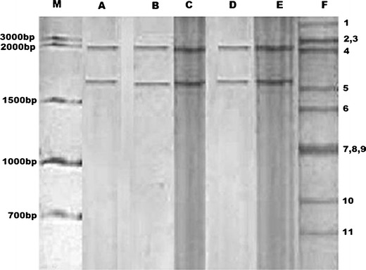
The PAGE analysis confirmed 17.64% (24/136) positivity for rotavirus (17.21% in buffalo calves (21/122) and 21.42% in cow calves (3/14)), showing a typical group A pattern (4:2:3:2), while 3.67% (5/136) presented two discrete bands that could be stained by silver impregnation. The position of PBV segments was found between segments 3 and 5 of group A rotavirus (Fig. 1). Moreover, the RNA profile of PBV obtained by PAGE did not provide information for grouping the PBV strains by animal species, since they all were found to have genomic segments of similar size (Fig. 1). All the five samples showed the same migration pattern on the polyacrylamide gel. The size of the bands was approximately 2.4 and 1.7 kbps for the larger and smaller segments, respectively, when compared to bovine group A rotavirus and 1 kbps DNA ladder (Fig. 1). Among the five PBV positive samples identified, three were from buffalo calves with clinical signs of acute diarrhoea (belonged to the herds in which the presence of rotavirus was also detected, data not shown). One sample from non-diarrhoeic buffalo calf also showed the presence of PBV pattern, whereas none of the faecal sample from healthy bovine and buffalo calves yielded positivity for group A bovine rotavirus. The buffalo calf samples were from subtropical parts of Central India. One diarrhoeic cow calf sample from temperate Western Himalayan part was also PBV-positive. None of the samples showed concurrent infection of rotavirus and PBV, whereas the simultaneous detection of rotavirus and PBV infecting calves has been reported by Vanopdenbosch and Wellemansm (1989). Though investigators have demonstrated the presence of PBV in various hosts with certain frequency, conclusive data regarding the pathogenicity of this virus are still lacking, and their role in the clinical manifestation remains to be better defined.
Fig. 1.

PBV electrophoretic genomic pattern obtained in faecal samples of bovine and buffalo calves. Lane indicates: M 1 kbps DNA ladder, A–E PBVs (with genomic segments of 2.4 and 1.7 kbps size), and F bovine group A rotavirus (genome segments marked 1–11 as per their gene size, segment no. 2, 3 and 7, 8, and 9 are co-migratory)
As out of 136 samples, 131 yielded PBV-negative results, it is pertinent here to point out that species which gave PBV-negative results could not be classified as non-susceptible ones, because it could not be ruled out that they were never infected with PBV or were infected but did not excrete PBV at the time of sample collection. These results confirm the circulation of PBV among bovine population of subtropical parts of central and temperate western Himalayan parts of India. The only available study that matches our results is that reported by Ghosh et al. (2009) from Eastern India, which described PBV in diarrhoeic calves. We further propose that more number of samples and hosts must be surveyed to reach at a better conclusion regarding the emergence of PBV and their association with diarrhoea in farm animals. There is urgent need for better diagnostics for the detection of PBVs at an early stage of infection.
Acknowledgments
Authors wish to thank MP Council of Science and Technology (MPCOST) and Director, Indian Veterinary Research Institute for providing grant to conduct this study. The financial help in the form of Junior Research Fellowship from Indian Council of Agricultural Research, New Delhi, India to second author is also acknowledged.
References
- Banyai K, Jakab F, Reuter G, Bene J, Uj M, Melegh B, Szucs G. Sequence heterogeneity among human picobirnaviruses detected in a gastroenteritis outbreak. Archives of Virology. 2003;148:2281–2291. doi: 10.1007/s00705-003-0200-z. [DOI] [PubMed] [Google Scholar]
- Bhattacharya R, Sahoo GC, Nayak MK, Rajendran K, Dutta P, Mitra U, Bhattacharya MK, Naik TN, Bhattacharya SK, Krishnan T. Detection of Genogroup I and II human picobirnaviruses showing small genomic RNA profile causing acute watery diarrhoea among children in Kolkata, India. Infection, Genetics and Evolution. 2007;7:229–38. doi: 10.1016/j.meegid.2006.09.005. [DOI] [PubMed] [Google Scholar]
- Buzinaro MG, Freitas PP, Kisiellus JJ, Ueda M, Jerez JA. Identification of a bisegmented double-stranded RNA virus (picobirnavirus) in calf faeces. Veterinary Journal. 2003;166:185–187. doi: 10.1016/S1090-0233(03)00031-5. [DOI] [PubMed] [Google Scholar]
- Carruyo GM, Mateu G, Martínez LC, Pujol FH, Nates SV, Liprandi F, Ludert JE. Molecular characterization of porcine picobirnaviruses and development of a specific reverse transcription-PCR assay. Journal of Clinical Microbiology. 2008;46:2402–2405. doi: 10.1128/JCM.00655-08. [DOI] [PMC free article] [PubMed] [Google Scholar]
- Fregolente MC, de Castro-Dias E, Martins SS, Spilki FR, Allegretti SM, Gatti MS. Molecular characterization of picobirnaviruses from new hosts. Virus Research. 2009;143:134–136. doi: 10.1016/j.virusres.2009.03.006. [DOI] [PubMed] [Google Scholar]
- Ghosh S, Kobayashi N, Nagashima S, Naik TN. Molecular characterization of full-length genomic segment 2 of a bovine picobirnavirus (PBV) strain: evidence for high genetic diversity with genogroup IPBVs. Journal of General Virology. 2009;90:2519–2524. doi: 10.1099/vir.0.013987-0. [DOI] [PubMed] [Google Scholar]
- Giordano M, Martınez L, Rinaldi D, Espul C, Martınez N, Isa M, Depetris A, Medeot S, Nates S. Diarrhea and enteric emerging viruses in HIV-infected patients. AIDS Research and Human Retroviruses. 1999;15:1427–1432. doi: 10.1089/088922299309937. [DOI] [PubMed] [Google Scholar]
- Herring AJ, Inglis NF, Ojeh CK, Snodgrass DR, Menzies JD. Rapid diagnosis of rotavirus infection by direct detection of viral nucleic acid in silver-stained polyacrylamide gels. Journal of Clinical Microbiology. 1982;16:473–477. doi: 10.1128/jcm.16.3.473-477.1982. [DOI] [PMC free article] [PubMed] [Google Scholar]
- Kusumakar AL, Savita, Minakshi YPS, Prasad G. Genomic diversity among group A rotaviruses from diarrhoeic children, piglets, buffalo and cow calves of Madhya Pradesh. Indian Journal of Microbiology. 2010;50:83–88. doi: 10.1007/s12088-010-0016-y. [DOI] [PMC free article] [PubMed] [Google Scholar]
- Masachessi G, Martınez LC, Giordano MO, Barril PA, Isa BM, Ferreyra L, Villareal D, Carello M, Asis C, Nates SV. Picobirnavirus (PBV) natural hosts in captivity and virus excretion pattern in infected animals. Archives of Virology. 2007;152:989–998. doi: 10.1007/s00705-006-0900-2. [DOI] [PubMed] [Google Scholar]
- Novikova NA, Epifanova NV, Fedorova OF, Golitsyna LN, Kupriianova NV. Detection of picobirnaviruses by electrophoresis of RNA in polyacrylamide gel. Vopr Virusol. 2003;48:41–43. [PubMed] [Google Scholar]
- Pereira HG, Fialho AM, Flewett TH, Teixeira JMS, Andrade ZP. Novel virus in human faeces. Lancet. 1988;9:103–104. doi: 10.1016/S0140-6736(88)90032-3. [DOI] [PubMed] [Google Scholar]
- Tamehiro CY, Alfieri AF, Medici KC, Alfieri AA. Segmented double segmented genomic RNA viruses in fecal samples from broiler chicken. Brazilian Journal of Microbiology. 2003;34:349–355. doi: 10.1590/S1517-83822003000400013. [DOI] [Google Scholar]
- Vanopdenbosch E, Wellemansm G. Birna-type virus in diarrhoeic calf faeces. Veterinary Record. 1989;125:610. [PubMed] [Google Scholar]
- Wang Y, Tu X, Humphrey C, McClure H, Jiang X, Qin C, Glass RI, Jiang B. Detection of viral agents in fecal specimens of monkeys with diarrhea. Journal of Medical Primatology. 2007;36:101–107. doi: 10.1111/j.1600-0684.2006.00167.x. [DOI] [PMC free article] [PubMed] [Google Scholar]


