Severe acute respiratory syndrome-related coronavirus (SARS-CoV-2) has rapidly spread throughout China and as of 8 March 2020 has spread to over 100 countries with 105,000 confirmed cases of coronavirus-related disease (COVID-19).1 The high infectivity and mortality of COVID-19 makes this a serious public health threat.2 Recent studies have confirmed that fever, dry cough, and fatigue are the main manifestations.3 Some patients have other symptoms, such as nasal congestion, runny nose, sore throat, myalgia, and diarrhea. Seriously-ill patients may develop dyspnea and/or hypoxemia one week after the onset of symptoms, and critically-ill patients can quickly progress to acute respiratory distress syndrome, septic shock, severe metabolic acidosis, coagulopathy, and multiple organ dysfunction syndrome.3
Figure.
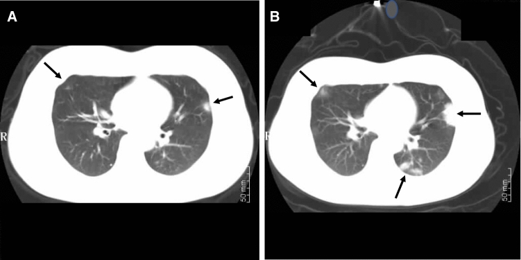
Chest computed tomography (CT) scan at the time of admission (A) of a 27-yr-old 36-week pregnant woman with coronavirus disease (COVID-19). The CT scan shows the characteristic peripheral (and/or subpleural) ground-glass opacities. These are seen in the left lower lobe/lingula junction and in the right middle lobe (arrows). Two days after admission (B), the size, density, and distribution of these opacities had progressed (arrows)
We report a 27-yr-old pregnant woman at 36 weeks gestation who was admitted to the hospital with fever, dry cough, and fatigue as the main manifestations. Her SARS-CoV-2 reverse transcriptase polymerase chain reaction (RT-PCR) test was positive and although she developed tachypnea, she did not develop significant hypoxemia. After admission, a computed tomography (CT) scan (Figure A) revealed the typical COVID-19 findings of patchy peripheral and subpleural ground-glass opacities4 in the left lower lobe/lingula junction. The right middle lobe of the lung also showed a small subpleural opacity of uneven density and blurred margins. Two days after admission, a repeat CT scan showed (Figure B) the number, density, and size of the lesions. Because of concern about potential further progression of the COVID-19 pulmonary manifestations, an uncomplicated elective Cesarean delivery was performed. The RT-PCR for SARS-CoV-2 was negative in the neonate.
Acknowledgments
Conflicts of interest
None.
Funding statement
None.
Editorial responsibility
This submission was handled by Dr. Hilary P. Grocott, Editor-in-Chief, Canadian Journal of Anesthesia.
Footnotes
Publisher's Note
Springer Nature remains neutral with regard to jurisdictional claims in published maps and institutional affiliations.
Rong Chen and Jun Chen authors contributed equally to this work
References
- 1.World Health Organization. Coronavirus disease 2019 (COVID-19) Situation Report – 48. Available from URL: https://www.who.int/docs/default-source/coronaviruse/situation-reports/20200308-sitrep-48-covid-19.pdf?sfvrsn=16f7ccef_4 (accessed March 2020).
- 2.Yang X, Yu Y, Xu J, et al. Clinical course and outcomes of critically ill patients with SARS-CoV-2 pneumonia in Wuhan, China: a single-centered, retrospective, observational study. Lancet Respir Med. 2020 doi: 10.1016/s2213-2600(20)30079-5. [DOI] [PMC free article] [PubMed] [Google Scholar]
- 3.Huang C, Wang Y, Li X, et al. Clinical features of patients infected with 2019 novel coronavirus in Wuhan, China. Lancet. 2020;395:497–506. doi: 10.1016/S0140-6736(20)30183-5. [DOI] [PMC free article] [PubMed] [Google Scholar]
- 4.Li Y, Xia L. Coronavirus Disease 2019 (COVID-19): Role of chest CT in diagnosis and management. AJR Am J Roentgenol. 2020 doi: 10.2214/AJR.20.22954. [DOI] [PubMed] [Google Scholar]


