Abstract
Imaging techniques allow for the conduct of noninvasive, in vivo longitudinal small-animal studies, but also require access to expensive and complex equipment, and personnel who are properly trained in their use. The authors describe their planning and staffing of the NIH Mouse Imaging Facility, and highlight important issues to consider when designing a similar facility.
Main
State-of-the-art biomedical research often uses rodents and other small animals for disease modeling. A recurring issue for many investigators is the desire to obtain anatomical and physiological information from valuable research animals without sacrificing them. In vivo imaging is a noninvasive way to gain insight into the animal's anatomy and physiology1; however, the unit cost and complexity of many such methods may preclude an investigator's ability to gain access to such devices. Location of the imaging equipment in a shared facility can overcome these obstacles. The vision of the NIH Mouse Imaging Facility (MIF) is to offer various state-of-the-art, in vivo small-animal imaging techniques in one facility. The MIF is a shared resource for the NIH intramural community, and currently has more than 70 active animal protocols. The MIF has three magnetic resonance imaging (MRI) scanners, a micro X-ray computed tomography (CT) scanner, two ultrasound scanners, a combined luciferase/GFP imager, and a laser Doppler imager. In addition to administrative personnel, the staff consists of a veterinarian, three imaging scientists, an electrical engineer, and three animal technicians. Setting up this facility took planning and intellectual contributions from experts in many fields. We provide resources for a wide variety of investigators from many of the various institutes within the NIH. This is not a 'how-to' manual, but we will discuss some of the issues that the principal players considered when designing and staffing the MIF.
Prior Considerations
A small-animal imaging facility can represent an enormous investment of capital and personnel. To obtain the maximum usefulness and ensure success, a certain amount of planning must take place before an instrument is ordered or facility construction begins. Planners should recruit the advice of experts in various imaging fields and involve as many people as necessary to share in the decision-making processes, so that everyone has a voice. Knowledge of the needs of the research community is one of the most important priorities in setting up any facility. A consultation with your research community will help to determine which imaging modalities are most needed. Consultation with experienced operators for each of the imaging modes chosen will elicit useful advice on special needs for each instrument. Veterinarians and animal care staff should also have input because they will have a considerable impact on traffic patterns, animal care requirements, and other features. If possible, one should visit other small-animal imaging facilities. What imaging modalities do they have? What problems have they encountered, and how did they solve them?
One must consider what types of animals and models could come to the facility for imaging, because these considerations will impact staffing choices, housing availability, and imaging modalities. One should also consider the health status of the animals; a facility that can accommodate immune-compromised animals has more stringent requirements. The biosafety level that the facility will maintain is also a consideration; animals carrying pathogens or treated with radiolabeled agents will require additional restrictions. The MIF operates as a clean conventional facility that excludes the following specific pathogens: coronaviruses, pneumovirus of mice (PVM), Sendai virus, endoparasites, and ectoparasites. To prevent cross-contamination between rodents, disposable, absorbent material covers all surfaces. All surgical instruments are steam-sterilized for survival procedures, and devices such as nose cones are either placed in cold sterilization or cleaned with a bleach-based disinfectant.
Planners should decide whether to design and staff the facility so that researchers can be trained to run the instruments independently, or whether to offer complete service, in which researchers can deliver animals to the facility and then return for images. This choice will have a profound effect on staffing requirements. The MIF is designed to be a resource, not a service, so that investigators are encouraged to participate fully. Even if the investigator has no desire to learn to run the equipment, it is our requirement that someone on the protocol be present and responsible for the animal during scanning.
Working Space
An important initial decision is whether or not the animals will be housed in the facility. The number of animals housed and the duration of housing will have an impact on space usage. Animal welfare guidelines dictate space requirements that must be carefully considered. Moreover, one must also consider space for traffic between housing, preparation rooms, and instruments. At the MIF, we provide temporary housing (Monday–Friday) for mice only. We have one 60-ft2 animal room with one ventilated rack that can hold 45 shoebox-sized cages. Additionally, we have 400 ft2 of animal husbandry support area for storage and cage washing. Animals involved in long-term studies that must come to the MIF periodically for imaging are usually returned to a quarantine facility.
Human and animal safety should be the primary consideration when planning an imaging facility design. In addition to excluding specific rodent pathogens, the MIF operates at Animal Biosafety Level-1 (ABSL-1). Some imaging animals may receive agents (chemotherapeutics, infectious organisms) in higher biosafety levels in other facilities. Those animals cannot be imaged until they satisfy ABSL-1 requirements.
Planners should determine the need for a preparation room. It is possible to perform simple procedures such as anesthesia induction and tail vein catheter placement adjacent to the imaging instrument. More complicated preparations that require sterile or aseptic procedures will require a dedicated room. In the MIF, there is a small preparation area adjacent to each instrument. In addition, we have a preparation room set aside for rodent procedures more complex than anesthesia induction and tail vein intravenous catheter placement.
Each preparation area is set up with anesthesia and physiological monitoring equipment, as well as a surgical microscope for microsurgery. We use inhalant anesthesia (isofluorane) as much as possible, and each imaging device preparation area is set up identically. All preparation areas have central gas supplies for oxygen, medical air, and nitrogen, as well as a vacuum system for scavenging anesthetic gases. Anesthesia and monitoring equipment must comply with magnet safety. Anything that enters the scanner room must be nonmagnetic, and any equipment within the fringe field must operate correctly.
Imaging Modes
Once there is an understanding of the goals of the research community, the needs of the investigators can determine the imaging techniques to be made available. Some techniques, such as optical imaging, are relatively inexpensive in terms of equipment, personnel, and space. Individual laboratories may have the financial capability to purchase these types of devices, but more elaborate techniques, such as MRI or CT scanning, require a more substantial investment of resources. Individual laboratories rarely have the funds to purchase such equipment or the means to maintain them; therefore, MRI and CT are usually the core of an imaging facility. Please refer to Table 1 for a summary of imaging modalities, their applications, and their estimated costs.
Table 1.
Summary of imaging modes, applications, complexity, and cost
| Imaging technology | Uses | Imaging speed | Maintenance level | Cost |
|---|---|---|---|---|
| MRI | Soft tissue, tumor, brain, cardiovascular, functional studies | Minutes to hours | High | $800,000–$2,000,000 |
| CT | Anatomical screening, bone, fat, soft tissue, tumors | Minutes | Modest | $250,000–$400,000 |
| Ultrasound | Cardiovascular, embryo-genesis,tumor, guided injections | Immediate | Low | $150,000 |
| GFP/luciferase | Cell trafficking, tumor growth | Minutes | Low | $200,000 |
| Laser Doppler | Capillary blood flow | Minutes | Low | $50,000 |
| PET | Tumors, organ function, cell metabolism | Minutes | Modest | $450,000–$900,000 |
MRI
MRI is one of the most powerful noninvasive imaging methods currently available to research. This technology uses radio waves and powerful magnets to generate radio-graph-like images of tissue. A strong magnetic field partially aligns the hydrogen atoms on water molecules in the tissue. A radio wave then disturbs the built-up magnetization, and radio waves are in turn emitted as the magnetization returns to its starting place. These radio waves can be detected and used to construct an image (Fig. 1A). Unlike radiographs, the MRI patient is not exposed to X-ray radiation, so repeated imaging is not a risky procedure2.
Figure 1. (A) Coronal MRI of a normal mouse.
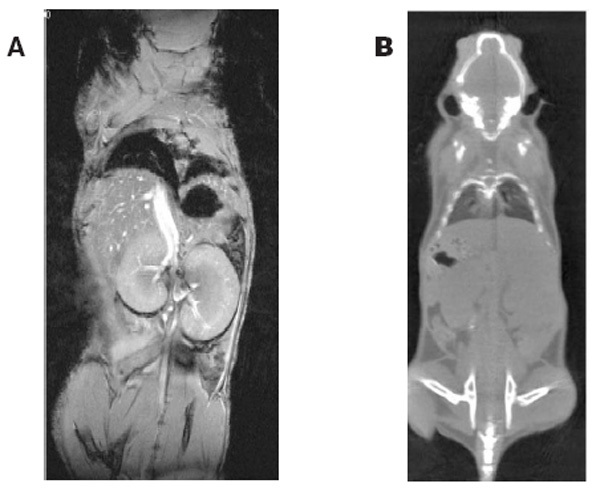
MRI uses radio waves and magnets to generate radiograph-like images of soft tissue. (B) Coronal CT view of a normal mouse. X-ray CT uses multiple radiographic views of a subject to construct an image.
MRI is excellent for imaging different types of soft tissue with high contrast3,4,5,6. Researchers can use it for anatomical and functional studies. Some applications include tumor growth and treatment, brain function and stroke, and cardiovascular disease. Contrast agents, which are drugs that function like histological stains, increase the usefulness of this imaging method. For example, Magnevist (Berlex Imaging, Wayne, NJ), a contrast agent in wide clinical use, intensifies the MR images of vascular tumors. The time required for an image acquisition can vary from a few minutes to >2 hours, depending on the size, type, and resolution of the data set.
Because of the surrounding magnetic field, an MRI system requires a large area (≥700 ft2). The magnet itself requires ∼200 ft2 of surrounding clear area. The fringe field is the distance that the surrounding magnetic field extends before it drops to the level of 5 gauss. The fringe field can occupy an area up to 250 ft2. The MRI suite must have a design that prevents casual visitors from entering the fringe field. A magnetic field >15 gauss can adversely affect persons with pacemakers or other metallic implants. The distance to the 15 gauss line varies widely with field strength and magnet type. There are magnets available that have a fringe field limited to a foot or less, but these cost more. MR scanners use extremely heavy magnets; therefore, most facilities locate the MR imager on the ground floor. Additionally, because the magnets are cooled by cryogens, good ventilation must be present. Rapid boil-off of the cryogens could lead to asphyxiation. The instrument manufacturer can provide a detailed description of space requirements and suggested architectural layouts. A facility will need to plan for additional space for the electronics and console for the scanner as well as a preparation area outside the magnetic field. This can amount to another 250 ft2.
Researchers currently consider a 7-Tesla MRI system an optimal field for imaging rodents. At the MIF we have several Bruker Avance MRI scanners (Bruker-Biospin, Billerica, MA). Some other manufacturers of MRI consoles are Varian (Varian Medical Systems, Inc., Palo Alto, CA), Tecmag (Tecmag, Houston, TX), and MRRS (MR Research Systems, Guildford, UK). A basic setup for the MRI includes the superconducting magnet, the imaging console, and several probes for approximately $800,000–$2,000,000, depending on the options. It is ideal to enclose the magnet in a room shielded from radiofrequency, but this construction would generate additional costs. The magnet itself comes with end caps that adequately shield it; however, passing anesthesia and monitoring lines becomes awkward.
Because of its complexity, the MRI requires a scientist with not only the necessary academic qualifications, but also a technical background for operation. There are many MRI measurement methods, each with many control variables, designed for specific purposes. The MR operator must be familiar with the basic physics of the MR measurement and the effect of the controls on the image outcome. If new methods are going to be developed or implemented, a postdoctoral-level scientist will be necessary. New methods require sophisticated knowledge of the hardware and computer programming. This individual will be responsible for running the scanner and maintaining the system. MRI maintenance requirements include both hardware and software.
Hardware maintenance includes the associated magnetic field. The magnet requires liquid nitrogen and liquid helium to maintain the superconducting magnetic field. Allowing for a safety margin, most magnets need liquid nitrogen once per week and liquid helium approximately twice per year. If the cryogen level is allowed to become too low, the magnetic field can spontaneously quench (i.e., lose its magnetic force). Quality assurance includes regular test images of a standard sample. A small loss in performance can result in unusable images.
The operating computer and software also require regular maintenance. System software requires periodic updates to install security patches, repair bugs, and add features. These updates are particularly important if the computers are on a network with outside access. Despite hardware and software designed to limit external access, new security holes appear on a regular basis. A malicious attack could destroy valuable work and render the instrument useless until the base software can be reinstalled.
Archiving and removing old data are important parts of computer maintenance. If the number and size of the stored image files grow too large, system performance can slow or even become blocked. A policy for data management can help prevent major problems, but periodic enforcement is still necessary. This policy can range from a simple principle of moving the oldest data first to a more complicated formula based on size and age. At the MIF, we encourage everyone to transfer data from the instrument as soon as possible. When data space becomes an issue, the users with the largest amount of data must export or delete files before they can resume scanning.
X-Ray CT
X-ray CT is an imaging method that uses multiple radiographic views of a subject to construct an image (Fig. 1B). In our system, an X-ray source and detector rotate around the subject 360 degrees while generating a number of projections or views. The MIF has a MicroCat II CT scanner built by ImTek, Inc. (Knoxville, TN). Other manufacturers of micro-CT equipment include GE Medical Systems (London, Ontario, Canada), Scanco Medical (Scanco USA, Inc., Southeastern, PA), and SkyScan (Aartselaar, Belgium). Each projection is the equivalent of a radiographic image. A method called 'filtered back projection' permits these projections to be reconstructed into a three-dimensional (3D) data set. A micro-CT scanner for rodents typically uses less X-ray power than a conventional clinical CT scanner, so the effective radiation exposure per scan is reduced. The highest radiation dose we have measured was 35 rad for skin, but typical scans are much less (9 rad). The radiation dose is ultimately dependent on the scan parameters (i.e., number of projections, exposure time, X-ray beam strength).
CT is excellent for studying the skeletal system, certain internal organs, and fat distribution7,8,9,10. CT can also be used for tumor studies, depending on the location of the tumor. A typical 3D image with 100-μm resolution takes about 10 minutes to acquire; higher resolution images may take up to 25 minutes. Image reconstruction can take as little as 5 extra minutes or up to 24 hours depending on hardware and software options. As with MRI, a contrast agent can be used to enhance the appearance of some organs. In rodents, the half-life of most clinical CT contrast agents is short, so some experimentation is required to find the optimum dose and administration regimen.
A micro-CT scanner requires ∼200 ft2 of space for the scanner and preparation area. The manufacturer can provide exact measurements of the machine footprint. There are several micro-CT manufacturers, and the price ranges from $250,000 to $400,000.
The instrument will require one technician for operation and maintenance. This individual should have a sophisticated education in biology or a related field and be comfortable with computers. The CT scanner relies on several computers on a small network. The CT operator will be responsible for maintaining the computer system software, managing the accumulated data, and quality assurance. A good knowledge of anatomy is vital for data interpretation, and the person should be able to interpret radiographs.
Optical Imaging
Reporter gene technology has revolutionized the ability to track cell populations in vivo11. Optical imaging devices, which allow the user to monitor gene expression, are easy to operate and have no special environmental requirements. There are several different types of optical imaging. Bioluminescence is an enzymatic biochemical process that produces light. The luciferase gene is first inserted into the targeted cell population. Image acquisition requires live cells to process the substrate and produce light. Advantages of bioluminescence include high sensitivity, and virtually no background noise; disadvantages include the need for cofactors (i.e., oxygen, magnesium, and ATP) and expensive substrates (luciferin). Fluorescence imaging also requires genetic manipulation; however, the technology does not require a living system to produce an image. Fluorescence imaging differs from bioluminescence in that light must be applied to the tissue to generate a signal. One advantage of fluorescence imaging is the commercial availability of numerous markers (GFP, DsRed, ICG, and Cy5.5) with no need for substrate or cofactors; a disadvantage is the low signal/noise ratio due to autofluorescence.
Basic equipment for bioluminescence or fluorescence imaging consists of a light-tight chamber, a sensitive camera (charge-coupled device), and a computer. It is possible for the ambitious investigator to manufacture his or her own imaging device; however, commercial products are available for about $200,000. The MIF owns a Xenogen IVIS 100 (Xenogen Corp., Alameda, CA); other manufacturers include Eastman Kodak (Eastman Kodak Co., Scientific Imaging Systems, Rochester, NY), Roper Scientific (Trenton, NJ), and Advanced Research Technologies, Inc. (Saint-Laurent, Quebec, Canada). Our imaging device occupies a space measuring 16 ft2; additional benchtop space is necessary for preparing the animal. Image acquisition is simple, and it is not necessary to have a dedicated system operator. Investigators can easily learn to perform their own imaging study. Our system's operating software first acquires a black-and-white photographic image of the subject, then acquires the luminescent image, and finally overlays the image onto the photograph for anatomical signal mapping (Fig. 2). Both bioluminescence and fluorescence imaging are useful to detect tumor cells, monitor tumor growth, or track cell movement (e.g., metastasis). Optical imaging can be highly specific, but it has low spatial resolution. Optical data are typically two-dimensional (2D), but 3D methods are in development. A high throughput of animals is possible because imaging time is short, usually from 1 second to 10 minutes.
Figure 2. Luciferase image of lung tumors overlaid on a positional image.
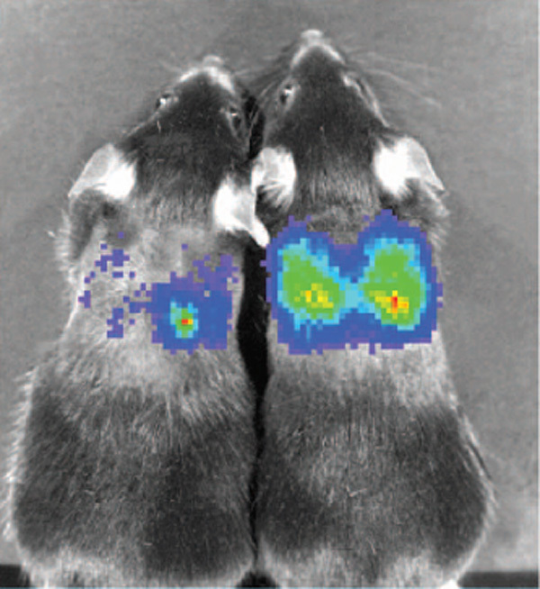
The intensity of the light detected correlates with the level of target gene expression.
Laser Doppler is another type of optical imaging that uses a laser light source and the Doppler effect to measure capillary blood flow (Fig. 3). The equipment consists of a laser source and computer, and costs less than $50,000. The footprint of the laser device is small (4 ft2 plus computer), but its sensitivity to movement warrants placement on an antivibration table. This machine is simple to use and is highly sensitive, but it is limited to small vessels within a 1-mm depth. The MIF owns a laser Doppler imager from Moor Instruments, Inc. (Wilmington, DE). Another manufacturer is Perimed, Inc. (North Royalton, OH).
Figure 3. Laser Doppler image of a human hand.
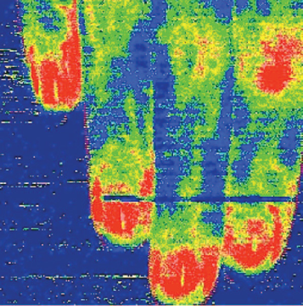
Doppler imagers use the Doppler effect to measure capillary blood flow.
Ultrasound Imaging
Ultrasound imaging is a rapid, real-time in vivo technique. An ultrasound transducer broadcasts sound waves beyond the audible range into tissue. As the sound waves encounter the interfaces between various types of tissue, they are reflected. The transducer detects the reflected sound waves and uses them to construct an image (Fig. 4). The depth of penetration depends on the frequency of the sound wave. The image-processing software produces an image in real time, and the various tissues within the image display different 'echogenic' properties.
Figure 4. B-mode ultrasound image of a mouse fetus.
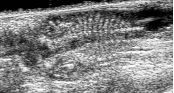
Ultrasound imagers measure reflected sound waves and use them to construct an image.
Ultrasound machines used for rodent imaging are similar to those used clinically; however, the larger field of view offered by a clinical scanner is not optimal for imaging smaller subjects. Resolution increases (and field of view decreases) as the ultrasound frequency increases; therefore, high-frequency ultrasound is ideal for rodents. Ultrasound is excellent for cardiac studies and evaluation of embryonic development12,13. It may also be used for various organ evaluations, tumors, and guided injections. Advantages of ultrasound include rapid image acquisition and ease of use; however, time is required for the operator to develop proficiency in the acquisition techniques and image interpretation. Knowledge of anatomy is useful for positioning the transducers, as well as evaluating the resulting images. A well-trained operator should be able to distinguish normal and abnormal anatomy. Scattering of the sound waves in the tissue often cause ultrasound images to appear 'noisy'. Also, most ultrasound images are 2D, although some 3D-capable instruments are available. Most ultrasound machines are built on portable carts for easy relocation. The image display is most clearly visible in low ambient light, so this should be a consideration when determining the location for ultrasound imaging.
The MIF owns two ultrasound machines: a Siemens Acuson Sequoia (Siemens Medical Solutions, Malvern, PA) and a Visualsonics Vevo 660 (VisualSonics, Inc., Toronto, Ontario, Canada). There are many other ultrasound manufacturers, including Philips (Philips Medical Systems, Andover, MA) and General Electric (GE Medical Systems, London, Ontario, Canada). A commercial ultrasound imager can cost about $150,000. Although one can perform data analysis on the scanner, this does take time away from available scan time. An additional data-processing workstation could cost as much as $60,000. The MIF does not have a dedicated ultrasonographer at this time. The investigators that use our Acuson ultrasound most frequently have experience in its operation and often collaborate with other investigators in need of their expertise. The MIF staff is developing proficiency in ultrasonography.
Positron Emission Tomography
Positron emission tomography (PET) is an excellent method to perform functional imaging in vivo14,15,16. Uptake of radioactive compounds can demonstrate the presence of tumor, abnormal cell function, or metabolic changes. This imaging technique can generate 3D data. PET imaging is highly sensitive but suffers from low spatial specificity and needs to be superimposed on an anatomical image for signal location (Fig. 5). The footprint of a micro-PET system may only be 16 ft2, but it is ideal to locate the micro-PET adjacent to a micro-CT or MRI because an anatomical image will be required for co-registration. This allows convenient transport of an anesthetized animal from one machine to another.
Figure 5.
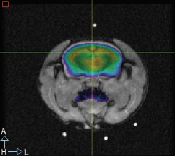
PET scan overlaid on an axial MR image, showing uptake of the radioactive glucose analog 2-fluor-2-deoxy-D-glucose (FDG).
Micro-PET imaging presents its own set of unique considerations. The use of radioactivity dictates its own specifications. The imaging area should be in a location that allows the environment to be properly controlled and appropriate precautions taken. Your institution should be able to provide a detailed description of the rules and regulations that govern the use of radionuclides. The imaging animals will remain radioactive for some time after imaging, so housing for these 'hot' animals must be considered. The half-lives of radionuclides in the radioactive compounds are typically short (e.g., 18 hours), so housing animals can be a temporary situation. The availability of radioactive compounds will have the greatest impact on the success of a micro-PET program. Radiolabeled glucose, oxygen, and ammonia are commercially available; however, many studies may need custom-made radioactive compounds. The ideal arrangement is an imaging suite located adjacent to the radiopharmacy that will produce the radionuclides. Production of these reagents can represent the largest expense because of the need for highly specific equipment and personnel. Manufacturers of micro-Pet imagers include General Electric and Philips Medical Systems. Micro-PET operation will also require a postdoctorate-level scientist to assemble, operate, and maintain the equipment.
Data Processing
It is easy to generate an enormous amount of image data in a relatively short amount of time. A single data set can range from a few kilobytes to over two gigabytes for high-resolution 3D data. The amount of image data generated over time can be a problem. Mass storage devices must be available for data storage during analysis. If backup and archiving are required, the amount of equipment needed can be considerable. A room will need to be dedicated to computer storage and workstations for post processing images. A minimal room will have space for a rack containing data servers and network equipment. Qualified information technology personnel should be able to provide additional advice.
In addition to data storage, extra workstations for data processing are almost a necessity. The alternative is allowing data to be analyzed on the acquisition instruments. This reduces the need for extra computers but takes time away from the instruments. The time required for data analysis can be considerable.
Some consideration must be given to who will analyze data. Often, users of the instrument will not have the experience or skill needed to gain the most information from the images. One solution is for image analysis to be part of the imaging service. Another approach is to train the investigators to analyze their own data; this reduces the facility burden, but reduces the usefulness of the service to new users. At the MIF, we train investigators to perform simple data analysis. For more complex data analysis, MIF staff collaborates with the investigators. This system works well because, having trained an investigator, the technician is freed up for other duties; the investigator can often then pass that training on to new individuals in his or her group as well as other collaborators.
Support Staffing
Once the user base is established, equipment needs are defined, and the facility floor plan is designed, one must consider the staffing needs. A variety of personnel are needed for a successful imaging facility. Diversity of experience will enhance the resources as well as expand the knowledge base of the entire staff. Certain base criteria must be met for each unique position, but it is our experience that personnel experienced in imaging laboratory animals are difficult to find. Many of the necessary technical procedures may be learned on site. We believe that a strong desire to learn may be more important than experience and formal education. It is important to have at least one person on the staff that is skilled in the routine procedures and can provide training for new staff members.
Animal Care and Preparation
As with any animal facility, husbandry staff is necessary. Ideally, housing for imaging animals should be on site and accessible. The size of the animal housing facility will determine the number of necessary personnel. Routine husbandry duties include daily health checks, cage changing, cage washing, room sanitation, sentinel maintenance, quality control, etc. Husbandry staff is the foundation of all successful animal research programs. The qualifications of the animal care staff are dependent on how many people will be required to run the facility and in what capacity the people are expected to function. If you can have dedicated husbandry staff, their qualifications will be less stringent, and a person with AALAS ALAT or LAT experience would be sufficient. Staff members who will be assisting with imaging experiments are expected to work independently, so it is ideal to have personnel with AALAS LATG certification or equivalent experience. If the technical staff is required to assist with (or perform) husbandry duties, they should understand that it is part of their job description so that no misunderstandings occur later.
Laboratory animals must be immobilized for imaging procedures. It is important for the laboratory animal technical staff to maintain a working knowledge of anesthesia and physiological monitoring for a variety of species. Additionally, basic procedure skills such as venipuncture are required for the administration of imaging contrast agents and other pharmaceuticals. Familiarity with aseptic techniques and rodent colony health management techniques will aid in maintaining the level of health standards for the facility. Additional useful procedures include intubation of rodents, placement of rodent femoral, jugular, and carotid catheters.
Facility and Equipment Maintenance
The computers acquiring the images and the networks they are connected to can be complicated. Many of the personnel operating the imaging devices have sufficient knowledge of computer operation and software programming to repair problems as they occur; however, given the number of computers needed for imaging and personnel support, it is a good idea to have someone dedicated to their maintenance. It is important that these networks are secure and protected from any outside abuse. The MIF has shared information technology personnel that maintain computer software security and upgrades.
The imaging instruments require regular hardware maintenance as well as periodic repair. The best solution is to have an engineer on the staff to address these issues. It is possible to maintain a service contract with an outside company, but an internal person can aid in quicker diagnosis and repair. The MIF has maintenance contract on all equipment, but the level of each maintenance plan varies from a 'parts only' plan to a full-service plan that includes yearly preventative maintenance. The MIF is fortunate to have MRI scientists who are knowledgable in MRI maintenance and repair, as well as an electrical engineer who assists with MRI and CT maintenance and repair.
The imaging facility also needs personnel to attend to nonscientific tasks. Administrators must handle daily paperwork such as budget management and supplies, as well as provide other organizational services. These people are critical, because if the facility is successful, none of the scientific personnel will have time to attend to administrative duties. The MIF has shared personnel for these tasks.
Facility Procedures
Any facility that conducts animal research should be accessible by authorized personnel only. We cannot overemphasize this because of magnet safety, radiation safety, and animal health. Traffic patterns for animals entering the facility may be necessary if the animal health level varies in different areas.
The amount of personal protective equipment (PPE) needed will be defined by the types of animals in the facility, as well as the health standard. The minimum PPE for handling animals in the MIF includes a disposable lab coat and gloves. Because animal studies are ongoing, and we temporarily house mice, personnel must don a lab coat upon entering our facility, regardless of their animal contact. Studies with nonhuman primates require a higher level of PPE that includes mucous membrane protection. Standard PPE worn around the MRI scanners must be nonmagnetic.
An Institutional Animal Care and Use Committee (IACUC) should approve every study conducted within the facility. New investigators may seek the assistance of facility personnel in writing their protocols. This gives the imaging facility personnel a chance to suggest feasible imaging modalities, preparation methods, and anesthesia regimes. The MIF has a committee of experienced researchers that review experiments for feasibility, safety, and time requirement.
Imaging time may be scheduled on a first-come first-served basis, or otherwise. Instruments with short imaging times allow for a larger number of studies in any given period of time. When demands exceed the available scan time, one solution is to assign blocks of time for specific groups. Then individuals within the group can work out their own priority for imaging. In our experience, it is easiest to have the equipment operators schedule the investigators. The operators will be most experienced with the requests of the prospective study and can schedule time accordingly. In the MIF, magnet scan time during regular working hours is assigned to institutes in blocks; the remaining scan time is assigned on a first-come first-served basis. The other imaging devices require less time per scan, so it has not been necessary to assign blocks of time yet; each investigator is assigned time on a first-come first-served basis. Scan time is charged back to the institute by the hour, and the dollar amount is calculated with a budget-driven formula.
Summary
Noninvasive imaging of rodents and other small animals is a powerful tool for biomedical research. Setting up an imaging facility is a complex process that involves many decisions affecting everything from available instruments to staff composition. Careful planning should help prevent operational snags. The advice of experienced people will be the most valuable asset. It is our opinion that the facility should be designed around a primary MRI scanner because MR offers versatility for many applications. Acquisition of other devices will be determined by the needs of the research community. Growth of the facility is limited only by usage and executive decisions. Staff must include a few experienced personnel, but inexperienced eager personnel can be trained to be experts. We have tried to provide an outline of some of the important considerations that went into the creation of the MIF.
Acknowledgements
The authors thank Stasia Anderson of Laboratory of Diagnostic Radiology Research, NIH, for providing an MR image, Dan Schimel of the NIH Mouse Imaging Facility for providing a CT image, Cecilia Lo of the Laboratory of Developmental Biology, NHLBI, NIH, for providing an ultrasound image, Takashi Murakami and Sam Hwang of the Dermatology Branch, NCI, NIH, for providing a luciferase image, Michael Green and the Imaging Physics Laboratory, NIH, for providing a PET image, and Afonso Silva of the Laboratory for Functional and Molecular Imaging, NINDS, NIH, for providing images for this paper. We would also like to thank Alan Koretsky of the Laboratory for Functional and Molecular Imaging for helpful discussions.
References
- 1.Balaban RS, Hampshire VA. Challenges in small animal noninvasive imaging. ILAR J. 2001;42(3):248–262. doi: 10.1093/ilar.42.3.248. [DOI] [PubMed] [Google Scholar]
- 2.Brent RL, Gordon WE, Bennett WR, Beckman DA. Reproductive and teratologic effects of electromagnetic fields. Reprod. Toxicol. 1993;7(6):535–580. doi: 10.1016/0890-6238(93)90033-4. [DOI] [PubMed] [Google Scholar]
- 3.Benveniste H, Blackband S. MR microscopy and high resolution small animal MRI: applications in neuroscience research. Prog. Neurobiol. 2002;67(5):393–420. doi: 10.1016/S0301-0082(02)00020-5. [DOI] [PubMed] [Google Scholar]
- 4.Budinger TF, Benaron DA, Koretsky AP. Imaging transgenic animals. Annu. Rev. Biomed. Eng. 1999;1:611–648. doi: 10.1146/annurev.bioeng.1.1.611. [DOI] [PubMed] [Google Scholar]
- 5.Allport JR, Weissleder R. In vivo imaging of gene and cell therapies. Exp. Hematol. 2001;29(11):1237–1246. doi: 10.1016/S0301-472X(01)00739-1. [DOI] [PubMed] [Google Scholar]
- 6.Krishna MC, Devasahayam N, Cook JA, Subramanian S, Kuppusamy P, Mitchell JB. Electron paramagnetic resonance for small animal imaging applications. ILAR J. 2001;42(3):209–218. doi: 10.1093/ilar.42.3.209. [DOI] [PubMed] [Google Scholar]
- 7.Ritman EL. Molecular imaging in small animals—roles for micro-CT. J. Cell. Biochem. Suppl. 2002;39:116–124. doi: 10.1002/jcb.10415. [DOI] [PubMed] [Google Scholar]
- 8.Bentley MD, Ortiz MC, Ritman EL, Romero JC. The use of microcomputed tomography to study microvasculature in small rodents. Am. J. Physiol. Regul. Integr. Comp. Physiol. 2002;282(5):R1267–R1279. doi: 10.1152/ajpregu.00560.2001. [DOI] [PubMed] [Google Scholar]
- 9.Paulus MJ, Gleason SS, Easterly ME, Foltz CJ. A review of high-resolution X-ray computed tomography and other imaging modalities for small animal research. Lab. Anim. (NY) 2001;30(3):36–45. [PubMed] [Google Scholar]
- 10.Paulus MJ, Gleason SS, Kennel SJ, Hunsicker PR, Johnson DK. High resolution X-ray computed tomography: an emerging tool for small animal cancer research. Neoplasia. 2000;2(1–2):62–70. doi: 10.1038/sj.neo.7900069. [DOI] [PMC free article] [PubMed] [Google Scholar]
- 11.Contag CH, Bachmann MH. Advances in in vivo bioluminescence imaging of gene expression. Annu. Rev. Biomed. Eng. 2002;4:235–260. doi: 10.1146/annurev.bioeng.4.111901.093336. [DOI] [PubMed] [Google Scholar]
- 12.Foster FS, Pavlin CJ, Harasiewicz KA, Christopher DA, Turnbull DH. Advances in ultrasound biomicroscopy. Ultrasound Med. Biol. 2000;26(1):1–27. doi: 10.1016/S0301-5629(99)00096-4. [DOI] [PubMed] [Google Scholar]
- 13.Hartley CJ, Taffet GE, Reddy AK, Entman ML, Michael LH. Noninvasive cardiovascular phenotyping in mice. ILAR J. 2002;43(3):147–158. doi: 10.1093/ilar.43.3.147. [DOI] [PubMed] [Google Scholar]
- 14.Chatziioannou AF. Molecular imaging of small animals with dedicated PET tomographs. Eur. J. Nucl. Med. Mol. Imaging. 2002;29(1):98–114. doi: 10.1007/s00259-001-0683-3. [DOI] [PubMed] [Google Scholar]
- 15.Wirrwar A, Schramm N, Vosberg H, Muller-Gartner HW. High resolution SPECT in small animal research. Rev. Neurosci. 2001;12(2):187–193. doi: 10.1515/revneuro.2001.12.2.187. [DOI] [PubMed] [Google Scholar]
- 16.Myers R, Hume S, Bloomfield P, Jones T. Radio-imaging in small animals. J. Psychopharmacol. 1999;13(4):352–357. doi: 10.1177/026988119901300404. [DOI] [PubMed] [Google Scholar]


