Abstract
Background
V. parahaemolyticus is autochthonous to the marine environment and causes seafood-borne gastroenteritis in humans. Generally, V. parahaemolyticus recovered from the environment and/or seafood is thought to be non-pathogenic and the relationship between environmental isolates and acute diarrhoeal disease is poorly understood. In this study, we explored the virulence potential of environmental V. parahaemolyticus isolated from water, plankton and assorted seafood samples collected from the Indian coast.
Results
Twenty-two V. parahaemolyticus isolates from seafood harboured virulence associated genes encoding the thermostable-direct haemolysin (TDH), TDH-related haemolysin (TRH), and Type 3 secretion systems (T3SS) and 95.5% of the toxigenic isolates had pandemic strain attributes (toxRS/new+). Nine serovars, with pandemic strain traits were newly identified and an O4:K36 tdh−trh+V. parahaemolyticus bearing pandemic marker gene was recognised for the first time. Results obtained by reverse transcription PCR showed trh, T3SS1 and T3SS2β to be functional in the seafood isolates. Moreover, the environmental strains were cytotoxic and could invade Caco-2 cells upon infection as well as induce changes to the tight junction protein, ZO-1 and the actin cytoskeleton.
Conclusion
Our study provides evidence that environmental isolates of V. parahaemolyticus are potentially invasive and capable of eliciting pathogenic characteristics typical of clinical strains and present a potential health risk. We also demonstrate that virulence of this pathogen is highly complex and hence draws attention for the need to investigate more reliable virulence markers in order to distinguish the environmental and clinical isolates, which will be crucial for the pathogenomics and control of this pathogen.
Keywords: V. parahaemolyticus, Seafood, Pandemic traits, Type 3 secretion system, Pulsed-field gel electrophoresis, Cytotoxicity, Invasion
Background
Vibrio parahaemolyticus is a Gram-negative, halophilic bacterium that inhabits marine and estuarine environments. Since its discovery in 1950 as the causative agent of gastroenteritis in Japan [1], V. parahaemolyticus has become one of the leading causes of food-borne illness in humans. Infection is closely associated with the consumption of raw or undercooked seafood, resulting in self-limiting diarrhoea [2]. Rarely, V. parahaemolyticus also causes wound infections and septicaemia [2]. The virulence of V. parahaemolyticus is associated with the production of thermostable-direct haemolysin (TDH) (encoded by tdh gene) and/or TDH-related haemolysin (TRH, encoded by trh gene) as well as two type III secretion systems (T3SS) [3–5]. Two sets of the genes T3SS1 and T3SS2 are present on chromosomes 1 and 2, respectively [5].
Though a majority of clinical V. parahaemolyticus generally carry tdh and/or trh, only a small proportion of environmental isolates have been found to harbour the hemolysin genes [6–8]. T3SS1 that produce cytotoxicity is present in all V. parahaemolyticus isolates irrespective of their source. T3SS2, on the other hand, is both cytotoxic and enterotoxic and has two phylogroups; T3SS2α that co-localise with tdh and T3SS2β found in association with trh [9]. The environmental population of V. parahaemolyticus is increasingly acquiring the virulence-related genes that classically define a clinical isolate [10, 11]. However, the true pathogenic potential of such strains has not been evaluated at large.
After the emergence of the V. parahaemolyticus serovar O3:K6 in 1996 from Kolkata, India [12], infections caused by several serovariants, collectively called pandemic strains, have increased due to their global spread with several diarrhoeal epidemics [13]. The pandemic clone has the typical tdh+trh−, toxRS/new+, orf8+/− genotype, which is identified by Group-specific PCR (GS-PCR) that detects the sequence variation in toxRS gene (toxRS/new) [14, 15]. The burden of V. parahaemolyticus diarrhoea is very high in Asian countries [16–18] and the outbreaks are related to raw shellfish consumption in the United States and Canada [19, 20]. In addition, sporadic outbreaks have been reported in coastal Europe [21, 22]. Although the pandemic strains are still prevalent in diarrhoeal cases in India [23], there is a paucity of information on V. parahaemolyticus from different environmental sources.
The aim of this study was to examine V. parahaemolyticus from environmental and seafood samples collected along the southern Indian coast for serogroup, putative virulence and their pathogenic potential on intestinal epithelial cell line.
Results
Identification of V. parahaemolyticus from the environment
Four hundred and seventeen V. parahaemolyticus isolates were identified from environmental and seafood samples collected through five districts of the coastal belt of Kerala during the sampling period. The highest recovery of the organism was from seafood (225/417, 53.9%) followed by water (152/417, 36.5%) and plankton (40/417, 9.6%). Environmental parameters such as temperature, pH and salinity were not checked during sampling, as the objective was focused mainly towards genetic profiling and pathogenic potential of V. parahaemolyticus.
Distribution of virulence genes
The isolates were screened for 22 virulence markers, including genes encoding haemolysins, T3SS1, T3SS2α, and T3SS2β and the pandemic strain marker of ORF-8 and group-specific PCR (GS-PCR). Twenty two (5.3%) isolates were found to contain either of the haemolysins and were potentially toxigenic (Table 1); 19 had tdh (tdh+trh−) while 3 had trh (tdh−trh+) and all the isolates represented seafood (22/225, 9.8%). None were found to be tdh+trh+.
Table 1.
Characteristics of 22 environmental V. parahaemolyticus isolates collected from the south-west coast of India
| Strain Id | Serotype | toxR | tlh | tdh | trh | toxRS/new | orf8 | T3SS2 genes | |
|---|---|---|---|---|---|---|---|---|---|
| H1 | O1:K17 | + | + | + | – | + | – | vopC, vopT, vscC2, vopA, vopB2, vopL | T3SS2α |
| H2 | OUT:KUT | + | + | + | – | + | – | vopC, vopZ, vopL | |
| H3 | O5:KUT | + | + | + | – | + | – | vopC, vopT, vscC2, vopA, vopB2 | |
| H4 | O5:K17 | + | + | + | – | + | – | vopC, vopZ, vopA, vopL | |
| H5 | O1:K19 | + | + | + | – | + | – | vopC, vopT | |
| H6 | O1:K25 | + | + | + | – | + | – | vopC, vopT, vopZ | |
| H7 | O1:K25 | + | + | + | – | – | – | vopC, vopT, vopZ | |
| H8 | O1:K23 | + | + | + | – | + | – | vopC, vscC2, vopB2, vopL | |
| H9 | O1:K25 | + | + | + | – | + | – | vopC, vscC2, vopB2, vopL | |
| H10 | O5:K17 | + | + | + | – | + | – | vopC, vopZ, vscC2, vopA, vopB2 | |
| H11 | O1:K25 | + | + | + | – | + | – | vopC, vopZ, vscC2 | |
| H12 | O10:K24 | + | + | + | – | + | – | vopC, vopZ, vopB2 | |
| H13 | O2:KUT | + | + | + | – | + | – | vopC, vopZ, vopB2, vopL | |
| H14 | O4:K29 | + | + | + | – | + | – | vopC, vopZ, vscC2, vopB2, vopL | |
| H15 | O5:K17 | + | + | + | – | + | – | vopC, vopB2 | |
| H16 | O1:KUT | + | + | + | – | + | – | vopC, vopB2, vopL | |
| H17 | O5:K20 | + | + | + | – | + | – | vopC, vopT, vopB2 | |
| H18 | O5:K20 | + | + | + | – | + | – | vopC, vopA, vopB2 | |
| H19 | O4:K42 | + | + | + | – | + | – | vopC, vopA, vopB2, vopL | |
| C12 | O3:KUT | + | + | – | + | + | – | vscC2, vopB2, vscS2, vopC, vopA, vopL | T3SS2β |
| C13 | O3:KUT | + | + | – | + | + | – | vscC2, vopB2, vscS2, vopC, vopA, vopL | |
| K23 | O4:K36 | + | + | – | + | + | – | vscC2, vopB2, vscS2, vopC, vopA, vopL |
The toxigenic isolates were then checked for T3SS genes coding for effectors and apparatus proteins. T3SS1 (vscP, vopS, vscK, vscF and VPA0450) was present in all the 22 isolates. Several genetic elements of T3SS2α were detected with isolates harbouring more than three genes. While vopC, thought to mediate invasion of non-phagocytic cells, was present in 19 tdh+trh− isolates, vopB2 was detected in 13, vscC2 in 7, vopT, vopL and vopA/P in 5, 9 and 6 isolates, respectively. Six of the isolates also harboured vopZ, the gene responsible for intestinal colonisation and enterotoxocity. Genes encoding the T3SS2β (vscC2, vopB2, vscS2, vopC, vopA/P and vopL) were present in all the three tdh−trh+ isolates (Table 1). Twenty one isolates (18 tdh+trh−, 3 tdh−trh+) belonged to the pandemic strain type (toxRS/new+).
PFGE analysis of toxigenic strains
The 22 toxigenic isolates consisted of 15 serovars with combinations of six O groups (O1, O2, O3, O4, O5 and O10) and nine different K types (K17, K19, K20, K23, K24, K25, K29, K36 and K42). The predominant serovar was O1:K25 (n = 4). Two tdh−trh+ isolates belonged to O3:KUT while the other was O4:K36.
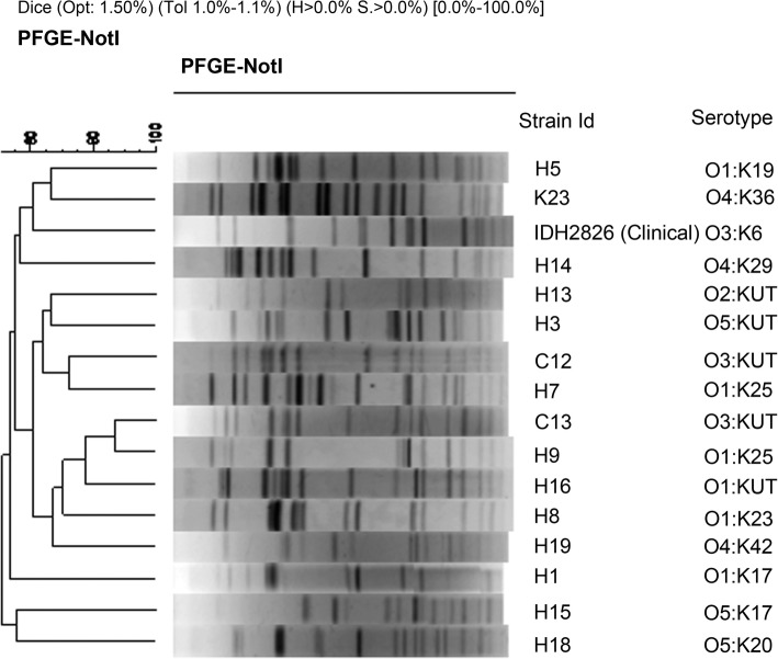
Based on the serovar profile, a total of 15 isolates were analysed by PFGE and the pattern compared to an O3:K6 clinical isolate (NICED, Kolkata, India). The minimum genetic similarity of the isolates was 55% and fell into four distinct clusters (Fig. 1). Those that clustered together at 60% similarity with the O3:K6 were isolates of O1:K19, O4:K29 (tdh+trh−) and O4:K36 (tdh−trh+) serovars. It was observed that isolates of the same O1:K25 serovar clustered differently. Likewise, the three tdh−trh+ isolates possessing identical virulence profiles were present in three different clusters. Interestingly, the GS-PCR positive and negative isolates clustered together. Overall, the results revealed a high genetic variability in environmental isolates of V. parahaemolyticus.
Fig. 1.
NotI digested PFGE profile of V. parahaemolyticus with dendrogram. Clustering was performed using the unweighted pair group method (UPGMA) and the Dice correlation coefficient with a position tolerance of 1.0%
Selection of strains for pathogenicity studies
A total of 12 isolates of V. parahaemolyticus including 8 environmental and 4 clinical were included in downstream assays (Table 2). The environmental strains were selected based on the T3SS profile and serovar type.
Table 2.
V. parahaemolyticus strains used for pathogenicity studies
| Group | Strain Id | Serogroup | Toxin Profile | Source |
|---|---|---|---|---|
| Environmental, toxigenic | H10 | O5:K17 | tdh+trh− | Present study, seafood |
| H12 | O10:K24 | tdh+trh− | ||
| H14 | O4:K29 | tdh+trh− | ||
| K23 | O4:K36 | tdh−trh+ | ||
| Environmental, non-toxigenic | AJM1 | O5:K17 | tdh−trh− | Present study, water |
| ME1 | O10:KUT | tdh−trh− | ||
| ME4 | O1:K32 | tdh−trh− | ||
| ME8 | O1:K32 | tdh−trh− | ||
| Clinical | RIMD2210633 | O3:K6 | tdh+trh− | Reference pandemic isolate, Japan |
| AP11243 | O1:KUT | tdh+trh− | Bangladesh | |
| IDH03525 | O3:K6 | tdh+trh− | India | |
| ATCC17802 | O1:K1 | tdh−trh+ | Japan |
Expression of virulence genes
Expression of virulence genes tdh, trh, effectors of T3SS1 (vopS, VPA0450), T3SS2α (vopC, vopT, vopZ, vopA/P, vopL) and T3SS2β (vopC, vopA/P, vopL) was determined by RT-PCR in relation to gyrB, which is a constitutively expressed housekeeping gene. The tdh gene was transcribed by all the tdh+trh− clinical isolates, whereas inconsistent results were observed with the seafood isolates H10, H12 and H14. Either the bands were very faint or not detected at all in repeated PCRs. It was concluded that the tdh gene was not functional in the seafood isolates. In contrast, the tdh−trh+ isolate K23 and its clinical counterpart ATCC17802 transcribed trh gene with the production of the corresponding amplified cDNA (Additional file 1: Figure S1).
The expression results were verified by the haemolytic activity of isolates on human RBC (Additional file 1: Figure S2). Both the tdh- and trh- containing seafood strains showed extremely weak haemolytic activity (≤5%) after 6 h of incubation, whereas the clinical isolates were 50% haemolytic after 6 h, except ATCC17802 that could lyse only 3% of RBC. The haemolytic action of the seafood isolates showed only a modest increase when incubation was extended till 12 h while a dramatic rise (> 90%) was seen with tdh+trh− clinical isolates except tdh−trh+ ATCC17802 (20%).
All the selected seafood isolates irrespective of the toxin type, transcribed T3SS1 effectors indicating they are functional (Additional file 1: Figure S3). Prominent amplification of T3SS2β genes was also detected in the isolate K23 (Additional file 1: Figure S3). On the other hand, T3SS2α effectors were not transcribed by any of the tdh+trh− seafood isolates. Surprisingly, a faint band was detected for vopV in the isolate H14, which was absent in the control RNA (Additional file 1: Figure S3). As expected, the clinical control isolates functionally expressed the T3SS genes.
Cytotoxicity of environmental V. parahaemolyticus
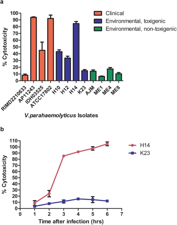
Cytotoxicity was tested by measuring the levels of cytoplasmic LDH released into the medium during infection of Caco-2. This release indicated the degree to which the integrity of host cell membrane was compromised. Only two clinical isolates, AP11243 (tdh+trh−) and ATCC17802 (tdh−trh+) were highly cytotoxic to Caco-2 cells (94% and 93%, respectively) while the reference strain RIMD2210633 showed a mean cytotoxicity of 8% (Fig. 2a). Among tdh+trh− seafood isolates, H10 and H12 showed good cytotoxicity (43% and 34%, respectively), whereas the isolate H14 was cytotoxic to 85% of cells, almost comparable to that of AP11243. The tdh−trh+ isolate K23 that had a fully functional T3SS exhibited low cytotoxic activity (15%). Very low cytotoxic activity, ranging from 6 to 17%, was shown by the four non-toxigenic isolates. However, when compared, the results between clinical, environmental toxigenic and non-toxigenic isolates were not statistically significant (p > 0.05, one-way ANOVA). A time course analysis of LDH release by tdh+trh− H14 and tdh−trh+ K23 was done up to 6 h post-infection. There was a constant increase in cytotoxicity for H14 with 100% cell lysis at 6 h while the cytotoxicity induced by K23 reached a peak at 4 h (15%) and slightly decreased thereafter (Fig. 2b).
Fig. 2.

LDH cytotoxicity assay. a Percentage of cytotoxic activity, as measured by LDH released from Caco-2 cells infected with clinical and environmental V.parahaemolyticus strains. The results represent the means of three independent determinations, performed six times ± SE. b Time course analysis of cytotoxicity induced by H14 and K23. The results represent the means of two independent determinations ± SE, each performed in triplicate
Adhesion and invasion
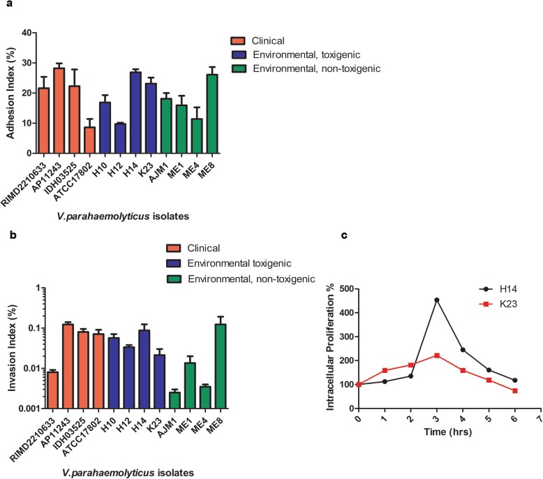
V. parahaemolyticus contains several adhesins to promote attachment and subsequent colonization of host cells. Since it is a key event for the successful establishment of infection, ability of the environmental isolates to adhere to Caco-2 cells were determined and compared to the clinical isolates. The adhesion of the V. parahaemolyticus isolates to Caco-2 cells ranged from 8 to 28% (Table 3, Fig. 3a). All the tdh+trh− clinical isolates showed good adhesion ability of ≥22%, except ATCC17802 (9%). Irrespective of the toxigenic profile, the environmental isolates exhibited more than 15% adhesion to Caco-2 cell with K23, H14 and ME8 (non-toxigenic) showing the maximum adhesion potential of 23, 27 and 26%, respectively. This shows that environmental isolates adhered firmly to intestinal epithelial cells. There was no difference in adhesion among tdh+ and trh+ pathotypes (p > 0.05, Student’s t-test) as well as between the different groups (p > 0.05, one-way ANOVA).
Table 3.
Adhesion and invasion indices of V. parahaemolyticus isolates
| Strain ID | Average ± SE (%) | |
|---|---|---|
| Adhesion Index | Invasion Index | |
| RIMD2210633 | 21.6 ± 3.79 | 0.008 ± 0.001 |
| AP11243 | 28.2 ± 1.66 | 0.12 ± 0.018 |
| IDH03525 | 22.3 ± 5.54 | 0.08 ± 0.015 |
| ATCC17802 | 8.62 ± 2.81 | 0.071 ± 10.02 |
| H10 | 16.9 ± 2.41 | 0.06 ± 0.015 |
| H12 | 9.75 ± 0.25 | 0.034 ± 0.005 |
| H14 | 26.9 ± 1.02 | 0.087 ± 0.037 |
| K23 | 23.1 ± 2.03 | 0.021 ± 0.009 |
| AJM | 18.1 ± 1.9 | 0.003 ± 0.001 |
| ME1 | 15.95 ± 3.15 | 0.014 ± 0.007 |
| ME4 | 11.4 ± 3.9 | 0.004 ± 0.001 |
| ME8 | 26.1 ± 2.55 | 0.12 ± 0.069 |
| S. Typhimurium | 30.8 ± 2.85 | 1.185 ± 0.665 |
| E. coli JM109 | 6.19 ± 1.89 | 0.004 ± 0.0005 |
Fig. 3.
Adhesion (a) and invasion (b) index of V. parahaemolyticus in Caco-2 cells. The values are means ± SE of three independent experiments, each performed in triplicate. Adhesion and invasion indices between groups are not statistically significant (p > 0.05). c Intracellular proliferation assay of V. parahaemolyticus H14 and K23 in Caco-2 cells
The ability of the selected V. parahaemolyticus isolates to invade Caco-2 cells was determined using the classical gentamicin protection assay. Before the experiment, gentamicin MIC of all the isolates was determined using E-test (MIC ≤4 μg/ml).
Among the clinical isolates, AP11243 displayed the highest mean invasion index (0.12%) followed by ATCC17802 (0.071%) and IDH03525 (0.08%) while RIMD2210633 showed very low invasion potential of 0.008% (Table 3, Fig. 3b). Of the toxigenic environmental isolates, 0.087% of H14 could invade Caco-2 cells followed by H10 and H12. Again, K23, with a fully functional vopC (mediates invasion of non-phagocytic cells) displayed a lower invasion potential (0.021%) compared to tdh+trh− counterparts. Among the tdh−trh− isolates, it was surprising to see the invasion index of ME8 was equal to that of AP11243 (0.12%). The rest of the isolates were not invasive, except for ME1, which showed a slight invasion, when compared with the non-invasive control E. coli JM109 (Table 3). Here also, the results were not statistically significant between the various groups under study (p > 0.05).
An intracellular proliferation assay was done subsequently to know whether the toxigenic environmental isolates could persist and replicate intracellularly following invasion into the Caco-2 cells. The experiment was done with the highly cytotoxic, adherent and invasive H14 (tdh+trh−) and K23 (tdh−trh+), which has shown a lesser pathogenic potential in spite of having a complete T3SS. H14, and to a lesser extent, K23 rapidly proliferated inside Caco-2 cells (Fig. 3c). Peak numbers of H14 (450% of initial intracellular CFU) was observed in about 3 h. This number decreased rapidly and intracellular bacteria were still visible at 6 h of infection even though the levels were similar to that detected upon initial enumeration (~ 100%). Intracellular K23 also peaked at 3 h of infection, but the number of bacteria was lower (220% of initial intracellular cfu) and at the end of 6 h, the numbers declined. The decrease in intracellular bacteria could be attributed to the lysis of eukaryotic cells and subsequent killing in the gentamicin containing medium. We did observe rounding of cells by 2 h of incubation and detachment of cells from the 24-well plates by 6 h, which made it impossible to prolong the experiment any further.
Alteration of ZO-1 and F-actin organisation by environmental V. parahaemolyticus
V. parahaemolyticus has been known to compromise epithelial barrier integrity and associated disruption of actin and the tight junction proteins [24, 25]. The effect of H14 and K23 on the localization of ZO-1 (a peripheral tight junction protein), and filamentous actin was examined using confocal microscopy. AP11243 was used as positive control strain as it showed more cytotoxicity and invasion potential than RIMD2210633. In uninfected cells, ZO-1 was located at the periphery and appeared as a continuous brightly stained band with a typical ‘honey-comb structure’ or ‘chicken-wire pattern’ revealing intact boundaries (Fig. 4). In cells infected with AP11243, there was clear disruption of the monolayer, with visible strand breaks and punctate discontinuity. Infection with tdh+trh− H14 isolate elicited a stronger disruption of ZO-1; large areas were seen where ZO-1 was completely displaced. In contrast, the tdh−trh+ K23 infected cells showed smaller but visible breaks, although the cell membrane integrity was preserved.
Fig. 4.
ZO-1 redistribution in Caco-2 cells caused by V. parahaemolyticus environmental strains after 2 h of infection. Micrographs are representative of two separate experiments
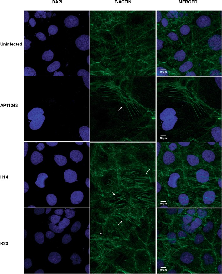
Actin filaments were visualised by fluorescently labelled bicyclic peptide, phalloidin that selectively binds to F-actin. Uninfected cells had branched, reticular actin network without any particular orientation. In contrast, AP11243 induced bundling of actin into long, parallel stress fibres. H14 infected cells had more number of such parallel arrays of F-actin. Surprisingly, cells infected with K23 depicted more prominent actin bundles that spanned the entire cell (Fig. 5).
Fig. 5.
Reorganization of filamentous actin following infection with V. parahaemolyticus. Confluent Caco-2 cells were infected with V. parahaemolyticus AP11243, H14 and K23 for 2 h and double labelled with Alexa Fluor 488 Phalloidin (F-actin, green) and DAPI (nucleus, blue). Micrographs are representative of two different experiments
Discussion
The present study examined the distribution of virulence related genes in 417 V. parahaemolyticus isolated from the environment and seafood and focused on tdh and/or trh isolates to determine the pathogenicity. An abundance of V. parahaemolyticus was seen in the water compared to plankton. This was in contrast to other studies where higher densities of V. parahaemolyticus were obtained from plankton [26, 27]. However, higher numbers of the bacterium were found in the Georgian coastal waters [28]. More than 5% of the V. parahaemolyticus isolates were potentially toxigenic, based on the presence of tdh and trh, of which tdh was detected in a higher number of isolates (4.6%). Previous studies from the south-west coast of India have reported a higher detection of trh-positive V. parahaemolyticus especially after 18 h of enrichment [29, 30]. The toxigenic V. parahaemolyticus isolates obtained in this study were identified exclusively from seafood and none of the water and plankton isolates harboured the haemolysin genes. Hemolysin gene identification is partly dependent on the detection technique employed. The use of modified primers could recover 48 and 8% tdh and trh bearing isolates respectively from an estuarine system [31]. In another study, DePaola et al., tested a large number of colonies from oysters by DNA probes against tlh and tdh and found that 21.8% of them were pathogenic [6]. Similarly, real time PCR could detect 20 and 40% more tdh and trh positive V. parahaemolyticus from oyster samples and 13 and 40% from water samples compared to the conventional techniques [32]. Thus, detection of toxigenic V. parahaemolyticus from environmental samples appears to be influenced by several factors.
T3SSs are an important determinant of pathogenicity of V. parahaemolyticus. All the toxigenic V. parahaemolyticus strains in this study carried the T3SS1 genes, consistent with previous reports [17, 33]. The distribution of T3SS2α genes was highly diverse with the loss of at least one gene from the tdh+trh− isolates. Some of the isolates showed weak amplification with the T3SS2α primer pair or additional primers designed. Jones et al. had also reported anomalies in the T3SS2α gene amplification [33]. This suggests high sequence variability in environmental isolates or an absence of these genes due to partial uptake of VPaI during horizontal transfer. Further, vopB2 was present in 68.4% of tdh+trh− isolates in contrast to earlier findings that it is associated with clinical and not environmental isolates [33, 34]. Some recent reports have documented the presence of vopB2 in environmental isolates [35, 36]. Hence, vopB2 may not be a good indicator of virulence as originally suggested [34]. About 33% of tdh+trh− isolates harboured vopZ gene that encodes the effector, VopZ, known for V. parahaemolyticus induced diarrhoea and intestinal pathology. To the best of our knowledge, this is the first study to document the presence of vopZ (VPA1336) in V. parahaemolyticus isolated from seafood.
Majority of the toxigenic isolates (95.5%) had pandemic strain features (toxRS/new+). Of these, nine serovars (O1:K17, O1:K19, O1:K23, O10:K24, O2:KUT, O4:K29, O5:K20, O4:K42 and O4:K36) were newly identified with pandemic strain traits. Another striking observation is the appearance of tdh−trh+ isolates carrying the toxRS/new+ pandemic marker. Pandemic strains of V. parahaemolyticus are mostly associated with the tdh gene with few reports of GS-positive trh-containing isolates from clinical cases [23, 37, 38]. Data pertaining to the pandemic status of environmental trh-positive V. parahaemolyticus is lacking. Thus, this study is the first to report the isolation of a new serovar (O4:K36) of tdh−trh+V. parahaemolyticus from seafood with potential pandemic traits. Currently the pandemic group is defined by the presence of tdh and toxRS/new genes. So, the pandemic status and evolution of the three tdh−trh+ isolates identified in this study should be confirmed employing other genetic markers including genomic islands VPaI-1, VPaI-4 and VPaI-5 that are specific for the pandemic group [39–41] as well as whole genome sequence analysis. Besides, our finding of tdh−trh+ O3:KUT serovar and its recent isolation from acute diarrhoea in India [23] underscores better cognizance of an epidemiological association between environmental and clinical strains.
Though considered a “gold standard”, it is only recently that PFGE has been extensively employed for subtyping environmental strains of V. parahaemolyticus [8, 42, 43]. The PFGE pattern of the 15 isolates of this study showed considerable diversity and the new serovars having pandemic attributes were not related to the pandemic O3:K6 isolate. Further, it was noted that strains with identical serotype and/or genotype had different PFGE profiles, as in the case of tdh+trh− isolates. Moreover, one of the O1:K25 isolate which was not positive in the GS-PCR, clustered along with the isolates having pandemic strain traits. A similar pattern was observed during molecular typing of V. parahemolyticus isolates in China, where seafood isolates having a varying combination of tdh, trh and toxRS/new genes clustered together with pandemic isolates whereas a tdh+trh−, toxRS/new+ clinical isolate was not genetically related to the pandemic clones [44]. Comparison of MLST datasets of different bacterial species revealed environmental isolates of marine and estuarine V. parahaemolyticus and V. vulnificus have very high homologous recombination rate as they frequently adapt for survival [45]. It was proposed that V. parahaemolyticus may have an epidemic population structure [46] where the isolates undergo frequent genetic recombination resulting in well-adapted clones [47]. Considering the increase in environmental strains acquiring virulence and pandemic attributes, it is important to know the genetics of this thriving population to reduce potential human health risk if newer virulent clones emerge.
To determine the pathogenic potential of environmental V. parahaemolyticus isolates, we used Caco-2 cell line assays. As an initial step, the transcriptional activity of the haemolysin and T3SS2 effector genes were analysed in the selected isolates. While all the clinical strains transcribed both the haemolysin genes, only trh gene was functional in the environmental isolates. The expression results were substantiated by low haemolytic activity of the environmental isolates on human RBC. It is shown that poor expression of tdh gene corresponds to two point mutations in the promoter region (substitution of A to G in the − 35 sequence, G to A at − 3 nucleotide position from the − 10 sequence) [48, 49]. We presume that these mutations may be one of the reasons for the observed reduced transcription. Moreover, we found the isolates carried a single copy of tdh (Additional file 1: Figure S4), which is generally responsible for weak and intermediate haemolysis due a weak promoter compared to tdh2 [48]. The existence of a second copy of tdh or mutations in the promoter needs to be further determined. Similarly, the low haemolytic activity of clinical as well as environmental tdh−trh+ isolates was not surprising as both contain the trh2 allele (Additional file 1: Figure S4), which exhibit reduced hemolysis, not due to reduced gene expression, rather, owing to the differences in the amino acid molecular conformation and binding capacity of RBC by the trh2 product [50, 51]. Further, the tdh-trh+ environmental isolate K23 transcribed the genes vopC, vopA and vopL encoding effectors that promote invasion and modulate host cell behaviour, making it a possible pathogen. A similar functional T3SS2β was seen in trh-positive V. parahaemolyticus elsewhere in India [52].
Even though T3SS2α was not functional in tdh+trh− environmental isolates, they were included in downstream analysis to know if the isolates could still induce any pathogenic effect. To begin with, we found all the isolates to be cytotoxic to Caco-2 cells without discernible differences in cytotoxicity between clinical and environmental isolates, as shown previously [53, 54]. Therefore, cytotoxicity may not be a reliable indicator of pathogenesis. It was also found that the ability to induce cytotoxicity was not dependent on the virulence genes tested. Interestingly, in the present investigation, the cytotoxicity of tdh−trh+ isolate K23 was very low compared to its clinical counterpart with a similar genetic profile. It is well established that T3SSs are responsible for host cell cytotoxic damage, with T3SS1 being the dominant contributor [55]. Perhaps the isolate might have a varied architecture and integrity of T3SS1 gene cluster causing it to assemble a defective translocon and a reduced delivery of T3SS1 effectors responsible for cytotoxicity. It is also possible that the clinical strain might harbour some exclusive genes not present in environmental isolate genome that can induce cytotoxic damage. Meanwhile, the cytotoxicity of the isolates that do not harbour virulence genes was noteworthy. There may be other yet unidentified genes contributing to host cytotoxicty and isolate-specific variations in the virulence of V. parahaemolyticus.
The environmental V. parahaemolyticus obtained in the study could adhere to and invade intestinal cell line, irrespective of the virulence profile. The ability of the representative tdh-positive (H14) and trh-positive (K23) isolates to invade was followed by survival and replication within the intracellular environment. Further, infection with H14 and K23 caused disruption of tight junctions and re-organisation of actin cytoskeleton, both of which can compromise the epithelial integrity. Recent research classifies V. parahaemolyticus as a facultative intracellular pathogen and T3SS2 effectors, VopC and VopL promote invasion and intracellular survival [56] in addition to inducing dramatic changes in the actin cytoskeleton along with VopV [57–59]. It was found that in spite of a functional vopC and a weakly transcribed vopL, H14 could still gain access to Caco-2 cell, persist inside and induce changes on tight junctions and actin cytoskeleton. Burdette et al. [60] showed that T3SS1 also causes changes in actin cytoskeleton. The effects seen with the environmental strains may be due to an active T3SS1 albeit the contribution of T3SS2 effectors cannot be ignored, especially with K23, which can emerge as a potential pathogen and elicit a full blown pathogenic response under a favorable environment of host intestine. Moreover, the study detected a non-toxigenic isolate, ME8, from estuarine water exhibiting an invasive phenotype. Perhaps it may be the result of high levels of adhesion rather than active invasion. Recent studies have identified tdh−trh−V. parahaemolyticus causing gastroenteritis [61–63], indicating the highly complex nature of the virulence of this pathogen.
Conclusion
This study is the first to extensively characterise the virulence, genetic diversity and elucidate the in-vitro pathogenic effects of V. parahaemolyticus isolated from Indian sub-continent. The results indicate the presence of new V. parahaemolyticus having pandemic traits from seafood that can adhere to, invade, replicate and cause structural changes in intestinal cell lines. In vivo studies are required to confirm enterotoxicity of these strains; yet, the demonstrated effects on intestinal cell line indicate their enhanced virulence potential. Attributing pathogenic potential for environmental isolates is an important aspect of understanding health risks associated with seafood borne V. parahaemolyticus. Although there have been significant progress in the virulence of V. parahaemolyticus, our data clearly indicate the difficulty in discriminating pathogenic and non-pathogenic V. parahaemolyticus with the current set of virulence genes, particularly when various pathotypes exist. We believe the concept of virulence in V. parahaemolyticus needs to be revisited and investigated for each pathotype. The threat of evolving pandemic and potentially pathogenic V. parahaemolyticus “environmental” strains looms on seafood consumers and thus, the need for dedicated V. parahaemolyticus surveillance programs in Indian marine and estuarine environment. Moreover, this is a reminder to strongly consider the bacterium in the event of a gastroenteritis outbreak.
Methods
Sample collection, processing and identification of V. parahaemolyticus
Environmental samples consisting of marine and estuarine water, plankton and different seafood were collected from several sampling sites during the period from 2012 to 2013, covering the coastal areas of Kerala. Samples were processed according to the American Public Health Association protocols [64] (see Additional file 1). For each sample, growth in alkaline peptone water (APW) was sub-cultured on thio-sulfate citrate bile salt sucrose (TCBS) agar (HiMedia, Mumbai, India), incubated for 18-24 h at 37 °C. Presumptive isolates (non-sucrose fermenting green colonies) were identified by PCRs for species- specific toxR [65] and tlh [66] genes.
Detection of virulence-associated genes
The identified isolates were tested for the haemolysin genes tdh and trh [66]. The pandemic strain marker genes were verified by GS- and ORF8-PCR [14, 15]. All the tdh- and/or trh-positive isolates were further tested for the T3SS genes specific to T3SS1, T3SS2α and T3SS2β (Additional file 1: Table S1). V. parahaemolyticus RIMD2210633 served as positive control for toxR, tlh, tdh, T3SS and T3SS2α while ATCC17802 was used as the control for trh and T3SS2β.
Serogrouping
Serological analysis of lipopolysaccharide (O) and capsular (K) antigens of V. parahaemolyticus was done using commercially available V. parahaemolyticus antisera kit (Denka Seiken, Tokyo, Japan) according to manufacturer’s instructions.
Pulsed-filed gel electrophoresis (PFGE)
PFGE of 15 V. parahaemolyticus toxigenic isolates belonging to diverse serovars was performed according to the standardised PulseNet protocol [67]. Salmonella enterica serovar Braenderup strain H9812 was used as a molecular weight marker. The PFGE profile of a clinical O3:K6 pandemic strain IDH2826 was included in the clonal comparison. The PFGE image was captured using a Gel Doc XR system (Bio-Rad Laboratories, Hercules, CA), and the gel image was normalized by aligning the peaks of the XbaI (Takara, Shiga, Japan) size standards of S. Braenderup in each gel and was analyzed by using BioNumerics software version 4.0 (Applied Maths, Sint-Martens-Latem, Belgium). The similarities between isolates were evaluated by cluster analysis with the UPGMA method and the Dice correlation coefficient with a position tolerance of 1.5%.
Reverse transcriptase (RT) PCR
Bacteria were grown in LB broth with 3% NaCl to their mid-logarithmic phase and the total RNA extracted with RNeasy mini kit (Qiagen, Valencia, CA, USA). The extracted RNA was reverse transcribed using High-Capacity cDNA Reverse Transcription Kit (Applied Biosystems, Foster City, CA, USA). The cDNA generated was used as a template for the PCR using respective T3SS primer pairs.
Caco-2 cell culture
Caco-2 (ATCC HTB37, RIKEN BioResource Center, Tsukuba, Japan) cells were grown in Dulbecco’s Modified Eagle Medium (DMEM, Gibco, Carlsbad, CA, USA) comprising 10% (v/v) Fetal Bovine Serum (FBS, PAN-Biotech, Aidenbach, Germany) and 100 μg/ml penicillin-streptomycin (Gibco). The cells were grown at 37 °C with 5% CO2 for 5–7 days.
Haemolysis assay
Haemolysis assay of the V. parahaemolyticus isolates was performed as described previously [53] with slight modifications. Briefly, freshly drawn human erythrocytes were rinsed three times with sterile phosphate buffered saline (PBS) and resuspended in PBS to 4% (v/v). About 100 μl of the suspension was mixed with an overnight culture of bacteria in LB broth-3% NaCl (3 X 10⁸ cfu/ml) and incubated at 37 °C for 6 h and 12 h. Subsequently, the plates were centrifuged and the haemoglobin released was estimated by measuring the absorbance of the supernatant at 415 nm (OD415). PBS and 20% (v/v) Triton-X 100 were used to determine the maximum and spontaneous haemoglobin release. The experiment was performed twice with six technical replicates. Percentage of haemolysis was expressed by the formula given below.
Cytotoxicity test
Bacteria were grown in LB-3% NaCl at 37 °C overnight with shaking. Caco-2 cells were seeded in 96-well plates and grown until confluent. Three different wells with Caco-2 cell monolayers were co-cultured with PBS-washed bacteria at a multiplicity of infection (MOI) of 100:1 for 4 h. The release of lactate dehydrogenase (LDH) into the medium was quantified using Cytotox96 non-radioactive cytotoxicity kit (Promega, Madison, WI, USA), following manufacturer’s instructions. The LDH release (% cytotoxicity) was calculated according to the formula:
The spontaneous release was the amount of LDH released from the cytoplasm of uninfected cells, whereas the maximum release was the amount of LDH released by total lysis of uninfected cells. The assay was replicated three times. A time course analysis of H14 and K23 cytotoxicity was also made at 1, 2, 3, 4, 5 and 6 h post infection.
Adhesion and invasion assays
Adherence and invasion of V. parahaemolyticus were determined simultaneously [68] with modifications. In brief, 5 × 10⁵ Caco-2 cells were seeded and grown to confluence in 24-well tissue culture plates. PBS washed bacteria was added at an MOI of 10:1 to the confluent monolayer and incubated at 37 °C for 2 h at 5% CO2. Infected monolayers were washed thrice with PBS and lysed using 0.1% Triton X-100 (Sigma-Aldrich, St Louis, MO, USA). Aliquots of the appropriate dilutions of the lysate were plated on LB with or without 3% NaCl and incubated overnight at 37 °C to count total adherent bacteria (CFU ml− 1). Adhesion efficiency was calculated as the total number of bacteria adhered expressed as a percentage of the number of bacteria in the original inoculum. For determining bacterial invasion, the infected monolayers were washed with PBS and further incubated for 1 h in DMEM containing 50 μg/ml gentamicin (Sigma) to kill the extracellular bacteria. After washing thrice, the monolayers were lysed and bacterial cells quantified. For each isolate, invasiveness was measured as the invasion index, which is the number of invaded organisms taken as a percentage of the number of adhered bacterial cells. The experiments were repeated three times and SEM was calculated. S. enterica serovar Typhimurium and E. coli JM109 were used as positive and negative controls. Isolates were scored as adherent or invasive if their index was more than that of E. coli JM109.
For the measurement of bacterial intracellular proliferation, invasion assay was carried out as above. After killing extracellular bacteria, the DMEM + 50 μg/ml gentamicin was removed and DMEM + 10 μg/ml gentamicin was added. At time points following the addition of DMEM-gentamicin mixture, cells were lysed and bacteria quantified. Proliferation was determined by calculating the percentage of the initial colony forming units (the point at which DMEM - gentamicin mixture was added) present at each time point.
Fluorescence microscopy
Caco-2 cells were seeded onto type I collagen-coated (0.01%, Sigma) coverslips in 6-well plate and grown till confluent. Cells were infected with an overnight culture of bacteria grown in LB broth with 3% NaCl at an MOI of 10:1 and incubated at 37 °C for 2 h. Following infection, coverslips were washed with PBS and fixed in 4% para-formaldehyde, after which the cells were rinsed with PBS supplemented with 0.1% Triton X-100 before being blocked for 1 h with 10% FBS and 0.1% Triton X-100. The cells were incubated overnight with rabbit anti zonula occludin protein 1 (ZO-1, N-term) (Invitrogen, Carlsbad, CA, USA) in blocking buffer. The cells were washed with PBS and labelled with Alexa-Fluor 568 goat anti-rabbit antibody (Molecular Probes, ThermoFisher Scientific, Waltham, MA, USA) in blocking buffer for 1 h. The coverslips were then stained with 4′,6-diamidino-2-phenylindole (DAPI) for 10 min, dried and coated with Fluoromount-G (EMS, Hatfield, PA, USA). For visualizing F-actin, cells were stained with Alexa-Fluor 488 phalloidin (Molecular Probes) after permeabilisation, and counterstained with DAPI. Images were captured with Leica TCS WLL SP8 confocal laser scanning microscope at 40X or 60X magnification with voltage and intensity kept constant and are presented as maximum intensity projections from Z-stacks. Images were processed using ImageJ (NIH).
Statistics
The significance of differences between groups was assessed using Student’s t-test or one way analysis of variance (ANOVA) with Tukey’s multiple comparison test using GraphPad Prism version 5.0 (GraphPad Software, San-Diego, CA, USA). p < 0.05 was considered statistically significant.
Supplementary information
Additional file 1: Supplementary method. Table S1. Primers and annealing temperatures (Ta) used to characterize V. parahaemolyticus isolates. Figure S1. Transcription of trh. Figure S2. Hemolytic activity of V. parahaemolyticus isolates on human RBC. Figure S3. Transcription of T3SS genes. Figure S4. Partial amino acid sequence alignment of TDH (A) and TRH (B).
Acknowledgements
DMP acknowledges the Indian Council of Medical Research, Government of India for the fellowship provided. We thank the team of National Institute of Cholera and Enteric Diseases (NICED), Kolkata, India for assistance with PFGE. We also thank Anurup K.G., RGCB for technical assistance with confocal microscopy. We are grateful to Prof. M. Radhakrishna Pillai, Director, RGCB for the facilities provided.
Abbreviations
- ANOVA
Analysis of variance
- CFU
Colony forming units
- GS-PCR
Group specific polymerase chain reaction
- LDH
Lactate dehydrogenase
- NICED
National Institute of Cholera and Enteric Diseases
- OD
Optical density
- ORF
Open reading frame
- PFGE
Pulsed-field gel electrophoresis
- SE
Standard error
- T3SS
Type III secretion system
- TDH
Thermostable-direct hemolysin
- TRH
TDH-related hemolysin
- UPGMA
Unweighted pair group method with arithmetic mean
- ZO-1
Zona occludens 1
Authors’ contributions
DMP and ST conceived and designed the experiments. DMP performed the experiments and drafted the manuscript. DMP and ST analysed the results. TR and ST critically reviewed the manuscript. All authors read and approved the final manuscript.
Funding
The authors received no specific funding for this work.
Availability of data and materials
The datasets used and/or analysed during the current study are available from the corresponding author on reasonable request.
Ethics approval and consent to participate
Not applicable.
Consent for publication
Not applicable.
Competing interests
The authors declare that they have no competing interests.
Footnotes
Publisher’s Note
Springer Nature remains neutral with regard to jurisdictional claims in published maps and institutional affiliations.
Supplementary information
Supplementary information accompanies this paper at 10.1186/s12866-020-01746-2.
References
- 1.Fujino T. On the bacteriological examination of shirasu food poisoning. Med J Osaka Univ. 1953;4:299–304. [Google Scholar]
- 2.Daniels NA, MacKinnon L, Bishop R, Altekruse S, Ray B, Hammond RM, et al. Vibrio parahaemolyticus infections in the United States, 1973–1998. J Infect Dis. 2000;181:1661–1666. doi: 10.1086/315459. [DOI] [PubMed] [Google Scholar]
- 3.Honda T, Iida T. The pathogenicity of Vibrio parahaemolyticus and the role of the thermostable direct haemolysin and related haemolysins. Rev Med Microbiol. 1993;4:106–113. doi: 10.1097/00013542-199304000-00006. [DOI] [Google Scholar]
- 4.Shirai H, Ito H, Hirayama T, Nakamoto Y, Nakabayashi N, Kumagai K, et al. Molecular epidemiologic evidence for association of thermostable direct hemolysin (TDH) and TDH-related hemolysin of Vibrio parahaemolyticus with gastroenteritis. Infect Immun. 1990;58:3568–3573. doi: 10.1128/IAI.58.11.3568-3573.1990. [DOI] [PMC free article] [PubMed] [Google Scholar]
- 5.Makino K, Oshima K, Kurokawa K, Yokoyama K, Uda T, Tagomori K, et al. Genome sequence of Vibrio parahaemolyticus: a pathogenic mechanism distinct from that of Vibrio cholerae. Lancet. 2003;361:743–749. doi: 10.1016/S0140-6736(03)12659-1. [DOI] [PubMed] [Google Scholar]
- 6.DePaola A, Ulaszek J, Kaysner CA, Tenge BJ, Nordstrom JL, Wells J, et al. Molecular, serological, and virulence characteristics of Vibrio parahaemolyticus isolated from environmental, food, and clinical sources in North America and Asia. Appl Environ Microbiol. 2003;69:3999–4005. doi: 10.1128/AEM.69.7.3999-4005.2003. [DOI] [PMC free article] [PubMed] [Google Scholar]
- 7.Nordstrom JL, Vickery MCL, Blackstone GM, Murray SL, DePaola A. Development of a multiplex real-time PCR assay with an internal amplification control for the detection of total and pathogenic Vibrio parahaemolyticus bacteria in oysters. Appl Environ Microbiol. 2007;73:5840–5847. doi: 10.1128/AEM.00460-07. [DOI] [PMC free article] [PubMed] [Google Scholar]
- 8.Chen AJ, Hasan NA, Haley BJ, Taviani E, Tarnowski M, Brohawn K, et al. Characterization of pathogenic Vibrio parahaemolyticus from the Chesapeake Bay, Maryland. Front Microbiol. 2017;8:2460. doi: 10.3389/fmicb.2017.02460. [DOI] [PMC free article] [PubMed] [Google Scholar]
- 9.Okada N, Iida T, Park KS, Goto N, Yasunaga T, Hiyoshi H, et al. Identification and characterization of a novel type III secretion system in trh-positive Vibrio parahaemolyticus strain TH3996 reveal genetic lineage and diversity of pathogenic machinery beyond the species level. Infect Immun. 2009;77:904–913. doi: 10.1128/IAI.01184-08. [DOI] [PMC free article] [PubMed] [Google Scholar]
- 10.Caburlotto G, Gennari M, Ghidini V, Tafi M, Lleo MM. Presence of T3SS2 and other virulence-related genes in tdh-negative Vibrio parahaemolyticus environmental strains isolated from marine samples in the area of the venetian lagoon, Italy. FEMS Microbiol Ecol. 2009;70:506–514. doi: 10.1111/j.1574-6941.2009.00764.x. [DOI] [PubMed] [Google Scholar]
- 11.Chen X, Zhu Q, Yu F, Zhang W, Wang R, Ye X, et al. Serology, virulence and molecular characteristics of Vibrio parahaemolyticus isolated from seafood in Zhejiang province. PLoS One. 2018;13:e0204892. doi: 10.1371/journal.pone.0204892. [DOI] [PMC free article] [PubMed] [Google Scholar]
- 12.Okuda J, Ishibashi M, Hayakawa E, Nishino T, Takeda Y, Mukhopadhyay AK, et al. Emergence of a unique O3:K6 clone of Vibrio parahaemolyticus in Calcutta, India, and isolation of strains from the same clonal group from southeast Asian travelers arriving in Japan. J Clin Microbiol. 1997;35:3150–3155. doi: 10.1128/JCM.35.12.3150-3155.1997. [DOI] [PMC free article] [PubMed] [Google Scholar]
- 13.Nair GB, Ramamurthy T, Bhattacharya SK, Dutta B, Takeda Y, Sack DA. Global dissemination of Vibrio parahaemolyticus serotype O3:K6 and its serovariants. Clin Microbiol Rev. 2007;20:39–48. doi: 10.1128/CMR.00025-06. [DOI] [PMC free article] [PubMed] [Google Scholar]
- 14.Matsumoto C, Okuda J, Ishibashi M, Iwanaga M, Garg P, Rammamurthy T, et al. Pandemic spread of an O3: K6 clone of Vibrio parahaemolyticus and emergence of related strains evidenced by arbitrarily primed PCR and toxRS sequence analyses. J Clin Microbiol. 2000;38:578–585. doi: 10.1128/JCM.38.2.578-585.2000. [DOI] [PMC free article] [PubMed] [Google Scholar]
- 15.Laohaprertthisan V, Chowdhury A, Kongmuang U, Kalnauwakul S, Ishibashi M, Matsumoto C, et al. Prevalence and serodiversity of the pandemic clone among the clinical strains of Vibrio parahaemolyticus isolated in southern Thailand. Epidemiol Infect. 2003;130:395–406. doi: 10.1017/S0950268803008458. [DOI] [PMC free article] [PubMed] [Google Scholar]
- 16.Chen J, Zhang R, Qi X, Zhou B, Wang J, Chen Y, et al. Epidemiology of foodborne disease outbreaks caused by Vibrio parahaemolyticus during 2010–2014 in Zhejiang Province, China. Food Control. 2017;77:110–115. doi: 10.1016/j.foodcont.2017.02.004. [DOI] [Google Scholar]
- 17.Li H, Tang R, Lou Y, Cui Z, Chen W, Hong Q, et al. A comprehensive epidemiological research for clinical Vibrio parahaemolyticus in Shanghai. Front Microbiol. 2017;8:1043. doi: 10.3389/fmicb.2017.01043. [DOI] [PMC free article] [PubMed] [Google Scholar]
- 18.Park K, Mok JS, Kwon JY, Ryu AR, Kim SH, Lee HJ. Food-borne outbreaks, distributions, virulence, and antibiotic resistance profiles of Vibrio parahaemolyticus in Korea from 2003 to 2016: a review. Fish Aquat Sci. 2018;21:3. doi: 10.1186/s41240-018-0081-4. [DOI] [Google Scholar]
- 19.Newton AE, Garrett N, Stroika S, Halpin JL, Turnsek M, Mody R. Increase in Vibrio parahaemolyticus infections associated with consumption of Atlantic Coast shellfish - 2013. MMWR Morb Mortal Wkly Rep. 2014;63:335–336. [PMC free article] [PubMed] [Google Scholar]
- 20.Taylor M, Cheng J, Sharma D, Bitzikos O, Gustafson R, Fyfe M, et al. Outbreak of Vibrio parahaemolyticus associated with consumption of raw oysters in Canada, 2015. Foodborne Pathog Dis. 2018;15:554–559. doi: 10.1089/fpd.2017.2415. [DOI] [PubMed] [Google Scholar]
- 21.Baker-Austin C, Stockley L, Rangdale R, Martinez-Urtaza J. Environmental occurrence and clinical impact of Vibrio vulnificus and Vibrio parahaemolyticus: a European perspective. Environ Microbiol Rep. 2010;2:7–18. doi: 10.1111/j.1758-2229.2009.00096.x. [DOI] [PubMed] [Google Scholar]
- 22.Martinez-Urtaza J, Powell A, Jansa J, Rey JLC, Montero OP, Campello MG, et al. Epidemiological investigation of a foodborne outbreak in Spain associated with U.S. West Coast genotypes of Vibrio parahaemolyticus. Springerplus. 2016;5:87. doi: 10.1186/s40064-016-1728-1. [DOI] [PMC free article] [PubMed] [Google Scholar]
- 23.Pazhani GP, Bhowmik SK, Ghosh S, Guin S, Dutta S, Rajendran K, et al. Trends in the epidemiology of pandemic and non-pandemic strains of Vibrio parahaemolyticus isolated from diarrheal patients in Kolkata, India. PLoS Negl Trop Dis. 2014;8:e2815. doi: 10.1371/journal.pntd.0002815. [DOI] [PMC free article] [PubMed] [Google Scholar]
- 24.Lynch T, Livingstone S, Buenaventura E, Fedwick J, Buret AG, Graham D, et al. Vibrio parahaemolyticus disruption of epithelial cell tight junctions occurs independently of toxin production. Infect Immun. 2005;73:1275–1283. doi: 10.1128/IAI.73.3.1275-1283.2005. [DOI] [PMC free article] [PubMed] [Google Scholar]
- 25.Ritchie JM, Rui H, Zhou X, Iida T, Kodoma T, Ito S, et al. Inflammation and disintegration of intestinal villi in an experimental model for Vibrio parahaemolyticus-induced diarrhea. PLoS Pathog. 2012;8:e1002593. doi: 10.1371/journal.ppat.1002593. [DOI] [PMC free article] [PubMed] [Google Scholar]
- 26.Sarkar BL, Nair GB, Banerjee AK, Pal SC. Seasonal distribution of Vibrio parahaemolyticus in freshwater environs and in association with freshwater fishes in Calcutta. Appl Environ Microbiol. 1985;49:132–136. doi: 10.1128/AEM.49.1.132-136.1985. [DOI] [PMC free article] [PubMed] [Google Scholar]
- 27.Chowdhury MAR, Yamanaka H, Miyoshi S, Shinoda S. Ecology and seasonal distribution of Vibrio parahaemolyticus in aquatic environments of a temperate region. FEMS Microbiol Lett. 1990;74:1–9. doi: 10.1111/j.1574-6968.1990.tb04046.x. [DOI] [Google Scholar]
- 28.Haley BJ, Kokashvili T, Tskshvediani A, Janelidze N, Mitaishvili N, Grim CJ, et al. Molecular diversity and predictability of Vibrio parahaemolyticus along the Georgian coastal zone of the Black Sea. Front Microbiol. 2014;5:45. doi: 10.3389/fmicb.2014.00045. [DOI] [PMC free article] [PubMed] [Google Scholar]
- 29.Deepanjali A, Kumar HS, Karunasagar I, Karunasagar I. Seasonal variation in abundance of total and pathogenic Vibrio parahaemolyticus bacteria in oysters along the southwest coast of India. Appl Environ Microbiol. 2005;71:3575–3580. doi: 10.1128/AEM.71.7.3575-3580.2005. [DOI] [PMC free article] [PubMed] [Google Scholar]
- 30.Parvathi A, Kumar HS, Bhanumathi A, Ishibashi M, Nishibuchi M, Karunasagar I, et al. Molecular characterization of thermostable direct haemolysin-related haemolysin (TRH)-positive Vibrio parahaemolyticus from oysters in Mangalore, India. Environ Microbiol. 2006;8:997–1004. doi: 10.1111/j.1462-2920.2006.00990.x. [DOI] [PubMed] [Google Scholar]
- 31.Gutierrez West CK, Klein SL, Lovell CR. High frequency of virulence factor genes tdh, trh, and tlh in Vibrio parahaemolyticus strains isolated from a pristine estuary. Appl Environ Microbiol. 2013;79:2247–2252. doi: 10.1128/AEM.03792-12. [DOI] [PMC free article] [PubMed] [Google Scholar]
- 32.Parveen S, Hettiarachchi KA, Bowers JC, Jones JL, Tamplin ML, McKay R, et al. Seasonal distribution of total and pathogenic Vibrio parahaemolyticus in Chesapeake Bay oysters and waters. Int J Food Microbiol. 2008;128:354–361. doi: 10.1016/j.ijfoodmicro.2008.09.019. [DOI] [PubMed] [Google Scholar]
- 33.Jones JL, Lüdeke CHM, Bowers JC, Garrett N, Fischer M, Parsons MB, et al. Biochemical, serological, and virulence characterization of clinical and oyster Vibrio parahaemolyticus isolates. J Clin Microbiol. 2012;50:2343–2352. doi: 10.1128/JCM.00196-12. [DOI] [PMC free article] [PubMed] [Google Scholar]
- 34.Noriea NF, Johnson CN, Griffitt KJ, Grimes DJ. Distribution of type III secretion systems in Vibrio parahaemolyticus from the northern Gulf of Mexico. J Appl Microbiol. 2010;109:953–962. doi: 10.1111/j.1365-2672.2010.04722.x. [DOI] [PubMed] [Google Scholar]
- 35.Paranjpye R, Hamel OS, Stojanovski A, Liermann M. Genetic diversity of clinical and environmental Vibrio parahaemolyticus strains from the Pacific northwest. Appl Environ Microbiol. 2012;78:8631–8638. doi: 10.1128/AEM.01531-12. [DOI] [PMC free article] [PubMed] [Google Scholar]
- 36.Gao L, Deng YQ, Chen C, Ke CW, Li BS, Long YY, et al. Population relationship of Vibrio parahaemolyticus isolates derived from aquaculture ponds, a seafood market, restaurants, and clinical samples. Foodborne Pathog Dis. 2016;13:333–342. doi: 10.1089/fpd.2015.2098. [DOI] [PubMed] [Google Scholar]
- 37.Meador CE, Parsons MM, Bopp CA, Gerner-Smidt P, Painter JA, Vora GJ. Virulence gene- and pandemic group-specific marker profiling of clinical Vibrio parahaemolyticus isolates. J Clin Microbiol. 2007;45:1133–1139. doi: 10.1128/JCM.00042-07. [DOI] [PMC free article] [PubMed] [Google Scholar]
- 38.Li J, Xue F, Yang Z, Zhang X, Zeng D, Chao G, et al. Vibrio parahaemolyticus strains of pandemic serotypes identified from clinical and environmental samples from Jiangsu, China. Front Microbiol. 2016;7:787. doi: 10.3389/fmicb.2016.00787. [DOI] [PMC free article] [PubMed] [Google Scholar]
- 39.Hurley CC, Quirke A, Reen FJ, Boyd EF. Four genomic islands that mark post-1995 pandemic Vibrio parahaemolyticus isolates. BMC Genomics. 2006;7:104. doi: 10.1186/1471-2164-7-104. [DOI] [PMC free article] [PubMed] [Google Scholar]
- 40.Chao G, Jiao X, Zhou X, Yang Z, Pan Z, Huang J, et al. Systematic functional pandemic strain-specific genes, three genomic islands, two T3SSs in foodborne, and clinical Vibrio parahaemolyticus isolates in China. Foodborne Pathog Dis. 2009;6:689–698. doi: 10.1089/fpd.2009.0274. [DOI] [PubMed] [Google Scholar]
- 41.Chao G, Jiao X, Zhou X, Wang F, Yang Z, Huang J, et al. Distribution of genes encoding four pathogenicity islands (VPaIs), T6SS, biofilm, and type i pilus in food and clinical strains of Vibrio parahaemolyticus in China. Foodborne Pathog Dis. 2010;7:649–658. doi: 10.1089/fpd.2009.0441. [DOI] [PubMed] [Google Scholar]
- 42.Terzi Gulel G, Martinez-Urtaza J. Molecular characterizations of Vibrio parahaemolyticus in seafood from the Black Sea, Turkey. Lett Appl Microbiol. 2016;62:494–500. doi: 10.1111/lam.12579. [DOI] [PubMed] [Google Scholar]
- 43.Lopatek M, Wieczorek K, Osek J. Antimicrobial resistance, virulence factors, and genetic profiles of Vibrio parahaemolyticus from seafood. Appl Environ Microbiol. 2018;84:e00537–e00518. doi: 10.1128/AEM.00537-18. [DOI] [PMC free article] [PubMed] [Google Scholar]
- 44.Chen W, Xie Y, Xu J, Wang Q, Gu M, Yang J, et al. Molecular typing of Vibrio parahaemolyticus isolates from the middle-east coastline of China. Int J Food Microbiol. 2012;153:402–412. doi: 10.1016/j.ijfoodmicro.2011.12.001. [DOI] [PubMed] [Google Scholar]
- 45.Vos M, Didelot X. A comparison of homologous recombination rates in bacteria and archaea. ISME J. 2009;3:199–208. doi: 10.1038/ismej.2008.93. [DOI] [PubMed] [Google Scholar]
- 46.Gonzalez-Escalona N, Martinez-Urtaza J, Romero J, Espejo RT, Jaykus L-A, DePaola A. Determination of molecular phylogenetics of Vibrio parahaemolyticus strains by multilocus sequence typing. J Bacteriol. 2008;190:2831–2840. doi: 10.1128/JB.01808-07. [DOI] [PMC free article] [PubMed] [Google Scholar]
- 47.Smith JM, Feil EJ, Smith NH. Population structure and evolutionary dynamics of pathogenic bacteria. BioEssays. 2000;22:1115–1122. doi: 10.1002/1521-1878(200012)22:12<1115::AID-BIES9>3.0.CO;2-R. [DOI] [PubMed] [Google Scholar]
- 48.Okuda J, Nishibuchi M. Manifestation of the Kanagawa phenomenon, the virulence-associated phenotype of Vibrio parahaemolyticus depends on a particular single base change in the promoter of the thermostable direct haemolysin gene. Mol Microbiol. 1998;30:499–511. doi: 10.1046/j.1365-2958.1998.01072.x. [DOI] [PubMed] [Google Scholar]
- 49.Shalu OA, Pisanov RV, Monakhova EV. Efficiency of Vibrio parahaemolyticus tdh gene expression depends upon two point mutations in its promoter region. Russ J Genet. 2012;48:1177–1183. doi: 10.1134/S1022795412120125. [DOI] [PubMed] [Google Scholar]
- 50.Kishishita M, Matsuoka N, Kumagai K, Yamasaki S, Takeda Y, Nishibuchi M. Sequence variation in the thermostable direct hemolysin-related hemolysin (trh) gene of Vibrio parahaemolyticus. Appl Environ Microbiol. 1992;58:2449–2457. doi: 10.1128/AEM.58.8.2449-2457.1992. [DOI] [PMC free article] [PubMed] [Google Scholar]
- 51.Bechlars S, Jäckel C, Diescher S, Wüstenhagen DA, Kubick S, Dieckmann R, et al. Characterization of trh2 harbouring Vibrio parahaemolyticus strains isolated in Germany. PLoS One. 2015;10:e0118559. doi: 10.1371/journal.pone.0118559. [DOI] [PMC free article] [PubMed] [Google Scholar]
- 52.Kumar BK, Deekshit VK, Rai P, Shekar M, Karunasagar I, Karunasagar I. Presence of T3SS2β genes in trh+Vibrio parahaemolyticus isolated from seafood harvested along Mangalore coast, India. Lett Appl Microbiol. 2014;58:440–446. doi: 10.1111/lam.12210. [DOI] [PubMed] [Google Scholar]
- 53.Mahoney JC, Gerding MJ, Jones SH, Whistler CA. Comparison of the pathogenic potentials of environmental and clinical Vibrio parahaemolyticus strains indicates a role for temperature regulation in virulence. Appl Environ Microbiol. 2010;76:7459–7465. doi: 10.1128/AEM.01450-10. [DOI] [PMC free article] [PubMed] [Google Scholar]
- 54.Caburlotto G, Lleò MM, Hilton T, Huq A, Colwell RR, Kaper JB. Effect on human cells of environmental Vibrio parahaemolyticus strains carrying type III secretion system 2. Infect Immun. 2010;78:3280–3287. doi: 10.1128/IAI.00050-10. [DOI] [PMC free article] [PubMed] [Google Scholar]
- 55.Hiyoshi H, Kodama T, Iida T, Honda T. Contribution of Vibrio parahaemolyticus virulence factors to cytotoxicity, enterotoxicity, and lethality in mice. Infect Immun. 2010;78:1772–1780. doi: 10.1128/IAI.01051-09. [DOI] [PMC free article] [PubMed] [Google Scholar]
- 56.de Souza SM, Orth K. Intracellular Vibrio parahaemolyticus escapes the vacuole and establishes a replicative niche in the cytosol of epithelial cells. MBio. 2014;5:e01506–e01514. doi: 10.1128/mBio.01506-14. [DOI] [PMC free article] [PubMed] [Google Scholar]
- 57.Liverman ADB, Cheng H-C, Trosky JE, Leung DW, Yarbrough ML, Burdette DL, et al. Arp2/3-independent assembly of actin by Vibrio type III effector VopL. Proc Natl Acad Sci U S A. 2007;104:17117–17122. doi: 10.1073/pnas.0703196104. [DOI] [PMC free article] [PubMed] [Google Scholar]
- 58.Hiyoshi H, Kodama T, Saito K, Gotoh K, Matsuda S, Akeda Y, et al. VopV, an F-actin-binding type III secretion effector, is required for Vibrio parahaemolyticus-induced enterotoxicity. Cell Host Microbe. 2011;10:401–409. doi: 10.1016/j.chom.2011.08.014. [DOI] [PubMed] [Google Scholar]
- 59.Zhang L, Krachler AM, Broberg CA, Li Y, Mirzaei H, Gilpin CJ, et al. Type III effector VopC mediates invasion for Vibrio species. Cell Rep. 2012;1:453–460. doi: 10.1016/j.celrep.2012.04.004. [DOI] [PMC free article] [PubMed] [Google Scholar]
- 60.Burdette DL, Yarbrough ML, Orvedahl A, Gilpin CJ, Orth K. Vibrio parahaemolyticus orchestrates a multifaceted host cell infection by induction of autophagy, cell rounding, and then cell lysis. Proc Natl Acad Sci U S A. 2008;105:12497–12502. doi: 10.1073/pnas.0802773105. [DOI] [PMC free article] [PubMed] [Google Scholar]
- 61.García K, Torres R, Uribe P, Hernández C, Rioseco ML, Romero J, et al. Dynamics of clinical and environmental Vibrio parahaemolyticus strains during seafood-related summer diarrhea outbreaks in southern Chile. Appl Environ Microbiol. 2009;75:7482–7487. doi: 10.1128/AEM.01662-09. [DOI] [PMC free article] [PubMed] [Google Scholar]
- 62.Ottaviani D, Leoni F, Serra R, Serracca L, Decastelli L, Rocchegiani E, et al. Nontoxigenic Vibrio parahaemolyticus strains causing acute gastroenteritis. J Clin Microbiol. 2012;50:4141–4143. doi: 10.1128/JCM.01993-12. [DOI] [PMC free article] [PubMed] [Google Scholar]
- 63.Banerjee SK, Kearney AK, Nadon CA, Peterson C-L, Tyler K, Bakouche L, et al. Phenotypic and genotypic characterization of Canadian clinical isolates of Vibrio parahaemolyticus collected from 2000 to 2009. J Clin Microbiol. 2014;52:1081–1088. doi: 10.1128/JCM.03047-13. [DOI] [PMC free article] [PubMed] [Google Scholar]
- 64.Lear DW. Field methodology. In: Greenberg AE, Hunt DA, editors. Laboratory procedures for the examination of seawater and shellfish. Washington DC: American Public Health Association; 1984.p. 18-36.
- 65.Kim YBU, Okuda JUN, Matsumoto C, Takahashi N, Hashimoto S, Nishibuchi M. Identification of Vibrio parahaemolyticus strains at the species level by PCR targeted to the toxR gene. 1999;37:1173–7. [DOI] [PMC free article] [PubMed]
- 66.Bej AK, Patterson DP, Brasher CW, Vickery MCL, Jones DD, Kaysner CA. Detection of total and hemolysin-producing Vibrio parahaemolyticus in shellfish using multiplex PCR amplification of tl, tdh and trh. J Microbiol Methods. 1999;36:215–25. [DOI] [PubMed]
- 67.Kam KM, Luey CKY, Parsons MB, Cooper KLF, Nair GB, Alam M, et al. Evaluation and validation of a PulseNet standardized pulsed-field gel electrophoresis protocol for subtyping Vibrio parahaemolyticus: an international multicenter collaborative study. J Clin Microbiol. 2008;46:2766–73. [DOI] [PMC free article] [PubMed]
- 68.Elsinghorst EA, Kopecko DJ. Molecular cloning of epithelial cell invasion determinants from enterotoxigenic Escherichia coli. Infect Immun. 1992;60:2409–17. [DOI] [PMC free article] [PubMed]
Associated Data
This section collects any data citations, data availability statements, or supplementary materials included in this article.
Supplementary Materials
Additional file 1: Supplementary method. Table S1. Primers and annealing temperatures (Ta) used to characterize V. parahaemolyticus isolates. Figure S1. Transcription of trh. Figure S2. Hemolytic activity of V. parahaemolyticus isolates on human RBC. Figure S3. Transcription of T3SS genes. Figure S4. Partial amino acid sequence alignment of TDH (A) and TRH (B).
Data Availability Statement
The datasets used and/or analysed during the current study are available from the corresponding author on reasonable request.


