

Abstract
DC-SIGN, a C-type lectin receptor expressed in dendritic cells (DCs), has been identified as a receptor for human immunodeficiency virus type 1, hepatitis C virus, Ebola virus, cytomegalovirus, dengue virus, and the SARS coronavirus. We used H5N1 pseudotyped and reverse-genetics (RG) virus particles to study their ability to bind with DC-SIGN. Electronic microscopy and functional assay results indicate that pseudotyped viruses containing both HA and NA proteins express hemagglutination and are capable of infecting cells expressing α-2,3-linked sialic acid receptors. Results from a capture assay show that DC-SIGN-expressing cells (including B-THP-1/DC-SIGN and T-THP-1/DC-SIGN) and peripheral blood dendritic cells are capable of transferring H5N1 pseudotyped and RG virus particles to target cells; this action can be blocked by anti-DC-SIGN monoclonal antibodies. In summary, (a) DC-SIGN acts as a capture or attachment molecule for avian H5N1 virus, and (b) DC-SIGN mediates infections in cis and in trans.
Keywords: H5N1, DC-SIGN, Pseudotyped virus, Dendritic cells
The global spread of highly pathogenic avian influenza A H5N1 viruses in poultry and sporadic human infections are viewed as parts of a pandemic threat. Between 2003 and April 2008 there have been 381 human cases and 240 deaths reported [1]. General influenza virus binding to cellular receptors is mediated by viral hemagglutinin and sialic acid (SA) receptors on cell surfaces. Avian flu viruses generally show a preference for binding to α-2,3-linked SA, whereas human influenza viruses prefer α-2,6-linked SA as a receptor [2].
The H5N1 virus triggers severe systemic infections resulting in a high mortality rate in humans, and its clinical manifestations differ from those associated with influenza A virus infection. H5N1-infected patients usually have high fever, few respiratory symptoms, pulmonary infiltrates, and lymphopenia; a small number have shown acute respiratory distress syndrome and sepsis [3]. Results from clinical laboratory diagnoses indicate the presence of H5N1 viral RNA in patient plasma, and the virus has been isolated from plasma specimens [4]. This suggests H5N1 dissemination from a primary infection site (the lungs) to other organs, where they cause systemic diseases [5]. The spreading mechanism and cells responsible for the systemic dissemination of the H5N1 influenza virus are still unclear.
Dendritic cells (DCs), a type of professional antigen presenting cell, play an important role in immune system homeostasis [6]. When pathogens invade a host, DCs sense and capture them via two pattern recognition receptors: toll-like receptors and C-type lectins [6]. A C-type lectin known as dendritic cell-specific ICAM-3 grabbing non-integrin (DC-SIGN)(CD209) is an important factor in DC immune regulation. DC-SIGN may be capable of capturing or internalizing pathogens as a means of mediating antigen presentation for T-cell activation and proliferation [6]. During interactions between DC-SIGN and pathogens, the latter would take advantage of this mechanism for immune escape. DC-SIGN was initially identified as an attachment molecule for the human immunodeficiency virus [7], but has since been identified as a receptor for several viruses [6]. In addition to enhancing viral infections in trans [8], it also serves as a receptor for virus entry and replication in DCs [9]. DC-SIGN binds distinct carbohydrate structure, such as mannose-containing glycoconjugates and fucose-containing Lewis blood group antigens, suggesting that it governs a broad pathogen-recognition pattern [10]. Since the HA protein of H5N1 is a glycoprotein with 9 potential N-linked glycosylation sites, we hypothesized that DC-SIGN interacts with the HA protein, consequently facilitating viral infection or dissemination.
We used H5N1 pseudotyped and reverse-genetics strains to elucidate their interactions with DC-SIGN, and found that pseudotyped viruses containing both H5N1 HA and NA proteins displayed hemagglutination and infection capability. In addition to enhancing H5N1 viral infectivity, we also observed that DC-SIGN expression facilitates virus transport to recipient cells via B-THP-1/DC-SIGN and human dendritic cells. These results suggest that DC-SIGN is an important attachment molecule mediating H5N1 virus infection in cis and in trans.
Materials and methods
Cell lines and plasmids. Seven cell lines were used in this study: HEK293T, MDCK, Vero, B-THP-1, B-THP-1/DC-SIGN, THP-1ATCC, and THP-1ATCC/DC-SIGN. The culture protocol was described in supplements. Plasmids with different genomic segments of H1N1 (A/PR8/34) and H5N1 (A/Vietnam/1204/03) influenza viruses (pHW191-PB2, pHW192-PB1, pHW193-PA, pHW194-HA, pHW195-NP, pHW196-NA, pHW197-M, pHW198-NS, pHW1203-HA, and pHW1203-NA) were generously provided by Dr. Robert G. Webster at the St. Jude Children’s Research Hospital in Memphis, USA. The plasmid pNL-Luc-E−R− contains an env-defective HIV-1 genome with a firefly luciferase reporter gene.
Pseudotyped H5 virus preparation. Pseudotyped viral particle production and concentration were performed as described previously [11], [12] and were described in supplements.
Infectivity assay. Following p24 quantification with a Coulter HIV-1 p24 antigen assay (Beckman Coulter), 20 or 100 ng of H5 or H5N1 pseudotyped and RG virus particles were collected and incubated with HEK293T, MDCK, or Vero cells. Luciferase activity was measured in cell lysate after 48 h of incubation at 37 °C. Quantities of pseudotyped H5N1 or RG strain particles were determined by real-time reverse transcription (RT)-PCR (for detecting H5N1 gene expression) [13] and luciferase activity (for the pseudotyped virus particles) [12]. The detail procedures were described in supplements.
Assessment of DC-SIGN-mediated infection in trans. DC-SIGN-mediated virus transfer efficiency was assessed by capture assay as described previously [8], [12] and details were described in supplements.
p24 binding assay. H5 pseudotyped viral binding to B-THP-1 DC-SIGN or THP-1 DC-SGN cells expressing DC-SIGN was measured via cell-associated p24 levels as described previously [14] and procedures were described in supplements.
Immunofluorescent staining. H5 pseudotyped virus binding to DC-SIGN-expressing cells was analyzed by immunofluorescence and laser-scanning confocal microscopy as described in [15] and the procedure was described in supplements.
Lectin staining. Briefly, combinations of either B-THP-1 and B-THP-1/DC-SIGN or THP-1 and THP-1/DC-SIGN were stained with lectins specific for α-2,3-linked SA (DIG-conjugated Maackia amurensis [MAA], 10 μg/ml; Roche) or α-2,6-linked SA (DIG-conjugated Sambucus nigra [SNA] 10 μg/ml; Roche).For a more detailed description of this procedure, see [16].
Preparation of monocyte-derived dendritic cells. Monocyte-derived dendritic cells were prepared as described previously [12] and the protocol was described in supplements.
Real-time RT-PCR evaluation. For H5N1 viral RNA detection, H5-specific primers and probes were used for real-time RT-PCR amplification [13] and described in supplements.
N-link glycosylation site prediction. Predictions of N-linked glycosylation on H5N1 hemagglutinin protein were performed using the NetNGlyc 1.0 Server and were described in supplements.
Results and discussion
The binding of DC-SIGN to pathogens depends on the presence of either high-mannose N-linked carbohydrate chains or fucosylated oligosaccharides on the envelope glycoproteins of the pathogens. We hypothesized interaction between the glycosylated envelope of avian influenza H5N1 viruses and the carbohydrate-recognition domain (CRD) of DC-SIGN. As show in Fig. 1 A, nine N-glycosylation sites on HA – seven in the HA1 domain (amino acid residues 26, 27, 39, 170, 181, 209, and 302) and two in the HA2 domain (residues 500 and 559) – were predicted using the NetNGlyc 1.0 program. The external globular structure is primarily composed of the HA1 domain; N-glycosylation sites 26, 27, 39, 170, 181, 209, and 302 are likely candidates for interaction with DC-SIGN (Fig. 1B).
Fig. 1.
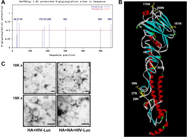
Prediction of N-linked glycosylation on hemagglutinin of H5N1 influenza strain A/Vietnam/1203/04 by NetNGlyc 1.0 Server, displayed on 3D structure with DS ViewerPro Trial program. (A) Of the 9 N-glycosylation sites, 7 were in the HA1 domain and 2 in the HA2 domain and the numbers represent a.a site in amino acid sequence. (B) Each N-glycosylation site on the hemagglutinin structure was labeled; numbers indicate relative amino acid site. (C) Different combinations of HIV-Luc plasmid with HA (H5N1) or NA (H5N1) plasmid were used to transfect HEK293T cells for H5 pseudotyped viral particle production. Images were taken from grids under a transmission electron microscope after coating and uranyl acetate staining. Upper panel: 10,000×; lower panel: 15,000×; proportional scale: 100 nm.
The pseudotyped virus system is useful for analyzing functional domains for the receptor-binding property of viral envelopes. Pseudotyped lentiviral particles expressing heterologous viral glycoproteins have been reported for SARS, HCV, Ebola, and dengue viruses [11], [17], [18]. It has also been reported that lentiviral particles pseudotyped with HA from H5N1 (H5) have entry characteristics (i.e., receptor usage, pH requirements, and neutralization) that are similar to those for the real H5N1 [11]. We therefore used a HIV-1-defective genome carrying a luciferase gene to generate two types of pseudotyped viruses: HA alone and HA/NA of the H5N1 virus. Results from TEM examinations revealed that both H5 and H5N1 pseudotyped virus particles contained spike-like HA structures on their surfaces (Fig. 1C). We also observed higher numbers of pseudotyped virus particles generated by HA/NA co-transfection compared to HA alone. Nefkens et al. [11] used a similar system to produce lentiviral pseudotyped viruses with HA from H5N1, adding neuraminidase to culture medium to harvest larger quantities. Their data are consistent with our observation that pseudotyped virus particles bearing both HA and NA proteins are much more easily released from cell surfaces compared to particles bearing the HA protein only.
We used a hemagglutinin test and cell susceptibility assay to compare the functional integrity of H5- and H5N1-pseudotyped viruses, and found that following TPCK treatment, both virus types expressed similar hemagglutinin activity, with titers ranging from 1:2 to 1:8. Both types displayed very low hemagglutinin activity without TPCK treatment (Supplementary Table 1). It has been reported that HA precursors must be cleaved into HA1 and HA2 in order to become capable of binding to cellular receptors [19].
Regarding cell susceptibility, three cell lines (HEK293T, MDCK, and Vero) were tested with equal amounts of H5- and H5N1-pseudotyped viruses standardized by p24 antigen. Our results indicate that judging by luciferase enzyme activity, the H5N1-pseudotyped virus had significantly higher infectivity than the H5-pseudotyped virus (p < 0.05). In addition, both MDCK and HEK293T cells were more susceptible to pseudotyped virus infection than the Vero cells (Supplementary Table 1). A possible explanation is that MDCK and HEK293T cells express more α-2,3-linked SA receptors than Vero cells [11]. This explains our rationale to use the MDCK and HEK293T cells in our experiments.
We used two cell lines—B-THP-1 and B-THP-1/DC-SIGN (a stable B-THP-1 clone expressing DC-SIGN molecules)—to investigate whether DC-SIGN is capable of mediating the binding and entry of H5N1 pseudotyped viral particles to target cells. Cells were incubated with H5N1 pseudotyped viruses; viral infectivity was determined with luciferase assays. As shown in Fig. 2 A, B-THP-1/DC-SIGN cells expressed twice as much luciferase activity as the B-THP-1 cells, and the ability of the H5N1 pseudotyped virus to infect B-THP-1/DC-SIGN cells was reduced when the cells were pre-treated with an anti-DC-SIGN monoclonal antibody. Similar results were observed using the combination of THP-1 and THP-1/DC-SIGN cell lines (data not shown). To rule out the possibility that enhanced infectivity was due to differential expression levels of avian influenza viruses that bind only to the α-2,3-linked SA receptor, both cell lines were stained with SNA and MAA. As shown in Fig. 2B-1 and -2, both cell lines had identical distributions of α-2,3- and α-2,6-linked SA receptors. To clarify whether DC-SIGN is a receptor that mediates the entry of H5N1 pseudotyped viral particles or if it simply enhances viral entry via SA receptors, we used vibrio cholerae sialidase to eliminate α-2,3-linked SA from the cells. Our results show that almost all of the α-2,3-linked SA was removed but DC-SIGN levels remained unchanged (Fig. 2B-3). Following treatment with sialidase, B-THP-1 and B-THP-1/DC-SIGN cells were incubated with H5N1 pseudotyped viral particles; we detected very low luminescence levels in the resulting cell lysates (data not shown). This strongly suggests that H5N1 pseudotyped viral particles interact with DC-SIGN and facilitate viral entry into target cells in cis via SA receptors. Transmission electron microscopy images demonstrate the attachment of many H5N1 pseudotyped viral particles to the surfaces of cells expressing DC-SIGN (Fig. 2C); indirect immunofluorescent antibody assay (IFA) results further indicate a colocalization between H5N1 pseudotyped particles and DC-SIGN (Fig. 2D). In addition to transmission electron microscopy and IFA colocalized staining, we also used HIV p24 quantification to study the binding of H5N1 pseudotyped virus particles to DC-SIGN, and found that p24 antigen levels in the cell lysates of DC-SIGN-expressing cells were significantly higher than those in the B-THP-1 and THP-1 control cells. Furthermore, p24 levels were reduced in the presence of anti-DC-SIGN monoclonal antibodies, with inhibition percentages of 23% and 28% for B-THP-1/DC-SIGN and THP-1/DC-SIGN cells, respectively (Table 1 ). These results are similar to those found in a previous report on HCV pseudotyped virus binding to DC-SIGN [14]; those authors reported that the anti-DC-SIGN monoclonal antibody (MAb 612X) had a 19–41% inhibitory effect on HCV pseudotyped virus binding to DC-SIGN.
Fig. 2.
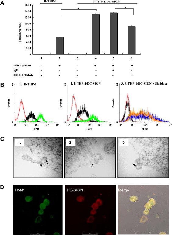
DC-SIGN as an attachment receptor for H5N1 pseudotyped virus and enhanced H5N1 pseudotyped virus infection in cis. (A) B-THP-1 cells and B-THP-1/DC-SIGN infected with the H5N1 pseudotyped virus (H5N1 p-virus), with or without anti-DC-SIGN MAb or IgG control preincubation, were used for infectivity assays. Bars represent B-THP-1/DC-SIGN pretreated with the following antibodies: 4, medium only; 5, IgG control; 6, anti-DC-SIGN MAb. (B) MAA and SNA were used to stain influenza virus receptor α-2,3- and α-2,6-linked SA on B-THP-1 and B-THP-1/DC-SIGN cells. Graphs 1 and 2: black and green lines represent MAA and SNA staining, respectively; red represents isotype control. Graph 3: black line represents MAA, blue and orange lines pre- and post-sialidase treatment, respectively. (C) H5N1 pseudotyped virus binding to DC-SIGN-expressing cells following the removal of the influenza receptor with sialidase. Images 1–3: arrows indicate H5N1 pseudotyped virus position in different sections of B-THP-1/DC-SIGN cells under TEM. (D) H5N1 pseudotyped virus and DC-SIGN colocalized staining under a confocal microscope. Green and red fluorescence represent H5N1 virus and DC-SIGN molecules on cell surfaces, respectively. Merged orange fluorescence on cell edges indicates their colocalization. Results shown are from one representative experiment out of three performed (∗p < 0.05).
Table 1.
DC-SIGN could specifically capture H5N1 pseudotyped viruses
| B-THP-1/DC-SIGN |
THP-1/DC-SIGN |
|||
|---|---|---|---|---|
| P24 (pg/ml) | Inhibition (%) | P24 (pg/ml) | Inhibition (%) | |
| IgG control | 340 | 0 | 366 | 0 |
| DC-SIGN MAb | 260 | 23.55 ± 2.3 | 262 | 28.65 ± 3.4 |
Note: Inhibition rate was calculated by using the formula 100 − [(P24 with anti-DC-SIGN/P24 with isotyped control) × 100%]. Each representative experiment of three independent experiments was shown. The percentages of viral binding were means of three independent experiments ± SD.
We used a standard capture assay to determine whether DC-SIGN is capable of mediating H5N1 pseudotyped viral infection in trans. As shown in Fig. 3 A (lanes 1, 2, 5, and 6), both B-THP-1/DC-SIGN and THP-1/DC-SIGN cells were capable of transferring H5N1 pseudotyped viruses to MDCK cells more efficiently than the B-THP-1 and THP-1 control cells. The differences in luciferase activity between cells with or without DC-SIGN expression were statistically significant (p < 0.05 and p < 0.01 for the B-THP-1 and THP-1 pairs, respectively). Furthermore, compared to cells treated with an IgG control antibody, luciferase activity decreased significantly when donor cells were pretreated with an anti-DC-SIGN monoclonal antibody (Fig. 3A, lanes 3, 4, 7, and 8). Combined, the data indicate that DC-SIGN binds with H5N1 virus particles and facilitates the viral infection of their target cells in trans.
Fig. 3.
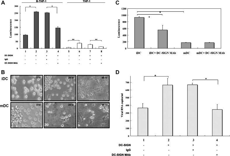
DC-SIGN mediates trans infection by H5N1 pseudotyped and reverse-genetics viruses. (A) The both sets of donor cells B-THP-1, B-THP-1/DC-SIGN and THP-1, THP-1/DC-SIGN were used in capture assay. The bars represent luciferase activity measured in MDCK cell lysates following co-culturing with donor cells in the presence or absence of MAb. (B) Immature and mature DCs were infected with H5N1 pseudotyped viruses for 0–48 h and cell morphology was observed under a light microscope. Upper and lower panels indicate iDCs and mDCs, respectively. (C) Immature and mature DCs with or without anti-DC-SIGN MAb treatment were used as donor cells to capture H5N1 pseudotyped virus particles and for co-culturing with MDCK cells. Luciferase activity was determined in MDCK lysates 48 h post-treatment. (D) The H5N1 reverse genetics strain A/Vietnam/1194/04 (105 copies/ml) was used for a capture assay. Virus-bound sialidase-treated B-THP-1/DC-SIGN and B-THP-1 donor cells were co-cultured with MDCK cells for 48 h; real-time RT-PCR was used to quantify H5N1 viral RNA in MDCK cell lysates. Results shown are from one representative experiment out of three performed (∗p < 0.05; ∗∗p < 0.01).
Interaction between DC-SIGN and H5N1 pseudotyped viruses was further confirmed using monocyte-derived DCs (MDDCs) purified from blood collected from healthy donors. Compared to immature DCs (iDCs), mature DCs (mDCs) expressed higher percentages of CD80, CD83, and CD86 cell markers (Supplementary Fig. 2A) and displayed both multiple pseudopods and irregular morphologies (Supplementary Fig. 2B); iDCs also expressed higher levels of DC-SIGN than mDCs (Supplementary Fig. 2A). Furthermore, the morphologies of both mDCs and iDCs changed dramatically 24 h post-infection with H5N1 pseudotyped viruses. At 48 h post-infection, both mDCs and iDCs adhered to the bottom of culture plates (Fig. 3B). Following incubation with the H5N1 pseudotyped virus at 4 °C for 1 h, DCs were co-cultured with MDCK cells and the resulting luciferase activity of MDCK lysates was measured. The results show that iDCs had a significantly higher capture ability compared to mDCs, and that this effect can be blocked by pre-treatment with anti-DC-SIGN monoclonal antibodies (Fig. 3C). Based on these findings, we conclude that iDCs play an important role in the dissemination of H5N1 pseudotyped virus infection due to its ability to capture and transfer viral infections to target cells.
Finally, we used H5N1 A/Vietnam/1194/04 reverse-genetics (RG) virus particles to confirm the phenomenon described in this study. As shown in Supplementary Fig. 1, B-THP-1/DC-SIGN cells had significantly higher copy numbers of H5N1 RG viruses than B-THP-1 cells 48 h post-infection (lanes 1 and 2, p < 0.05). Infection was significantly blocked when cells were pre-treated with anti-DC-SIGN monoclonal antibodies (lane 4). Results from a capture assay show that compared to the B-THP-1 control, the B-THP-1/DC-SIGN cells captured and transferred significantly higher numbers of H5N1 RG viruses to MDCK cells, and that the effect can be blocked by anti-DC-SIGN monoclonal antibodies (Fig. 3D). According to these results, DC-SIGN is capable of mediating H5N1 RG virus infection in cis and in trans.
We found evidence indicating that DC-SIGN does not function as a main entry receptor in the same manner as α-2,3-linked SA for the avian H5N1 virus, but instead acts as a capture or attachment molecule for the virus and as an infection mediating factor both in cis and in trans. Thitithanyanont et al. [20] previously showed that DCs express both α-2,3-linked and α-2,6-linked SA, and that H5N1 viruses are capable of escaping viral-specific immunity and dissemination to other organs through infection and replication in DCs. There are several existing reports indicating that iDCs and mDCs differ in viral infection susceptibility—for example, in HIV-1 and SARS CoV [21], [22]. Consistent with these studies, we found that iDCs had higher capture and transfer capabilities than mDCs.
Thitithanyanont et al. [20] also demonstrated that 24 h post-infection with the avian H5N1 virus, the majority of DCs died at a very low MOI. Our results indicate that the H5N1 pseudotyped virus induced extensive activation of iDCs and mDCs after 48 h of incubation, but mDCs showed lower susceptibility to the H5N1 virus. Our results also imply that a reduction in DC-SIGN expression may lead to the phenomenon described in this report. Based on our findings, we suggest that iDCs residing in the lower respiratory tract or deep lung compartments are susceptible to H5N1 virus infection via the α-2,3-linked SA receptor and DC-SIGN. Infected iDCs then migrate to lymphoid tissues and other organs and transfer the H5N1 virus to other target cells expressing α-2,3-linked SA receptors, resulting in a systemic infection. Another recently published study suggests that CD4+ T-cells may be an important target for H5N1 virus infection [23].
Acknowledgments
Our gratitude goes to the Vaccine Research and Development Center, National Health Research Institute, Taiwan for providing H5N1 A/Vietnam1194/04 RG strain and to members of Dr. Shie-Liang Hsieh’s lab for their advice and support for human DC isolation. This work was supported by a grant from the National Science Council, Republic of China (NSC 96-2321-B-010-007) and Aim for the Top University Plan.
Footnotes
Supplementary data associated with this article can be found, in the online version, at doi:10.1016/j.bbrc.2008.06.078.
Appendix A. Supplementary data
References
- 1.Gambotto A., Barratt-Boyes S.M., de Jong M.D., Neumann G., Kawaoka Y. Human infection with highly pathogenic H5N1 influenza virus. Lancet. 2008;371:1464–1475. doi: 10.1016/S0140-6736(08)60627-3. [DOI] [PubMed] [Google Scholar]
- 2.Yao L., Korteweg C., Hsueh W., Gu J. Avian influenza receptor expression in H5N1-infected and noninfected human tissues. FASEB J. 2008;22:733–740. doi: 10.1096/fj.06-7880com. [DOI] [PubMed] [Google Scholar]
- 3.Beigel J.H., Farrar J., Han A.M., Hayden F.G., Hyer R., de Jong M.D., Lochindarat S., Nguyen T.K., Nguyen T.H., Tran T.H., Nicoll A., Touch S., Yuen K.Y. Avian influenza A (H5N1) infection in humans. N. Engl. J. Med. 2005;353:1374–1385. doi: 10.1056/NEJMra052211. [DOI] [PubMed] [Google Scholar]
- 4.Chutinimitkul S., Bhattarakosol P., Srisuratanon S., Eiamudomkan A., Kongsomboon K., Damrongwatanapokin S., Chaisingh A., Suwannakarn K., Chieochansin T., Theamboonlers A., Poovorawan Y. H5N1 influenza A virus and infected human plasma. Emerg. Infect. Dis. 2006;12:1041–1043. doi: 10.3201/eid1206.060227. [DOI] [PMC free article] [PubMed] [Google Scholar]
- 5.Uiprasertkul M., Puthavathana P., Sangsiriwut K., Pooruk P., Srisook K., Peiris M., Nicholls J.M., Chokephaibulkit K., Vanprapar N., Auewarakul P. Influenza A H5N1 replication sites in humans. Emerg. Infect. Dis. 2005;11:1036–1041. doi: 10.3201/eid1107.041313. [DOI] [PMC free article] [PubMed] [Google Scholar]
- 6.Zhou T., Chen Y., Hao L., Zhang Y. DC-SIGN and immunoregulation. Cell. Mol. Immunol. 2006;3:279–283. [PubMed] [Google Scholar]
- 7.Curtis B.M., Scharnowske S., Watson A.J. Sequence and expression of a membrane-associated C-type lectin that exhibits CD4-independent binding of human immunodeficiency virus envelope glycoprotein gp120. Proc Natl Acad Sci USA. 1992;89:8356–8360. doi: 10.1073/pnas.89.17.8356. [DOI] [PMC free article] [PubMed] [Google Scholar]
- 8.Geijtenbeek T.B., Kwon D.S., Torensma R., van Vliet S.J., van Duijnhoven G.C., Middel J., Cornelissen I.L., Nottet H.S., KewalRamani V.N., Littman D.R., Figdor C.G., van Kooyk Y. DC-SIGN, a dendritic cell-specific HIV-1-binding protein that enhances trans-infection of T cells. Cell. 2000;100:587–597. doi: 10.1016/s0092-8674(00)80694-7. [DOI] [PubMed] [Google Scholar]
- 9.Tassaneetrithep B., Burgess T.H., Granelli-Piperno A., Trumpfheller C., Finke J., Sun W., Eller M.A., Pattanapanyasat K., Sarasombath S., Birx D.L., Steinman R.M., Schlesinger S., Marovich M.A. DC-SIGN (CD209) mediates dengue virus infection of human dendritic cells. J. Exp. Med. 2003;197:823–829. doi: 10.1084/jem.20021840. [DOI] [PMC free article] [PubMed] [Google Scholar]
- 10.van Kooyk Y., Geijtenbeek T.B. DC-SIGN: escape mechanism for pathogens. Nat. Rev. Immunol. 2003;3:697–709. doi: 10.1038/nri1182. [DOI] [PubMed] [Google Scholar]
- 11.Nefkens I., Garcia J.M., Ling C.S., Lagarde N., Nicholls J., Tang D.J., Peiris M., Buchy P., Altmeyer R. Hemagglutinin pseudotyped lentiviral particles: characterization of a new method for avian H5N1 influenza sero-diagnosis. J. Clin. Virol. 2007;39:27–33. doi: 10.1016/j.jcv.2007.02.005. [DOI] [PubMed] [Google Scholar]
- 12.Shih Y.P., Chen C.Y., Liu S.J., Chen K.H., Lee Y.M., Chao Y.C., Chen Y.M. Identifying epitopes responsible for neutralizing antibody and DC-SIGN binding on the spike glycoprotein of the severe acute respiratory syndrome coronavirus. J. Virol. 2006;80:10315–10324. doi: 10.1128/JVI.01138-06. [DOI] [PMC free article] [PubMed] [Google Scholar]
- 13.Payungporn S., Chutinimitkul S., Chaisingh A., Damrongwantanapokin S., Buranathai C., Amonsin A., Theamboonlers A., Poovorawan Y. Single step multiplex real-time RT-PCR for H5N1 influenza A virus detection. J. Virol. Methods. 2006;131:143–147. doi: 10.1016/j.jviromet.2005.08.004. [DOI] [PubMed] [Google Scholar]
- 14.Cormier E.G., Durso R.J., Tsamis F., Boussemart L., Manix C., Olson W.C., Gardner J.P., Dragic T. L-SIGN (CD209L) and DC-SIGN (CD209) mediate transinfection of liver cells by hepatitis C virus. Proc. Natl. Acad. Sci. USA. 2004;101:14067–14072. doi: 10.1073/pnas.0405695101. [DOI] [PMC free article] [PubMed] [Google Scholar]
- 15.Barth H., Ulsenheimer A., Pape G.R., Diepolder H.M., Hoffmann M., Neumann-Haefelin C., Thimme R., Henneke P., Klein R., Paranhos-Baccala G., Depla E., Liang T.J., Blum H.E., Baumert T.F. Uptake and presentation of hepatitis C virus-like particles by human dendritic cells. Blood. 2005;105:3605–3614. doi: 10.1182/blood-2004-05-1952. [DOI] [PubMed] [Google Scholar]
- 16.Liu C.K., Wei G., Atwood W.J. Infection of glial cells by the human polyomavirus JC is mediated by an N-linked glycoprotein containing terminal alpha(2-6)-linked sialic acids. J. Virol. 1998;72:4643–4649. doi: 10.1128/jvi.72.6.4643-4649.1998. [DOI] [PMC free article] [PubMed] [Google Scholar]
- 17.Alvarez C.P., Lasala F., Carrillo J., Muniz O., Corbi A.L., Delgado R. C-type lectins DC-SIGN and L-SIGN mediate cellular entry by Ebola virus in cis and in trans. J. Virol. 2002;76:6841–6844. doi: 10.1128/JVI.76.13.6841-6844.2002. [DOI] [PMC free article] [PubMed] [Google Scholar]
- 18.Hu H.P., Hsieh S.C., King C.C., Wang W.K. Characterization of retrovirus-based reporter viruses pseudotyped with the precursor membrane and envelope glycoproteins of four serotypes of dengue viruses. Virology. 2007;368:376–387. doi: 10.1016/j.virol.2007.06.026. [DOI] [PMC free article] [PubMed] [Google Scholar]
- 19.Elliot A.J., Steinhauer D.A., Daniels R.S., Oxford J.S. Functional and antigenic analyses of the 1918 influenza virus haemagglutinin using a recombinant vaccinia virus expression system. Virus Res. 2006;122:11–19. doi: 10.1016/j.virusres.2006.06.004. [DOI] [PubMed] [Google Scholar]
- 20.Thitithanyanont A., Engering A., Ekchariyawat P., Wiboon-ut S., Limsalakpetch A., Yongvanitchit K., Kum-Arb U., Kanchongkittiphon W., Utaisincharoen P., Sirisinha S., Puthavathana P., Fukuda M.M., Pichyangkul S. High susceptibility of human dendritic cells to avian influenza H5N1 virus infection and protection by IFN-alpha and TLR ligands. J. Immunol. 2007;179:5220–5227. doi: 10.4049/jimmunol.179.8.5220. [DOI] [PubMed] [Google Scholar]
- 21.Granelli-Piperno A., Delgado E., Finkel V., Paxton W., Steinman R.M. Immature dendritic cells selectively replicate macrophagetropic (M-tropic) human immunodeficiency virus type 1, while mature cells efficiently transmit both M- and T-tropic virus to T cells. J. Virol. 1998;72:2733–2737. doi: 10.1128/jvi.72.4.2733-2737.1998. [DOI] [PMC free article] [PubMed] [Google Scholar]
- 22.Spiegel M., Schneider K., Weber F., Weidmann M., Hufert F.T. Interaction of severe acute respiratory syndrome-associated coronavirus with dendritic cells. J. Gen. Virol. 2006;87:1953–1960. doi: 10.1099/vir.0.81624-0. [DOI] [PubMed] [Google Scholar]
- 23.Li Y.G., Thawatsupha P., Chittaganpitch M., Rungrojcharoenkit K., Li G.M., Nakaya T., Auwanit W., Ikuta K., Sawanpanyalert P. Higher in vitro susceptibility of human T cells to H5N1 than H1N1 influenza viruses. Biochem. Biophys. Res. Commun. 2008 doi: 10.1016/j.bbrc.2008.04.123. [DOI] [PubMed] [Google Scholar]
Associated Data
This section collects any data citations, data availability statements, or supplementary materials included in this article.