

Abstract
Interleukin-1 receptor activated kinases (IRAKs) play crucial roles in the Toll-like receptor (TLR) mediated signal transduction pathways that control host innate immune responses. Here we report the cloning of an IRAK1 cDNA (named ScIRAK1) from the mandarin fish. The predicted ScIRAK1 peptide contains a death domain and a serine/threonine-specific kinase domain. Quantitative RT-PCR showed that ScIRAK1 mRNA was primarily expressed in blood cells and posterior kidney. Seven days following infection with infectious spleen and kidney necrosis virus (ISKNV), the ScIRAK1 mRNA level was significantly higher in the blood cells of clinically symptomatic fish than in the blood cells of asymptomatic fish or control fish injected with phosphate-buffered saline. Additional experiments showed that overexpression of ScIRAK1 in the 293T cells could induce NF-κB activation. These results suggest that ScIRAK1 may play a role in the pathology of ISKNV infection in the mandarin fish.
Keywords: IRAK1, ISKNV, NF-κB, Infection, Mandarin fish
Introduction
The Toll-like receptors (TLRs) can detect specific features of evolutionarily distant pathogens, and the Interleukin-1 receptor (IL-1R) family proteins enable cells to initiate early host defense responses [1]. IL-1 receptor activated kinases (IRAKs) play crucial roles as mediators in the TLR/IL-1R signal transduction pathways [2]. Moreover, IRAK family member IRAK1 has been identified as a key component of the IL-1R signaling pathway in mammals [3]. Upon ligand binding to TLR/IL-1R, IRAK1 is recruited to the receptor complex to initiate the NF-κB signaling pathway, leading to induction of inflammatory target genes such as TNF-α, IL-6, IL-1β, and IL-12 [4]. Furthermore, IRAK1 is involved in activation and nuclear translocation of STAT3 (signal transducer and activator of transcription 3), STAT1 and IRF7 (interferon regulation factor 7), as well as in downstream gene expression [2], [5], [6]. Phosphorylated IRAK1 also undergoes ubiquitin-mediated degradation or sumoylation resulting in nuclear translocation and transcriptional activation of inflammatory target genes [7]. However, little is known about IRAK family member functions in fish, with the exception of zebrafish [8], [9].
The mandarin fish Siniperca chuatsi is a predominantly farmed fish species in China, with an annual output of 210,000 tons with a value of 1.5 billion US dollars in 2007. In the last decade, the spread of infectious spleen and kidney necrosis virus (ISKNV) has resulted in significant economic loss for many fish farms [10], [11]. ISKNV is classified in the Megalocystivirus genus of the Iridoviridae family. Thirteen cultured fish species and 39 wild fish species have been confirmed to be hosts of ISKNV-like viruses [12], including the mandarin fish. Infected fish develop serious systemic diseases [13], and ISKNV infection is capable of causing severe epizootics resulting in mass mortalities. Therefore, it is important to study the pathology and immunity of the mandarin fish in order to search for effective ways to protect the fish from diseases. In the present study, we cloned an IRAK1 cDNA from the mandarin fish (named ScIRAK1), and showed that after ISKNV infection, the ScIRAK1 mRNA level was significantly higher in fish with typical disease symptoms than in fish without symptoms or control fish injected with phosphate-buffered saline (PBS). Furthermore, overexpression of ScIRAK1 in the 293T cells could induce NF-κB activation. These results suggest that ScIRAK1 may play a role in the pathology of ISKNV infection in the mandarin fish.
Materials and methods
Experimental animals. Healthy mandarin fish (average weight = 250 g) were obtained from Nanhai Fish Culture Farm (Guangdong, China) and kept in aquaria at 28 °C. Fish were fed with healthy specimens at an average feeding rate of 10 g grass carp fry/kg/day. Before the experiments, the fish were maintained for at least 2 weeks and were determined to be free of ISKNV infections by PCR [14].
Cloning of ScIRAK1 cDNA by RT-PCR and rapid amplification of cDNA ends (RACE). Total RNA was extracted from the mandarin fish spleen using the RNeasy Mini Kit (Qiagen, Germany) as per the manufacturer’s instructions and then used to synthesize the 3′- and 5′-RACE cDNA templates with the BD SMART RACE cDNA amplification kit (Clontech, Japan) according to the user’s manual. Two degenerate oligonucleotide primers (F1 and R1, Table 1 ) were designed based on the conserved cDNA sequences of IRAK1 from human, mouse, dog, frog and zebrafish. Touchdown PCR reactions were performed using primers F1 and R1, with 1 cycle of denaturation at 94 °C for 3 min, 10 cycles of 94 °C for 30 s, 58-1 °C/cycle for 40 s, 72 °C for 40 s, and 28 cycles of 94 °C for 35 s, 48 °C for 40 s and 72 °C for 40 s, followed by a 5 min extension at 72 °C. PCR products were cloned into the pGEM-T Easy vector (Promega, USA) and sequenced. Based on the partial sequence obtained, specific primers (5′RACE and 3′RACE, Table 1) were designed to obtain the 3′- and 5′-end cDNA sequences of ScIRAK1 by RACE.
Table 1.
Summary of primers used in this study.
| Primers | Sequence (5′–3′)⁎ |
|---|---|
| For fragment amplification | |
| F1 | tgggmdmkggaggrttyggagtkg |
| R1 | accacdccraagctgyagahrtc |
| For RACE | |
| 5′RACE | gaacactggcgtacctgcctgat |
| 3′RACE | tcctccagcagagtccagtccag |
| Universal Primer a Mix (UPM) | ctaatacgactcactatagggcaagcagtggtatcaacgcagagt |
| Nest Universal Primer(NUP) | ctaatacgactcactatagggc |
| 3′-RACE cds primer A | aagcagtggtatcaacgcagagtac(t)30vn |
| For real-time PCR | |
| Real-5F | ccacggagacatcaagagttca |
| Real-3R | tttaccaaccgatgccgtc |
| 18S-F | atggtactttaggcgcctac |
| 18S-R | tatacgctattggagctgg |
| For plasmid construction | |
| F2 | ccggaattcagatgtcggcaggagacccgagg |
| R2 | cgggatccctatcagtcgtgttcagcgggaagataa |
D = not C; H = not G; M = A or C; N = A, C, G or T; R = A or G; V = not T; Y = C or T.
Expression profiles of ScIRAK1 in healthy and ISKNV-challenged mandarin fish. To determine expression of ScIRAK1 mRNA in healthy mandarin fish, the gills, anterior kidney, posterior kidney, spleen, liver, intestine, heart, brain, skin, muscle and stomach were dissected from three healthy mandarin fish and used for isolation of total RNA using the RNeasy Mini Kit (Qiagen, Germany). Total RNA from blood cells was also extracted from three healthy mandarin fish using TRIzol reagent (Invitrogen, USA). Individual total RNA samples from the same tissues were pooled and treated with RNase-free DNase I for quantitative RT-PCR.
ISKNV filtrates were prepared as described previously [11]. One milliliter of the filtrates was used for infection by intramuscular injection to each fish. Fifty fish were injected with ISKNV filtrates. For controls, 15 fish were injected with PBS (pH 7.2) (1 mL/fish). All the mandarin fish were maintained in 30 L aquaria (3 fish/aquarium) at 30 °C for 15 days. At 0, 1, and 4 days after ISKNV or PBS injection, RNA samples were isolated from the blood cells of three randomly selected mandarin fish from each group. On the 7th day after infection, three fish exhibiting lethargy, unresponsiveness to disturbances, pale body pigmentation, cessation of eating, and gill pallor were selected, and RNA was extracted from blood cells. Additionally, three fish without clinical symptoms and three fish from the PBS-injected control group were selected, and RNA was extracted from blood cells. On the 15th day after ISKNV infection, some fish recovered from the infection and showed no more clinical signs. Three of these recovered fish were selected, and RNA was extracted from blood cells. Three fish were also selected from the PBS-injected group 15 days post-injection, and RNA was extracted from blood cells.
RNA from the three fish in the same group was pooled and reverse transcribed with ReverTra Ace MMLV reverse transcriptase (TOYOBO, Japan). The resultant cDNAs were analyzed by quantitative RT-PCR.
Quantitative RT-PCR analysis. Primers Real-5F and Real-3R (Table 1) were used to amplify the ScIRAK1 cDNA fragment. Since 18S rRNA is one of the most reliable reference genes for quantitative RT-PCR of total RNA [15], primers 18S-F and 18S-R (Table 1) were used for amplification of 18S rRNA cDNA as an internal control. All amplifications and detections were carried out using the cDNAs described above as templates in the LightCycler 480 instrument (Roche, Sweden) with SYBR Green Realtime PCR Master Mix (QPK201) (TOYOBO, Shanghai, China) by using the following program: 95 °C for 30 s, 1 cycle; 95 °C for 5 s, 55 °C for 10 s, 72 °C for 15 s, 40 cycles, followed by a 30 min dissociation curve that was used to identify primer dimers. Statistical analyses were performed using LightCycler 480 Basic Software Version 1.2. All quantitative RT-PCR assays were repeated three times.
Construction of expression plasmids. The pCMV-Flag vector was prepared from the pEGFP-C3 vector (Clontech, Japan). pEGFP-C3 was digested with NheI and XhoI to remove the GFP sequence. Two primers (5′-gctagcgccgccatggattacaaggatgacgacgataaggccctcgag-3′; 5′-ctcgagggccttatcgtcgtcatccttgtaatccatggcggcgctagc-3′) were then synthesized and annealed to produce an adaptor. The adaptor was digested with NheI and XhoI and ligated into the NheI/XhoI digested pEGFP-C3 to prepare the pCMV-Flag vector. The entire open reading frame of ScIRAK1 was amplified using primers F2 and R2 (Table 1); the resultant PCR product was digested with EcoRI and BamHI and inserted into linearized pCMV-Flag to produce pCMV-IRAK1. Plasmid DNAs were prepared using Endo-free Plasmid Extraction Kit (Omega, China), and the sequences were confirmed by sequencing. HEK293T cells were transfected with pCMV-Flag or pCMV-IRAK1 plasmid using Lipofectamine 2000 reagent (Invitrogen, USA) according to the manufacturer’s instructions.
Cell transfection and NF-κB activity assay. HEK293T cells were maintained at 37 °C in Opti-MEM I medium (Invitrogen, USA) supplemented with 10% fetal bovine serum (Invitrogen, USA). For DNA transfection, cells were seeded in 96-well plates. When the cells were 70–90% confluent, they were co-transfected with 150 ng pCMV-Flag or pCMV-IRAK1, 50 ng pNF-κB-Luc (Promega, USA) and 1 ng pRL-TK Renilla luciferase plasmid (Promega, USA) as an internal control. At 24 or 48 h after transfection, firefly and Renilla luciferase activities were measured using the Dual-Luciferase Reporter Assay System (Promega, USA) according to the manufacturer’s instructions. Significance was analyzed by one-way ANOVA followed by Bonferroni’s post hoc adjustment. All statistics were performed using the SPSS program.
Results
Nucleotide and deduced amino acid sequences of ScIRAK1
By PCR amplification with degenerate primers and RACE, a full-length cDNA of the mandarin fish IRAK1 (ScIRAK1) was obtained (GenBank Accession No. FJ436359). The ScIRAK1 cDNA is 2676 bp with an open reading frame of 2265 bp, which encodes a putative protein of 754 amino acids. The calculated molecular mass of ScIRAK1 is 82.69 kDa, with a predicted pI of 5.93. Simple modular architecture research tool (SMART, http://smart.embl-heidelberg.de) [16] analysis shows that ScIRAK1 contains a death domain (residues 7–95, Fig. 1 ) and a serine/threonine-specific kinase domain (residues 196–485, Fig. 1).
Fig. 1.
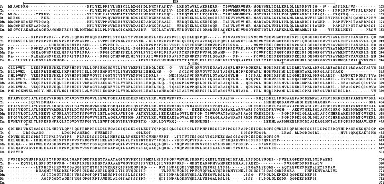
Comparison of mandarin fish ScIRAK1 with IRAK1 proteins from some other species. Dm, Drosophilamelanogaster Pelle (Accession No. AAA28750); Xt, Xenopus tropicalis (Accession No. AAH75439); Sc, Siniperca chuatsi (Accession No. FJ436359); Dr, Danio rerio (Accession No. XP_697688); Tn, Tetraodon nigroviridis (Accession No. CAF93411); Mm, Mus musculus (Accession No. NP_032389); Hs, Homo sapiens (Accession No. AAC41949); “*” indicates Thr-66 in the death domain of H. sapiens IRAK1, which is critical for interaction with signaling molecules. “+” indicates Thr-209 and Thr-387 in the kinase domain of human IRAK1 that are phosphorylated by IRAK4. “#” indicates Lys-239 and Asp-340 that are critical for the kinase function of human IRAK1. DD: death domain; S/T-Kc: serine/threonine protein kinase catalytic domain.
The deduced amino acid sequence of ScIRAK1 was compared with IRAK1 proteins from the zebrafish (Danio rerio), green spotted puffer (Tetraodon nigroviridis), western clawed frog (Xenopus tropicalis), mouse (Mus musculus), human (Homo sapiens), and Drosophila melanogaster (named Pelle) by using the Clustal V method of Megalign program of DNASTAR Lasergene software (Fig. 1). ScIRAK1 is most similar to IRAK1 of the green spotted puffer (40.2% identity), and it is also similar to IRAK1 of other species with 21.8–35.9% identities. The Thr-66 residue in the death domain of human IRAK1, which is critical for interaction with signaling molecules, is conserved in IRAK1 proteins of the mandarin fish, zebrafish, western clawed frog and mouse [17]. Furthermore, the Thr-209 and Thr-387 residues in the kinase domain of human IRAK1, which are phosphorylated by IRAK4 [18], as well as the Lys-239 and Asp-340 that are critical for the kinase function of IRAK1 [19] are all conserved in these animal species (Fig. 1).
Expression of ScIRAK1 mRNA in healthy mandarin fish
To study tissue distribution of ScIRAK1 mRNA in healthy mandarin fish, real-time RT-PCR was performed using equivalent amounts of RNAs from various mandarin fish tissues. The results showed that ScIRAK1 mRNA was detected in all the tissues tested (Fig. 2 A). ScIRAK1 mRNA level, when normalized to the expression level in the spleen (1-fold), was high in the blood cells (∼6.58-fold) and relatively high in the posterior kidney (∼1.92-fold), stomach (∼1.42-fold), brain (∼1.17-fold) and intestine (∼1.08-fold), but low in the liver (∼0.13-fold), skin (∼0.08-fold) and muscle (∼ 0.01-fold).
Fig. 2.
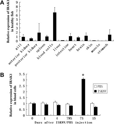
Expression of ScIRAK1 mRNA in mandarin fish. (A) Quantitative PCR analysis of ScIRAK1 mRNA expression in various tissues of healthy mandarin fish. Each column represents an average expression level of three healthy mandarin fish. ScIRAK1 mRNA levels were normalized to the 18S rRNA transcript. The expression level of ScIRAK1 mRNA in the spleen was used as the calibrator (set as 1). (B) Quantitative PCR analysis of ScIRAK1 mRNA expression in the blood cells after ISKNV or PBS injection. The expression level of ScIRAK1 mRNA in healthy blood cells (day 0) was used as the calibrator (set as 1). 7S indicates the fish with apparent symptoms 7 days after virus infection; 7NS indicates the fish without any symptoms 7 days after virus infection. “15” indicates the fish that recovered from the virus infection without any symptoms 15 days after virus infection. Asterisks indicate significant difference (P < 0.01) from the calibrator as determined by one-way AVONA.
Expression profiles of ScIRAK1 after ISKNV challenge
After ISKNV challenge, mandarin fish showed no visible clinical sign of infection during the first 4 days post-challenge, but they were all infected with ISKNV as confirmed by PCR [14]. At the 7th day post-challenge, 43 out of 50 infected fish showed apparent clinical symptoms such as lethargy, unresponsiveness to disturbances, pale body pigmentation, cessation of taking food and gill pallor, while the seven other infected fish did not show any symptoms. The expression of ScIRAK1 mRNA in blood cells (normalized to the expression level at day 0 of the healthy control group) was significantly upregulated (∼3.07-fold) at 7 days post-infection in the fish with apparent symptoms (Fig. 2B). However, expression levels of the ScIRAK1 transcript in the blood cells of infected fish without clinical symptoms were slightly lower (∼0.81-fold) than in control fish. At 15 days post-infection, some fish recovered from the virus infection, and the expression of ScIRAK1 transcript in the blood cells was slightly lower (∼0.71-fold) than in control fish, while expression of ScIRAK1 mRNA did not significantly change in the PBS-injected control fish. These results suggest that ScIRAK1 mRNA expression was induced in the blood cells of symptomatic fish after these fish were infected with ISKNV.
ScIRAK1 activates NF-κB in the 293T cells
To study regulation of NF-κB activation by ScIRAK1, we overexpressed ScIRAK1 in 293T cells by transiently transfecting the cells with pCMV-IRAK1 and assaying the activity of firefly luciferase under the control of NF-κB. Renilla luciferase expression was assayed as an internal control. We observed that at 24 h after transfection, the relative luciferase activity in cells transfected with pCMV-IRAK1 (∼416.92) was significantly higher than that in cells transfected with pCMV-Flag (∼79.42) (Fig. 3 , P < 0.01). At 48 h after transfection, the relative luciferase activity in cells transfected with pCMV-IRAK1 (∼823.85) was much higher than that in cells transfected with the control vector (∼115.38) (Fig. 3, P < 0.01).
Fig. 3.
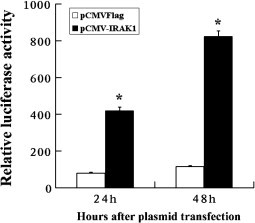
Relative induction of luciferase activity by ScIRAK1. The 293T cells were transfected with 150 ng of pCMV-IRAK1 or pCMV-Flag plasmid, 50 ng pNF-κB-Luc together with 1 ng pRL-TK Renilla luciferase plasmid (as an internal control, Promega, USA). At 24 or 48 h after transfection, cells were harvested, and firefly and Renilla luciferase activities were measured using Dual-Luciferase Reporter Assay System. Data are presented as means and SD from three independent biological samples (three cell cultures) with three replicates from each sample. Asterisks indicate significant differences (P < 0.01) as determined by one-way AVONA.
Discussion
IRAK1 is a critical signaling mediator of innate immunity. Although genes encoding IRAK1 proteins have been identified in the genomes of the zebrafish D. rerio, the Japanese pufferfish Takifugu rubripes and the green spotted puffer T. nigroviridis [8], little is known about the functions of IRAK1 in fish. In this study, we cloned a cDNA for ScIRAK1 from an economically important mandarin fish. We showed that at the 7th day after ISKNV infection, expression of ScIRAK1 mRNA in the blood cells was significantly higher in fish with apparent clinical symptoms than in fish without clinical symptoms (Fig. 2B). We also demonstrated that overexpression of ScIRAK1 in 293T cells could induce NF-κB activation. To our knowledge, this is the first report of a functional IRAK1 in fish.
IRAK1 is a key molecule in the TLR/IL-1R signaling pathways [20]. Prior to TLR/IL-1R receptor activation, IRAK1 is found in the cytosol. Upon ligand binding to TLR/IL-1R, IRAK1 binds to MyD88, which interacts with the receptor. IRAK1 is subsequently phosphorylated and activated by IRAK4. Hyperphosphorylated IRAK1 then leaves the receptor complex and forms a cytosolic IRAK1-TRAF6 (TNF receptor-associated factor 6) complex, resulting in eventual activation of the downstream pathways [2]. Phosphorylation of IRAK1 is a hallmark of IRAK activation. In humans, Thr-209 and Thr-387 in IRAK1 are phosphorylated [18]. These two threonine residues are conserved in ScIRAK1 (Thr-193 and Thr-373, Fig. 1). Therefore, we speculate that ScIRAK1 may be phosphorylated during TLR activation.
After pathogen infection, activation of NF-κB may create inappropriate inflammatory responses (a so-called “cytokine storm”) that can inadvertently damage the host to such a degree that it causes illness and even death [21]. Documented examples of this phenomenon include the following: infection with the West Nile Virus (WNV) leads to a cytokine storm and subsequent disruption of the blood–brain barrier, which facilitates entry of the virus into the brain [22]; reinfection with dengue virus results in dengue hemorrhagic fever and dengue shock syndrome, which has been linked to a cytokine storm, ultimately causing an increase in vascular permeability [23]; and infections with viruses such as poxvirus [21], severe acute respiratory syndrome (SARS) [24], [25], [26] and influenza virus [27] result in tissue damages as a result of a cytokine storm. IRAK1 has been reported to play important roles in tissue injury related to a cytokine storm. For example, IRAK1 was shown to be a central kinase involved in acute LPS-induced myocardial contractile dysfunction and septic shock [28], [29]. In this study, at the 7th day after ISKNV infection, most infected fish showed typical symptoms, and quantitative RT-PCR showed that the ScIRAK1 mRNA level was significantly higher in the diseased fish than in the fish without symptoms. Further work showed that ScIRAK1 could induce NF-κB activation. Thus, it is possible that upregulation of ScIRAK1 in the mandarin fish after ISKNV infection may activate NF-κB, resulting in a “cytokine storm” and subsequent tissue damage, which is related to disease progression and pathology.
Acknowledgments
This research was supported by the National Natural Science Foundation of China under Grant No. U0631008; the National Basic Research Program of China under Grant No. 2006CB101802; the National High Technology Research and Development Program of China (863 Program) under Grant Nos. 2006AA09Z445, 2006AA100309; the Guangdong Province Natural Science Foundation under Grant No. 20023002.
References
- 1.Trinchieri G., Sher A. Cooperation of Toll-like receptor signals in innate immune defence. Nat. Rev. Immunol. 2007;7:179–190. doi: 10.1038/nri2038. [DOI] [PubMed] [Google Scholar]
- 2.Gottipati S., Rao N.L., Fung-Leung W.P. IRAK1: a critical signaling mediator of innate immunity. Cell. Signal. 2008;20:269–276. doi: 10.1016/j.cellsig.2007.08.009. [DOI] [PubMed] [Google Scholar]
- 3.Cao Z., Henzel W.J., Gao X. IRAK: a kinase associated with the interleukin-1 receptor. Science. 1996;271:1128–1131. doi: 10.1126/science.271.5252.1128. [DOI] [PubMed] [Google Scholar]
- 4.Akira S., Takeda K. Toll-like receptor signalling. Nat. Rev. Immunol. 2004;4:499–511. doi: 10.1038/nri1391. [DOI] [PubMed] [Google Scholar]
- 5.Huang Y., Li T., Sane D.C., Li L. IRAK1 serves as a novel regulator essential for lipopolysaccharide-induced interleukin-10 gene expression. J. Biol. Chem. 2004;279:51697–51703. doi: 10.1074/jbc.M410369200. [DOI] [PubMed] [Google Scholar]
- 6.Nguyen H., Chatterjee-Kishore M., Jiang Z., Qing Y., Ramana C.V., Bayes J., Commane M., Li X., Stark G.R. IRAK-dependent phosphorylation of Stat1 on serine 727 in response to interleukin-1 and effects on gene expression. J. Interferon Cytokine Res. 2003;23:183–192. doi: 10.1089/107999003765027384. [DOI] [PubMed] [Google Scholar]
- 7.Su J., Richter K., Zhang C., Gu Q., Li L. Differential regulation of interleukin-1 receptor associated kinase 1 (IRAK1) splice variants. Mol. Immunol. 2007;44:900–905. doi: 10.1016/j.molimm.2006.03.021. [DOI] [PubMed] [Google Scholar]
- 8.Stein C., Caccamo M., Laird G., Leptin M. Conservation and divergence of gene families encoding components of innate immune response systems in zebrafish. Genome Biol. 2007;8:R251. doi: 10.1186/gb-2007-8-11-r251. [DOI] [PMC free article] [PubMed] [Google Scholar]
- 9.Phelan P.E., Mellon M.T., Kim C.H. Functional characterization of full-length TLR3, IRAK-4, and TRAF6 in zebrafish (Danio rerio) Mol. Immunol. 2005;42:1057–1071. doi: 10.1016/j.molimm.2004.11.005. [DOI] [PubMed] [Google Scholar]
- 10.He J.G., Weng S.P., Huang Z.J., Zeng K. Identification of outbreak and infectious diseases pathogen of Siniperca chuatsi. Acta Sci. Nat. Univ. Sunyatseni. 1998;5:74–77. (in Chinese, with English abstract) [Google Scholar]
- 11.He J.G., Weng S.P., Zeng K., Huang Z.J., Chan S.M. Systemic disease caused by an iridovirus-like agent in cultured mandarinfish, Siniperca chuatsi (Basilewsky), in China. J. Fish Dis. 2000;23:219–222. [Google Scholar]
- 12.Wang Y.Q., Lu L., Weng S.P., Huang J.N., Chan S.M., He J.G. Molecular epidemiology and phylogenetic analysis of a marine fish infectious spleen and kidney necrosis virus-like (ISKNV-like) virus. Arch. Virol. 2007;152:763–773. doi: 10.1007/s00705-006-0870-4. [DOI] [PubMed] [Google Scholar]
- 13.He J.G., Zeng K., Weng S.P., Chan S.M. Experimental transmission, pathogenicity and physical–chemical properties of infectious spleen and kidney necrosis virus (ISKNV) Aquaculture. 2002;204:11–24. [Google Scholar]
- 14.He W., Yinlt Z.X., Li Y., Huo W.L., Guan H.J., Weng S.P., Chan S.M., He J.G. Differential gene expression profile in spleen of mandarin fish Siniperca chuatsi infected with ISKNV, derived from suppression subtractive hybridization. Dis. Aquat. Organ. 2006;73:113–122. doi: 10.3354/dao073113. [DOI] [PubMed] [Google Scholar]
- 15.Kim B.R., Nam H.Y., Kim S.U., Kim S.I., Chang Y.J. Normalization of reverse transcription quantitative-PCR with housekeeping genes in rice. Biotechnol. Lett. 2003;25:1869–1872. doi: 10.1023/a:1026298032009. [DOI] [PubMed] [Google Scholar]
- 16.Letunic I., Copley R.R., Pils B., Pinkert S., Schultz J., Bork P. SMART 5: domains in the context of genomes and networks. Nucleic Acids Res. 2006;34:D257–D260. doi: 10.1093/nar/gkj079. [DOI] [PMC free article] [PubMed] [Google Scholar]
- 17.Neumann D., Kollewe C., Pich A., Cao P., Resch K., Martin M.U. Threonine 66 in the death domain of IRAK-1 is critical for interaction with signaling molecules but is not a target site for autophosphorylation. J. Leukoc. Biol. 2008;84:807–813. doi: 10.1189/jlb.0507290. [DOI] [PubMed] [Google Scholar]
- 18.Kollewe C., Mackensen A.C., Neumann D., Knop J., Cao P., Li S., Wesche H., Martin M.U. Sequential autophosphorylation steps in the interleukin-1 receptor-associated kinase-1 regulate its availability as an adapter in interleukin-1 signaling. J. Biol. Chem. 2004;279:5227–5236. doi: 10.1074/jbc.M309251200. [DOI] [PubMed] [Google Scholar]
- 19.Janssens S., Beyaert R. Functional diversity and regulation of different interleukin-1 receptor-associated kinase (IRAK) family members. Mol. Cell. 2003;11:293–302. doi: 10.1016/s1097-2765(03)00053-4. [DOI] [PubMed] [Google Scholar]
- 20.Martin M.U., Wesche H. Summary and comparison of the signaling mechanisms of the Toll/interleukin-1 receptor family. Biochim. Biophys. Acta. 2002;1592:265–280. doi: 10.1016/s0167-4889(02)00320-8. [DOI] [PubMed] [Google Scholar]
- 21.Stanford M.M., McFadden G., Karupiah G., Chaudhri G. Immunopathogenesis of poxvirus infections: forecasting the impending storm. Immunol. Cell Biol. 2007;85:93–102. doi: 10.1038/sj.icb.7100033. [DOI] [PubMed] [Google Scholar]
- 22.Wang T., Town T., Alexopoulou L., Anderson J.F., Fikrig E., Flavell R.A. Toll-like receptor 3 mediates West Nile virus entry into the brain causing lethal encephalitis. Nat. Med. 2004;10:1366–1373. doi: 10.1038/nm1140. [DOI] [PubMed] [Google Scholar]
- 23.Pang T., Cardosa M.J., Guzman M.G. Of cascades and perfect storms: the immunopathogenesis of dengue haemorrhagic fever–dengue shock syndrome (DHF/DSS) Immunol. Cell Biol. 2007;85:43–45. doi: 10.1038/sj.icb.7100008. [DOI] [PubMed] [Google Scholar]
- 24.Theron M., Huang K.J., Chen Y.W., Liu C.C., Lei H.Y. A probable role for IFN-gamma in the development of a lung immunopathology in SARS. Cytokine. 2005;32:30–38. doi: 10.1016/j.cyto.2005.07.007. [DOI] [PMC free article] [PubMed] [Google Scholar]
- 25.Huang K.J., Su I.J., Theron M., Wu Y.C., Lai S.K., Liu C.C., Lei H.Y. An interferon-gamma-related cytokine storm in SARS patients. J. Med. Virol. 2005;75:185–194. doi: 10.1002/jmv.20255. [DOI] [PMC free article] [PubMed] [Google Scholar]
- 26.Nagata N., Iwata N., Hasegawa H., Fukushi S., Harashima A., Sato Y., Saijo M., Taguchi F., Morikawa S., Sata T. Mouse-passaged severe acute respiratory syndrome-associated coronavirus leads to lethal pulmonary edema and diffuse alveolar damage in adult but not young mice. Am. J. Pathol. 2008;172:1625–1637. doi: 10.2353/ajpath.2008.071060. [DOI] [PMC free article] [PubMed] [Google Scholar]
- 27.Us D. Cytokine storm in avian influenza. Mikrobiyol. Bul. 2008;42:365–380. [PubMed] [Google Scholar]
- 28.Thomas J.A., Haudek S.B., Koroglu T., Tsen M.F., Bryant D.D., White D.J., Kusewitt D.F., Horton J.W., Giroir B.P. IRAK1 deletion disrupts cardiac Toll/IL-1 signaling and protects against contractile dysfunction. Am. J. Physiol. Heart Circ. Physiol. 2003;285:H597–H606. doi: 10.1152/ajpheart.0655.2001. [DOI] [PubMed] [Google Scholar]
- 29.Cuschieri J., Bulmus V., Gourlay D., Garcia I., Hoffman A., Stayton P., Maier R.V. Modulation of macrophage responsiveness to lipopolysaccharide by IRAK-1 manipulation. Shock. 2004;21:182–188. doi: 10.1097/01.shk.0000111828.07309.26. [DOI] [PubMed] [Google Scholar]