Abstract
Objective:
This study aims for molecular identification of naturally growing Bacillus cereus strain from a unique source, able to survive, and alleviate heavy metals from the nature.
Materials and Methods:
Pure isolate from Murrah buffalo milk was prepared in B. cereus selective Polymyxin pyruvate egg-yolk mannitol–bromothymol blue agar (PEMBA) medium through a cascade of contamination free subcultures. The morphological and biochemical tests were done prior to 16S rRNA gene sequencing for strain identification and further physiological tests. The test strain was inoculated in both solid and suspension culture medium supplemented individually with Cd, Cu, Ag, and Zn to reveal the qualitative and quantitative heavy metal tolerance properties, respectively. Finally, the data collected from the in vitro assessment was statistically analyzed
Results:
Molecular analysis revealed that the test strain was B. cereus BF2, which was motile, catalase positive and Gram positive rod. B. cereus BF2 was found significant at 0.3% bile salt tolerance [two-way analysis of variance (ANOVA)—p value is < 0.0001] where, t-test p value is < 0.0002 between Control Group (CG) and TGR-1; p < 0.037 between TGR-1 and 2; p < 0.0014 between CG and TGR-2. Similarly, B. cereus BF2 was significant in pH tolerant up to 8.0 with p < 0.0115 (in scale p < 0.05). The heavy metal tolerance test revealed that the test metals could not stop the growth of B. cereus BF2 even after 24 h of incubation but partially suppressed the growth kinetics for letting into stationary phase. Among the four heavy metals, Cd and Zn showed partial antagonism to the growth of B. cereus BF2. The survivability was highly significant in the medium supplemented with Zn (p < 0.0001) and Ag (p < 0.018).
Conclusion:
Bacillus cereus BF2 can survive in selective heavy metals with metal resistance and biodegradation capacity.
Keywords: Bacillus cereus BF2; PEMBA; 16S rRNA sequencing; selective heavy metals (Cd, Ag, Cu, Zn); in vitro assessment
Introduction
Heavy metals are naturally occurred metal groups that can work as contaminants for ecosystem if deposited in high amount in nature. Mining, surface finishing industries, air or water pollution, milling are the principal emergence of heavy metal pollution. Of late, excessive bioaccumulation of heavy metals can render massive adversity to the living beings [1]. They are toxic, mutagenic, and carcinogenic. Micro-organisms growth, metabolism, and differentiation outright or obliquely linked with metals. A myriad of bacteria showing its tolerance at different concentrations of heavy metals [2,3]. For the capacity of bioaccumulation and resistance property assessment on differentiated metal ions, isolation, identification, and necessary characterizations are required. Bacillus cereus has this type of marvelous retention. B. cereus strains are acquainted to dwell in soil and food as motile, facultative anaerobic, spore forming, Gram-positive rods; considered severe food spoiling pathogen which often consequence non-gastrointestinal-tract infections at diversified fatality range. Some Bacillus spp. occupy in extreme environment, namely, B. badius, Bacillus subtilis, and B. cereus [4]. Bacillus spp. has already proved potentially antagonistic to pathogens, such as Escherichia coli and Staphylococcus aureus [5].
Often the range of pH, bile salt concentration, and organic–inorganic entities affect heavy metal toxicity on microorganisms and even influenced with the facts of speciation [6]. Some probiotic bacteria can also tolerate heavy metal toxicity at different stressed conditions. Lactobacillus fermentum SN_4 and Lactobacillus rhamnosus SN_6 have exhibited survivability against heavy metals [7]. According to Kirillova et al. [8], few Lactobacillus strains showed their cadmium and lead removability. In the same way, L. fermentum and L. plantarum revealed the same property of bioremediation [8].
In our experiment, whether or not different entities and concentrations of metals (cadmium, copper, silver, and zinc) play roles on the test strain of B. cereus survivability was scrutinized and the metal fortitude level of B. cereus strain of our interest was analyzed. The ultimate aims of our investigation were regulated through isolation, identification, and characterization of a new B. cereus strain from a unique source and the profiling of heavy metal tolerance property of the strain so that a new micro-organism can unveil its potentiality to the list of established bioremediation agents and can be a good choice for the next generation bioremediation tool.
Materials and Methods
Isolation of presumptive B. cereus
Milk sample of a Murrah buffalo (from Haryana, India) was collected from Government Buffalo Farm, Bagerhat District, Bangladesh using Nordic Iceberg. Primary culture was prepared on Polymyxin pyruvate egg-yolk mannitol–bromothymol blue agar (PEMBA) medium selective for B. cereus [9] at 37°C for 24 to 72 h from the 11th step of serial dilution of the milk sample. Following that, seven times consecutive contamination-free subculture were commenced to prepare pure isolate. Finally, similar to different looking 10 single colonies from the final pure culture plate were taken for further characterization separately.
Morphological and biochemical tests
The morphological tests considered the study of the bacterial size, shape, and motility status, while gram staining and catalase test were for biochemical tests of the presumptive pure isolates [10] exhibiting probiotic properties. The best result showing colony was elected for 16S rRNA gene analysis to identify the exact strain embedded inside.
Molecular identification of the test strain
16S rRNA sequencing were undertaken through RNA extraction, 1.2% Agarose Gel Electrophoresis, isolated RNA amplification with Universal 16S rRNA Specific Primer 8F (AGAGTTTGATCCTGGCTCAG) and 1492R (AAGTCGTAACAAGGTAACC) using Veriti® 99 well Thermal Cycler (Model No. 9902). A single amplified polymerase chain reaction (PCR) band of ~1400 bp was obtained for enzymatically purified for Sanger Sequencing. Bi-directional DNA sequencing reaction of PCR amplification was carried out with 8F and 1401R primers using BDT v3.1 Cycle sequencing kit on ABI 3730xl Genetic Analyzer. Finally, the gene sequence of the target isolate of our interest was submitted to NCBI through Gene Bank and the new accession number was registered. Analysis of the evolutionary relationship using MEGA5 was done following Neighbor-Joining method as referred by Saitou and Nei [11].
Physiological tests
Bile salt and pH tolerance test at 10 different concentrations with two replications of procedure for each test were undertaken following [12].
Heavy metal tolerance test
The test strain was cultured in PEMBA medium supplemented with 3CdSO4.8H2O; Ag2SO4, ZnSO4.7H2O and CuSO4.5H2O (each with 0.05%, 0.15%, and 0.5% concentration) to observe bacterial growth patterns through naked observation. Besides, Polymyxin pyruvate egg-yolk mannitol–bromothymol blue (PEMB)-broth media containing the aforementioned metal salts at the same concentration were prepared separately and the target strain was inoculated for incubation at 37°C for 24 h. The optical density (OD) value was taken every 12 h interval by UV-Vis Spectrophotometer UV-1280 (Shimadzu) to identify the growth response of the test strain to the concentrations of heavy metals over time.
I0 = incident optical intensity; I = transmitted optical intensity
Statistical analysis
The statistical analysis of the data collected from OD observation was performed using statistical analysis system and GraphPad Prism 8.
Results and Discussion
The effect of bile salt concentration on B. cereus BF2
Based on the morphological and biochemical characteristics (catalase test, gram staining test, oxidase test etc.), the isolate was clearly identified as Bacillus spices. Different fluctuations were observed when employing a series of bile concentration at different times. As a control of the study, broth with 0% concentration of bile salt was selected. Table 1 illustrates the survival rate of bacteria at ten different bile salt concentrations (0.1%–1.0%). The highest survival capability for Treatment Group Replication-1 was seen at 0.5 and 0.8 concentration of bile salt. At higher concentration of bile salt (1.0), the lower survivability of B. cereus BF2 was depicted. In case of Treatment Group Replication-2, higher tolerance level was seen in the presence of 0.4 bile salt concentration. Gradual increment of bile salt concentrations resulted in gradual decrease of the bacterial growth rate as well as their tolerance level [13].
Table 1. Survivability of B. cereus BF2 at different concentrations of bile salt and various pH levels.
| Bile Salt (%)∞ | OD650 | pH∞∞ | OD650 | |||
|---|---|---|---|---|---|---|
| *** | CGa | TGR-1b | TGR-2c | ** | R1 | R2 |
| 0.1 | 0.129 | 0.133 | 0.144 | 1 | 0.797 | 0.799 |
| 0.2 | 0.133 | 0.14 | 0.156 | 2 | 0.432 | 0.43 |
| 0.3 | 0.133 | 0.147 | 0.164 | 3 | 0.333 | 0.334 |
| 0.4 | 0.132 | 0.147 | 0.166 | 4 | 0.356 | 0.356 |
| 0.5 | 0.133 | 0.148 | 0.161 | 5 | 0.359 | 0.361 |
| 0.6 | 0.134 | 0.143 | 0.154 | 6 | 0.366 | 0.369 |
| 0.7 | 0.134 | 0.142 | 0.151 | 7 | 0.38 | 0.381 |
| 0.8 | 0.133 | 0.148 | 0.139 | 8 | 0.398 | 0.399 |
| 0.9 | 0.133 | 0.143 | 0.137 | 9 | 0.169 | 0.17 |
| 1.0 | 0.135 | 0.135 | 0.132 | 10 | 0.148 | 0.149 |
Grouped data analysis of the two-way ANOVA reports-
p < 0.0001 (which is highly significant to the scale p < 0.05) for the bile salt tolerance test; a,b,c are all significant values (in scale p < 0.05)
p < 0.0002 between CG and Treatment Group Replication-1 (TGR-1)
p < 0.037 between the TGR-1and 2
p < 0.0014 between CG and TGR-2
t-test reports-
p < 0.0115 (significant to the scale p < 0.05) in the pH tolerance test data considering the two replications (R)
The effect of acid tolerance test
Various pH levels (1–10) were selected with B. cereus BF2 to check their growth and survival capacity in stressed condition. At pH 2 and 9, sudden fall in their growth was observed than the initial growth rate. At pH 10, the lower growth rate was recorded as growth decreased with the pH increment. The similar finding was noticed in the investigations of Thomassin et al. [14] regarding B. cereus proliferation. Browne and Dowds [15] experiment was also analogous with our finding.
In another experiment, the result recorded no bacterial growth below pH 5 and growth developed when the pH gradually increased [16]. Such a phenomenon also previously described by Everis and Betts [17] in case of B. polymyxa and Clostridium tyrobutyricum. B. thuringiensis was found to grow well at pH 4.0–7.0 [18]. Some experiments [19,20] deduced that the food acidity causing B. cereus better grew in the range of minimal pH (4.5%–5.15%).
Molecular identification and phylogenetic analysis
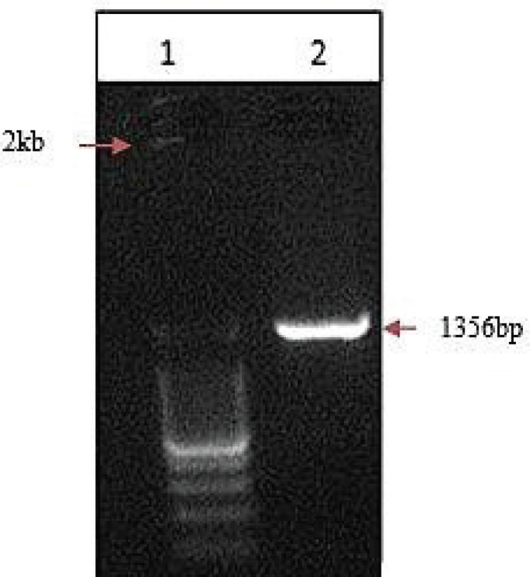
Agarose gel Electrophoresis was used for analysis of PCR result. The Figure 1 exhibits the band of 16S rRNA gene of B. cereus on 1.2% gel electrophoresis when observed under trans-illuminator. The size of the PCR product was 1,356 bp measuring with the ladder of 2 kb.
Figure 1. 1.2% Agarose gel electrophoresis showing 16S rRNA amplicon of Bacillus cereus BF2 at lane 2 where, lane 1 indicates 2kb ladder.

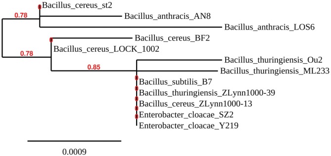
The targeted gene sequencing revealed that the strain was B. cereus BF2 (GenBank Accession No. MH569091.1). The phylogenetic tree has covered 12 different bacterial strains. Identification of the target strains was completed following its higher similarities to the reference strains in the Gene Bank. The phylogenetic lineage of B. cereus BF2 was compared with the sequence of B. cereus st2, B. anthracis AN8, B. cereus LOCK 1002, B. anthracis LOS6, B. thuringiensis Ou2, B. thuringiensis ML 233, B. cereus ZLynn1000-13, B. thuringiensis ZLynn1000-39, B. subtilis B7, Enterobacter cloacae SZ2, and E. cloacae Y219 from NCBI.
Three different strains of B. cereus were found with their maximum similarities with B. cereus BF2, including B. cereus st2, B. cereus LOCK 1002, B. cereus ZLynn 1000-13 (Fig. 2). B. anthracis LOS6 and B. thuringiensis Ou2 were also closely related to different species. In contrast, there is distant relationship between B. cereus BF2 and E. cloacae strains. Different researchers found similar relationship among different strains of B. cereus and B. thuringiensis in their phylogenetic tree analysis [21,22].
Figure 2. Evolutionary relationship of Bacillus cereus strain BF2 (Accession No. MH569091.1).
The phylogenetic analysis through the Neighbor-Joining method as referred by Saitou and Nei [11]. Following Felsenstein [23], the bootstrap consensus tree inferred from 1,000 replicates was taken to represent the evolutionary history of the taxa analyzed. The branches corresponding to partitions reproduced in less than 50% bootstrap replicates were collapsed [23].
Analyzing the survivability against heavy metals
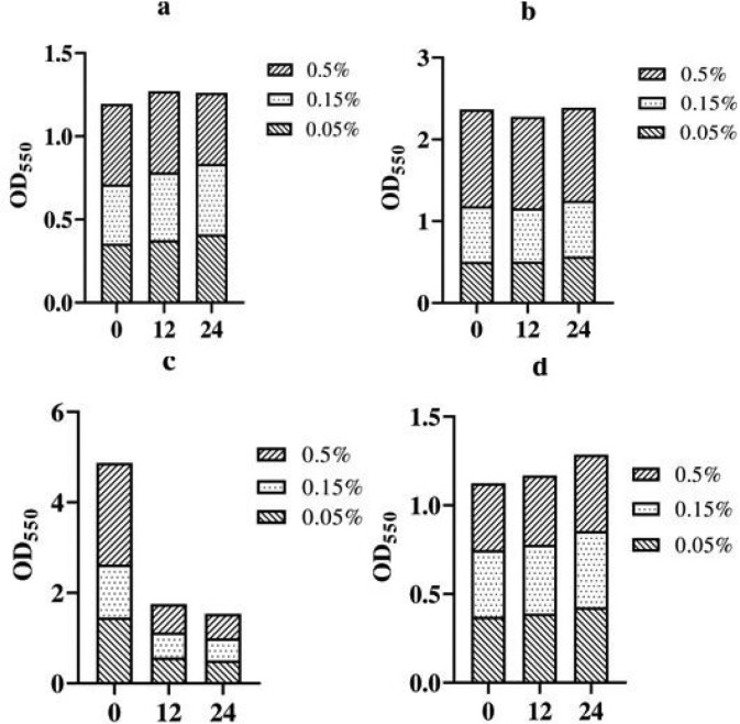
The evaluation of growth of B. cereus BF2 against different heavy metals was done in either solid and suspension culture conditions (at 0.05%, 0.15%, and 0.5%). For qualitative analysis, solid PEMBA media supplemented with heavy metals (Cd, Cu, Ag, and Zn) provided no distinguishable suppression on the bacterial growth except Cd and Zn supplementation. In contrast, the quantitative assessment by OD from the suspension culture showed different growth pattern in heavy metals survivability at 550 nm in different time interval (Fig. 3). The two-way ANOVA test revealed “p” values at 0.6737; 0.31; 0.018, and 0.0001 (in scale of significance p < 0.05) in 3CdSO4.8H2O (3a); CuSO4.5H2O (3b); AgSO4 (3c); and ZnSO4.7H2O (3d), respectively.
Figure 3. Effects of various concentrations of heavy metals on the growth of B. cereus BF2 over time.
The Figure 3a and d demonstrates that B. cereus BF2 continued its growth even after 12 h incubation when 0.05% of Cd and Zn were applied. At 24 h of incubation with 0.5% concentration of both of the heavy metals, the bacterial strain started entering slight stationary phase losing the growth kinetics. In the Figure 3b and c, B. cereus continued its growth after 12 h when subjected to Cu and Ag, while the growth pattern eventually increased after 24 h at various concentrations. Results of the experiment pointed out that the strain entirely survived on Cu and Ag and continued their growth on those heavy metals for longer periods. According to Behera et al. [24], Cd and Cu had more toxic effect on the B. cereus growth. The findings of Kalantar [25] were quite similar with us where they reported that when the concentration of Cd increases the Bacillus spp. growth declines. But, the result differed in some studies [26,27], where the B. cereus grew well in Cd comprising media.
In case of Zn, some studies on Bacillus spp. demonstrated their findings [28,29] that high concentration of Zn showed depletion on bacterial growth. The result of the experiment of Khande et al. [30] showed similarity to our findings. According to Ghahfarokhi et al. [31], Gram-negative bacteria showed significant survivability with Ag at different concentration which supports our outcomes.
According to Ghahfarokhi et al. [31] Ag showed good growth suppression on the B. cereus [31], which was dissimilar with our outcome. In case of Cu, inhibition of Bacillus spp. growth was reported in some experiments [32,33]. But a few studies indicated that the effect of Cu exposed no suppression on the B. cereus growth which was line with our investigation [34,35].
Conclusion
In this study, B. cereus BF2 has found significant survival at 0.3% bile salt and pH up to 8.0. The tolerance of B. cereus BF2 in culture medium supplemented with cadmium, copper, silver, and zinc was not found very distinguishable at qualitative assessment but diversified tolerance and viability response of the strain were observed in broth culture. Cd and Zn were found partial suppressive but could not stop the growth of B. cereus BF2. On the other hand, Cu and Ag were accumulated most significantly by Bacillus strain which was commensurate to our target of interest. To recapitulate, the results exhibited in this study indicate that B. cereus had phenomenal bioaccumulation and metal tolerant properties and it can clearly be manipulated regarding bioremediation purposes.
Acknowledgment
The research work was fully funded by The World Bank, which was arranged and monitored by Bangladesh Agricultural Research Council, Project Implementation Unit, phase 2, and project ID: 532. The authors are also very grateful to the animal cell culture Lab authority for providing logistic supports during the research activity.
Conflict of interest
The authors declared that they have no conflict of interest.
Authors’ contribution
Salauddin Al Azad prepared the synopsis and conducted the total lab work, collected, and conserved the data obtained from the research. The rest four authors performed all statistical analysis and prepared the manuscript according to the suggestions and authorization of Salauddin Al Azad and reviewed the manuscript individually.
References
- [1].Yigit S, Altindag A. Concentration of heavy metals in the food web of Lake Egirdir, Turkey. J Environ Biol. 2006;27(3):475–8. [PubMed] [Google Scholar]
- [2].de Silva AA, de Carvalho MA, de Souza SA, Dias PM, da Silva Filho RG, de Meirelles Saramago CS, et al. Heavy metal tolerance (Cr, Ag and Hg) in bacteria isolated from sewage. Braz J Microbiol. 2012;43(4):1620. doi: 10.1590/S1517-838220120004000047. https://doi.org/10.1590/s1517-83822012000400047. [DOI] [PMC free article] [PubMed] [Google Scholar]
- [3].Kim SU, Cheong YH, Seo DC, Hur JS, Heo JS, Cho JS. Characterisation of heavy metal tolerance and biosorption capacity of bacterium strain CPB4 (Bacillus spp.) Water Sci Technol. 2007;55(1–2):105–11. doi: 10.2166/wst.2007.007. https://doi.org/10.2166/wst.2007.007. [DOI] [PubMed] [Google Scholar]
- [4].Munna MS, Tahera J, Afrad MM, Nur IT, Noor R. Survival of Bacillus spp. SUBB01 at high temperatures and a preliminary assessment of its ability to protect heat-stressed Escherichia coli cells. BMC Res Notes. 2015;8(1):637. doi: 10.1186/s13104-015-1631-9. https://doi.org/10.1186/s13104-015-1631-9. [DOI] [PMC free article] [PubMed] [Google Scholar]
- [5].Rafiq A, Khan SA, Akbar A, Shafi M, Ali I, Rehman FU, et al. Isolation and identification of antibiotic producing microorganisms from soil. Int J Pharm Sci Res. 2018;9:1002. [Google Scholar]
- [6].Nwuche CO, Ugoji EO. Effects of heavy metal pollution on the soil microbial activity. Int J Environ Sci Technol. 2008;5(3):409–14. https://doi.org/10.1007/bf03326036. [Google Scholar]
- [7].Prasad N, Tripathi M, Shukla S, Ramteke PW, Chandra R. Functional properties of heavy metal tolerant probiotic strains isolated from curd. Annu Res Rev Biol. 2018:1–1. https://doi.org/10.9734/arrb/2018/43480. [Google Scholar]
- [8].Kirillova AV, Danilushkina AA, Irisov DS, Bruslik NL, Fakhrullin RF, Zakharov YA, et al. Heavy metal tolerance (Cr, Ag AND Hg) in bacteria isolated from s assessment of resistance and bioremediation ability of Lactobacillus strains to lead and cadmium. Int J Microbiol. 20172017:9869145. doi: 10.1155/2017/9869145. https://doi.org/10.1155/2017/9869145. [DOI] [PMC free article] [PubMed] [Google Scholar]
- [9].Holbrook R, Anderson JM. An improved selective and diagnostic medium for the isolation and enumeration of Bacillus cereus in foods. Can J Microbiol. 1980;26(7):753–9. https://doi.org/10.1139/m80-131. [Google Scholar]
- [10].Abdullah-Al-Mamun M, Jakir Hasan M, Al Azad S, Giash Uddin M, Shahriyar S, Jyoti Mondal K. Evaluation of potential probiotic characteristics of isolated lactic acid bacteria from goat milk. Biotechnol J Int. 2016;14(2):1–7. https://doi.org/10.9734/bbj/2016/26397. [Google Scholar]
- [11].Saitou N, Nei M. The neighbor-joining method: a new method for reconstructing phylogenetic trees. Mol Biol Evol. 1987;4(4):406–25. doi: 10.1093/oxfordjournals.molbev.a040454. https://doi.org/10.1093/oxfordjournals.molbev.a040454. [DOI] [PubMed] [Google Scholar]
- [12].Barai P, Hossain KM, Rahman SM, Al Mazid MF, Gazi MS. Antidiarrheal efficacy of probiotic bacteria in castor oil induced diarrheal mice. Prev Nutr Food Sci. 2018;23(4):294. doi: 10.3746/pnf.2018.23.4.294. https://doi.org/10.3746/pnf.2018.23.4.294. [DOI] [PMC free article] [PubMed] [Google Scholar]
- [13].Kristoffersen SM, Ravnum S, Tourasse NJ, Økstad OA, Kolstø AB, Davies W. Low concentrations of bile salts induce stress responses and reduce motility in Bacillus cereus ATCC 14570. J Bacteriol. 2007;189(14):5302–13. doi: 10.1128/JB.00239-07. https://doi.org/10.1128/jb.00239-07. [DOI] [PMC free article] [PubMed] [Google Scholar]
- [14].Thomassin S, Jobin MP, Schmitt P. The acid tolerance response of Bacillus cereus ATCC14579 is dependent on culture pH, growth rate and intracellular pH. Arch Microbiol. 2006;186(3):229–39. doi: 10.1007/s00203-006-0137-1. https://doi.org/10.1007/s00203-006-0137-1. [DOI] [PubMed] [Google Scholar]
- [15].Browne N, Dowds BC. Acid stress in the food pathogen Bacillus cereus. J Appl Microbiol. 2002;92(3):404–14. doi: 10.1046/j.1365-2672.2002.01541.x. https://doi.org/10.1046/j.1365-2672.2002.01541.x. [DOI] [PubMed] [Google Scholar]
- [16].Jobin MP, Clavel T, Carlin F, Schmitt P. Acid tolerance response is low-pH and late-stationary growth phase inducible in Bacillus cereus TZ415. Int J Food Microbiol. 2002;79(1–2):65–73. doi: 10.1016/s0168-1605(02)00180-0. https://doi.org/10.1016/s0168-1605(02)00180-0. [DOI] [PubMed] [Google Scholar]
- [17].Everis L, Betts G. pH stress can cause cell elongation in Bacillus and Clostridium species: a research note. Food Control. 2001;12(1):53–6. https://doi.org/10.1016/s0956-7135(00)00017-7. [Google Scholar]
- [18].Kweon C, Choi S, Kwon H, Kim E, Kang H, Moon J, et al. Isolation, characterization, and evaluation of Bacillus thuringiensis isolated from cow milk. Korean J Vet Res. 2012;52:169–76. [Google Scholar]
- [19].Mikolajcik EM, Kearney JW, Kristoffersen T. Fate of Bacillus cereus in cultured and direct acidified skimmilk and cheddar cheese. J Milk Food Technol. 1973;36(6):317–20. https://doi.org/10.4315/0022-2747-36.6.317. [Google Scholar]
- [20].Valero M, Fernandez PS, Salmeron MC. Influence of pH and temperature on growth of Bacillus cereus in vegetable substrates. Int J Food Microbiol. 2003;82(1):71–9. doi: 10.1016/s0168-1605(02)00265-9. https://doi.org/10.1016/s0168-1605(02)00265-9. [DOI] [PubMed] [Google Scholar]
- [21].Ko KS, Kim JM, Kim JW, Jung BY, Kim W, Kim IJ, et al. Identification of Bacillus anthracis by rpoB sequence analysis and multiplex PCR. J Clin Microbiol. 2003;41(7):2908–14. doi: 10.1128/JCM.41.7.2908-2914.2003. https://doi.org/10.1128/jcm.41.7.2908-2914.2003. [DOI] [PMC free article] [PubMed] [Google Scholar]
- [22].Mukhopadhyay S, Akmal A, Stewart AC, Hsia RC, Read TD. Identification of Bacillus anthracis spore component antigens conserved across diverse Bacillus cereus sensu lato strains. Mol Cell Proteomics. 2009;8(6):1174–91. doi: 10.1074/mcp.M800403-MCP200. https://doi.org/10.1074/mcp.m800403-mcp200. [DOI] [PMC free article] [PubMed] [Google Scholar]
- [23].Felsenstein J. Confidence limits on phylogenies: an approach using the bootstrap. Evolution. 1985;39(4):783–91. doi: 10.1111/j.1558-5646.1985.tb00420.x. https://doi.org/10.2307/2408678. [DOI] [PubMed] [Google Scholar]
- [24].Behera M, Dandapat J, Rath CC. Effect of heavy metals on growth response and antioxidant defense protection in Bacillus cereus. J Basic Microbiol. 2014;54(11):1201–9. doi: 10.1002/jobm.201300805. https://doi.org/10.1002/jobm.201300805. [DOI] [PubMed] [Google Scholar]
- [25].Kalantari N. Evaluation of toxicity of iron, chromium and cadmium on Bacillus cereus growth. Iranian J Basic Med Sci. 2008;10(4):222–8. [Google Scholar]
- [26].Doyle JJ, Marshall RT, Pfander WH. Effects of cadmium on the growth and uptake of microorganisms. Appl Environ Microbiol. 1975;29(4):562–4. doi: 10.1128/am.29.4.562-564.1975. [DOI] [PMC free article] [PubMed] [Google Scholar]
- [27].Akhter K, Ghous T, Andleeb S, Ejaz S, Khan BA, et al. Bioaccumulation of heavy metals by metal-resistant bacteria isolated from Tagetes minuta rhizosphere, growing in soil adjoining automobile workshops. Pak J Zool. 2017;49(5) https://doi.org/10.17582/journal.pjz/2017.49.5.1841.1846. [Google Scholar]
- [28].Nair S, Bharathi PL, Chandramohan D. Effect of heavy metals on marine Bacillus sp. and Flavobacterium sp. Ecotoxicology. 1993;2(3):220–9. doi: 10.1007/BF00116426. https://doi.org/10.1007/bf00116426. [DOI] [PubMed] [Google Scholar]
- [29].Ravikumar S, Williams GP, Shanthy S, Gracelin NA, Babu S, Parimala PS. Effect of heavy metals (Hg and Zn) on the growth and phosphate solubilising activity in halophilic phosphobacteria isolated from Manakudi mangrove. J Environ Biol. 2007;28(1):109–14. [PubMed] [Google Scholar]
- [30].Khande R, Sharma SK, Ramesh A, Sharma MP. Zinc solubilizing Bacillus strains that modulate growth, yield and zinc biofortification of soybean and wheat. Rhizosphere. 2017;4:126–38. https://doi.org/10.1016/j.rhisph.2017.09.002. [Google Scholar]
- [31].Ghahfarokhi SA, Naji T, Mazdapour M, Kazemi A, Tajehmiri A. Antibacterial effect of silver nanoparticles on Bacillus cereus. Int J Basic Biosci. 2014;2(2):6–11. [Google Scholar]
- [32].Otludil B, Otludil BA, Demir R, Tolan V, Temel H. The effects on extracellular and membrane in amylase production of the tetradentate schiff base, Its Mn (II), Ni (II), Cu (II) and Zn (II) complexes and metal ions in Bacillus subtilis. Biotechnol Biotec Eq. 2005;19(2):105–10. https://doi.org/10.1080/13102818.2005.10817199. [Google Scholar]
- [33].Rathnayake IVN, Megharaj M, Bolan N, Naidu R. Tolerance of heavy metals by gram positive soil bacteria. Environ Eng. 2010;4:191–5. [Google Scholar]
- [34].Bairagi H, Ghati A, Ray L. Biosorption of copper ions by Bacillus cereus M1 16 from aqueous solution. Indian Chem Eng. 2010;51(3):203–14. https://doi.org/10.1080/00194500903361348. [Google Scholar]
- [35].Trihadiningrum Y. Bioremoval of chromium, copper and cadmium by Bacillus cereus in simulated electroplating wastewater. IPTEK J Proc Ser. 2014;1(1) https://doi.org/10.12962/j23546026.y2014i1.406. [Google Scholar]


