Abstract
Background
Phlebotomus (Larroussius) perniciosus and Canis familiaris are respectively the only confirmed vector and reservoir for the transmission of Leishmania (L.) infantum MON-1 in Tunisia. However, the vector and reservoir hosts of the two other zymodemes, MON-24 and MON-80, are still unknown. The aim of this study was to analyze the L. infantum life cycle in a Tunisian leishmaniasis focus. For this purpose, we have focused on: i) the detection, quantification and identification of Leishmania among this sand fly population, and ii) the analysis of the blood meal preferences of Larroussius (Lar.) subgenus sand flies to identify the potential reservoirs.
Methodology and findings
A total of 3,831 sand flies were collected in seven locations from the center of Tunisia affected by human visceral leishmaniasis. The collected sand flies belonged to two genus Phlebotomus (Ph.) (five species) and Sergentomyia (four species). From the collected 1,029 Lar. subgenus female sand flies, 8.26% was positive to Leishmania by ITS1 nested PCR. Three Leishmania spp. were identified: L. infantum 28% (24/85), L. killicki 13% (11/85), and L. major 22% (19/85). To identify the blood meal sources in Ph. Lar. subgenus sand flies, engorged females were analyzed by PCR-sequencing targeting the vertebrate cytochrome b gene. Among the 177 analyzed blood-fed females, 169 samples were positive. Sequencing results showed seven blood sources: cattle, human, sheep, chicken, goat, donkey, and turkey. In addition, mixed blood meals were detected in twelve cases. Leishmania DNA was found in 21 engorged females, with a wide range of blood meal sources: cattle, chicken, goat, chicken/cattle, chicken/sheep, chicken/turkey and human/cattle. The parasite load was quantified in fed and unfed infected sand flies using a real time PCR targeting kinetoplast DNA. The average parasite load was 1,174 parasites/reaction and 90 parasites/reaction in unfed and fed flies, respectively.
Conclusion
Our results support the role of Ph. longicuspis, Ph. perfiliewi, and Ph. perniciosus in L. infantum transmission. Furthermore, these species could be involved in L. major and L. killicki life cycles. The combination of the parasite detection and the blood meal analysis in this study highlights the incrimination of the identified vertebrate in Leishmania transmission. In addition, we quantify for the first time the parasite load in naturally infected sand flies caught in Tunisia. These findings are relevant for a better understanding of L. infantum transmission cycle in the country. Further investigations and control measures are needed to manage L. infantum transmission and its spreading.
Author summary
Leishmania (L.) infantum is responsible for both visceral leishmaniasis and sporadic cutaneous leishmaniasis in Tunisia. The isoenzymatic typing of this taxon revealed three zymodemes and only one (L. infantum MON-1) presents the transmission cycle elucidated. In this study, we conducted an entomological survey using CDC light traps in central Tunisia wherein the three zymodemes of L. infantum coexist, to investigate the presence of L. infantum, to quantify the parasite load, and to analyze the blood meal sources in infected sand flies belonging to Larroussius (Lar.) subgenus. Our results demonstrate the role of Ph. Lar. species in L. infantum transmission and their potential role in L. major and L. killicki life cycles. The high parasite load observed in Ph. perfiliewi underline its incrimination in L. infantum transmission. Also, blood meal analysis showed that Lar. subgenus sand flies fed on cattle, goat, sheep, chicken, and human. Thus, in the light of the present results, further studies should be performed for a better understanding of L. infantum transmission cycles.
Introduction
Leishmaniases are vector-borne diseases caused by Leishmania (L.) protozoan parasites and are transmitted to humans by the bite of infected female sand flies. Leishmaniases are widespread across 98 countries and 3 territories on 5 continents, with more than 58,000 visceral leishmaniasis cases (VL) and 220,000 cutaneous leishmaniasis cases (CL) per year [1]. In the Mediterranean basin, these two clinico-epidemiological forms of leishmaniasis coexist. Tunisia is endemic for leishmaniases, presenting a higher prevalence for CL compared to VL [1]. Indeed, CL is characterized by a large spectrum of clinical forms and caused by three Leishmania species: L. major, L. infantum, and L. killicki (synonymous L. tropica) [2, 3]. Leishmania major has a zoonotic transmission cycle with Phlebotomus (Ph.) papatasi as vector. Psammomys obesus, Meriones shawi, and Meriones libycus are the described reservoirs for this parasite, while Mustela nivalis, Paraechinus aethiopicus, and Atelerix algirus are potential reservoirs [4–7]. Leishmania killicki, the agent of the chronic CL in Tunisia, shows also a zoonotic life cycle with Ph. sergenti and Ctenodactylus gundii as potential vector and reservoir, respectively [8–11]. Leishmania infantum is responsible for both sporadic CL and VL. The isoenzymatic typing of this species has revealed three zymodemes, MON-1, MON-24, and MON-80 [3]. Until date, only the life cycles of CL and VL caused by L. infantum zymodeme MON-1 have been elucidated. Thus, Ph. perniciosus has been reported to be the vector and the domestic dog has been described as reservoir [12, 13]. Nevertheless, the vector and the reservoir hosts of the other SCL and VL causative zymodemes are still unidentified [14]. Previous studies have described L. infantum zymodeme MON-24 and MON-80 isolated in some dogs (in Algeria and Tunisia). However, we have to highlight that no one of these reports has fulfilled all the criteria to conclude to the reservoir role of the suspected animal. These criteria include: i) high animal population density, ii) spatial proximity to transmission cycles and humans and iii) high prevalence of infection without acute disease signs and with parasite forms present in the skin or bloodstream [15, 16]. Since then, several epidemiological and entomological surveys have been conducted to identify the vectors and the reservoirs of these undefined cycles.
Studies carried out in Tunisia have reported sand flies specimens of Ph. langeroni, Ph. longicuspis, Ph. perfiliewi, Ph. perniciosus, Ph. papatasi, and Sergentomyia (Ser.) minuta infected with L. infantum DNA using conventional PCR [17–19]. At present, many studies have been performed for Leishmania detection and quantification by using real time PCR (qPCR) assay. Thus, parasite loads help to understand the persistence and the development of Leishmania in sand fly midgut since a high parasite load is correlated to strong evidence of Leishmania transmission [20, 21]. Indeed, qPCR for Leishmania detection and quantification was used for Ph. duboscqi, Ph. sergenti in Iran, Ph. papatasi, Ph. alexandri in Iraq, Ph. perfiliewi, Ph. perniciosus, Ph. neglectus, in Italy, Lutzomyia longipalpis, Lutzomyia migonei in Brazil, and Ph. perniciosus in Spain [20, 22–28]. In Tunisia, Benabid et al., have performed qPCR targeting the kinetoplast DNA (kDNA) to assess Leishmania infection in sand flies and only Ph. perniciosus species has been found infected by L. infantum [29]. However, parasite loads have not been determined, and their estimation would bring additional information in vector competence studies.
On the other hand, feeding and host preference are key factors in determining the suspected reservoirs. In this sense, there are several works focused on the study of the blood meal in engorged females of Larroussius subgenus. An entomological survey conducted in the Center East of Tunisia reported two blood meal sources: cattle and horse in Ph. perniciosus and Ph. longicuspis, respectively [19]. Moreover, cattle, sheep, and wild rabbits were identified in engorged Ph. perniciosus collected in the CL focus situated in the center of the country [30]. However, the conducted studies were limited to a small number of engorged sand flies belonging to Lar. subgenus. So it would be interesting to investigate in L. infantum foci to analyze a bigger number of sand flies and identify the potential reservoirs.
In the present study, an epidemiological investigation of leishmaniasis caused by L. infantum was conducted in an endemic area of both human CL and VL aiming to: i) identify Leishmania in sand flies belonging to Lar. subgenus, ii) quantify the parasite loads in infected sand flies using qPCR and iii) assess the blood meal feeding behaviors of sand flies belonging to Lar. subgenus to identify the vector feeding preferences and potential mammalian reservoirs.
Material and methods
Sand fly collection
The study was carried out in the governorate of Kairouan, center of Tunisia (between 35°40’ N and 10° 05’ E). A semi-arid climatic conditions weather conditions characterize this region. The mean temperatures for the entire region rise between 9°C and 22°C. During the summer, the temperature typically rises as high as 40°C [31]. The area is composed of hills and plains with the presence of different types of cultivation and farming irrigation, making it convenient for sand flies population and peridomestic animal spreading. Kairouan is known as a heterogeneous focus of both CL and VL. Indeed, since 1982, the annual incidence of cutaneous and visceral leishmaniasis in Kairouan region was about 1044 and 45 cases respectively [32, 33]. Besides, we considered that this region is the more suitable focus to study L. infantum life cycle since three zymodemes of L. infantum (MON-1, MON-24, and MON-80) were isolated and identified. Indeed, the isoenzymatic analysis of isolated strains causing VL in this region has shown that L. infantum MON-1 was the most identified zymodeme (61.11%) followed by MON-24 (33.33%) and MON-80 (5.55%) [3].
Our study was conducted for three years (2014–2016) in the vicinity of human VL cases houses. The collection was carried out one day per week, from July to October during the activity peak period of the vector. In 2014, seven sites were analyzed. However, in 2015 and 2016 only four sites were re-analyzed, corresponding to those locations where infected Larroussius species were abundant (Table 1). CDC miniature light traps were placed outdoors, close to houses and animal shelters. Five traps were placed in each site on the sunset and removed before the sunrise of the next day. Before collection, all landowners were contacted, and all traps were set up with their permission.
Table 1. Geographical and ecological characteristics of sand flies collection sites.
| Region code | GPS reference | Year of collection | Total number of traps | Animals at proximity |
|---|---|---|---|---|
| Region A | 35°23’49.6"N 10°03’00.1"E | 2014 | 5 | chicken, turkey, goat, sheep, dog, cat, horse, donkey, rabbit |
| 2015 | 5 | |||
| 2016 | 5 | |||
| Region B | 35° 56′ N, 10° 01′ E | 2014 | 5 | dog, horse, donkey, horse, sheep, cat, chicken, goat |
| 2015 | 5 | |||
| 2016 | 5 | |||
| Region C | 35°24’11.8"N 9°57’58.3"E | 2014 | 5 | cattle, goat, sheep, chicken, dog, cat, turkey, horse, donkey |
| 2015 | 5 | |||
| 2016 | 5 | |||
| Region D | 35° 37′ 07″ N, 9° 55′ 34″ E | 2014 | 5 | sheep, goat, chicken, horse, dog, cat, turkey, rabbit |
| 2015 | 5 | |||
| 2016 | 5 | |||
| Region E | 35° 50′ N, 9° 35′ E | 2014 | 5 | chicken, sheep, goat, cat, dog |
| Region F | 35° 21′ N, 9° 49′ E | 2014 | 5 | chicken, sheep, goat, cat, dog |
| Region G | 35° 38′ N, 9° 40′ E | 2014 | 5 | chicken, sheep, goat, donkey, cattle, cat, dog |
Sand fly dissection and identification
After collection, traps were transferred directly to the laboratory where sand flies were sorted using a mouth aspirator. Individual specimens were transferred to a glass slide, the head and the three posterior segments of the abdomen were dissected and mounted in Hoyer mounting medium as described before for taxonomic identification [34]. Morphological identification of phlebotomine species was carried out according to the differential characters described in the identification keys [35–37]. Remaining parts of the abdomen were stored in ethanol 70% at -20°C in a sterile microtube until DNA extraction. Female sand flies were classified into three categories according to their abdomen state (engorged, unfed, and gravid). Only females belonging to Lar. subgenus were included in the molecular study.
PCR detection and typing of the parasite
PCR detection of the parasite
DNA was extracted from sand flies belonging to Lar. subgenus using DNeasy Blood & Tissue extraction Kits (QIAGEN, Hilden, Germany) according to the manufacturer’s instructions. DNA samples were eluted in 100 μl of Tris EDTA buffer and stored at -20°C. DNA was quantified and its purity analysed using a Nanodrop 2000c spectrophotometer (Thermo Scientific). Screening for Leishmania infection was performed via PCR amplification of the 18S ribosomal RNA using primers previously described [38].
PCR typing of the parasite
In order to identify Leishmania species, nested PCR amplification of the ribosomal internal transcribed spacer 1 (ITS1) region was used as previously described with few modifications [39]. Two PCR reactions in separate tubes were conducted. In the first PCR, the ITS1 locus of the Leishmania ribosomal DNA was amplified using the following primers: LITSR-D, 5’CTGGATCATTTTCCGATG 3’, and L5.8S-R, 5’TGATACCACTTATCGCACTT 3’. The reaction was performed in a final volume of 50 μl containing 1× PCR buffer, 1.5 mM MgCl2, 200 μM deoxynucleotides, 0.5 pM of each primer, and 2U Taq DNA polymerase and 60 ng of extracted DNA. The amplification protocol is as follow: an initial denaturation at 94°C for 15 min followed by 34 cycles at 95°C for 20s, 53°C for 30s, 72 °C for 1 min and a final extension at 72 °C for 10 min. Then, 25 μl of the first PCR products were diluted in 1 ml of PCR-grade H2O, and 10 μl of these dilutions were used as template for the second PCR using the primers SAC-D, 5’CATTTTCCgATgATTACACC3’, and VAN2-R, 5’ gCgACACgTTATgTgAgCCg3’. Amplification was performed following the same conditions of the first PCR. Two negative controls for sample DNAs and PCR reagents were used to assess contamination in nested PCR. Positive samples to the nested PCR were digested with HaeIII enzyme, and PCR digestion products were visualized through a 12% polyacrylamide gel stained with ethidium bromide solution. Specific band patterns were observed under UV light exposure. To confirm PCR-RFLP results, some samples were verified by sequencing.
Blood meal analysis
The approach used is based on a PCR amplification of a 359 bp fragment of the vertebrate cytochrome b (cyt b) gene with universal primers cyto 1 and cyto 2 according to the protocol of Jiménez et al. [34]. PCR was performed with 50 ng of extracted DNA in a final volume of 25 μl. Amplicons were analyzed by electrophoresis in 1.5% agarose gel. For negative samples, a second PCR was performed using a pair of degenerated primers: cyt bb1 (5’-CCATCMAACATYTCADCATGATGAAA-3’) and cyt bb2 (5’-GCHCCTCAGAATGAYATT TGKCCTCA-3’) according to the protocol described by González et al. [40]. The PCR products were purified and sequenced.
Leishmania quantification in infected sand flies
Parasite loads were quantified in seventeen infected sand flies (those we had enough volume of DNA) by using a qPCR. Kinetoplast minicircle primers JW11 (5’-CCTATTTTACACCAACCCCCAGT-3’) and JW12 (5’-GGG TAGGGGCGTTCTGCGAAA-3’) were used according to the protocol described by González et al. [20, 41]. Two negative controls (non-template control (NTC) and negative control (NC) from reared sand flies) and two positive controls (103 and 106 promastigotes dilutions) were included in each PCR reaction. After amplification with the Corbett Rotor-Gene 6000 real-time PCR System (Qiagen), threshold cycle (Ct) values were calculated by Rotor-Gene Series Software version 1.7.
Sequencing and analysis methodologies
The PCR products were purified using a QIAquick Gel Extraction Kit (QIAGEN), and DNA concentration was quantified for each positive sample. The PCR products were sequenced with ABI PRISM 3730XL DNA Analyzer (Applied Biosystems, EEUU). Sequences were edited using the BioEdit v7.0.0.1. program. Nucleotide sequences obtained were analyzed with the DNASTAR (Lasergen v7.1, Madison, WI, USA). Homologies with the available sequences data in GenBank were carried out with the Blastn software (http://www.ncbi.nlm.nih.gov/BLAST).
Phylogenetic analysis
Partial ITS1 sequences obtained with nested PCR of infected sand flies and three Leishmania reference strains were analyzed. Reference strains were obtained either from “Centre National de Référence des leishmanioses, Montpellier France” (L. major code MHOM/MA/81/LEM265) or isolated in our laboratory for which the isoenzymatic identification was confirmed in the above-cited reference center (L. killicki code MHOM/TN/2005/LC05 and L. infantum code MHOM/TN/2003/23S). These strains were chosen to cover the three Leishmania species existing in Tunisia foci (L. major, L. killicki, and L. infantum). Besides, a partial ITS1 sequence of L. siamensis was recovered from Gene Bank data base (JX898938.1) and used as an outgroup to anchor the tree. Phylogenetic analysis was performed with MEGA X software version 10.0.4 using the Maximum Likelihood method and Kimura 2-parameter models. The tree topology was supported by 1000 bootstrap replicates.
Statistical analysis
Statistical analysis was performed with GraphPad Prism 8 software. The non parametric statistical Kruskal-Wallis method was used to calculate significant differences between sand flies species distribution and parasite infection. Fisher’s exact test was used to compare parasite loads. Results were considered statistically significant when p-values were less than 0.05.
Accession numbers
Accession numbers of sequences used in this study: MK474646, MK474647, MK474648, MK474649, MK474650, MK474651, MK474652, MK474653, MK474640, MK474641, MK474642, MK463621, MK463622, MK463623, MK463624, MK463625, MK463626, MK463627, MK463628, MK463629. Accession numbers of sequences discussed in this study: MG980399, KP691596, KY963132.
Results
Phlebotomine sand fly collection
Sand flies were collected at seven different locations where at least one case of human VL has been recorded before. A total of 3,831 sand flies were caught (2,049 males and 1,782 females), being the sex ratio 1.14. Details of the collected specimens are shown in Table 2. The study of phlebotomine sand fly fauna in these regions revealed the presence of nine species belonging to two genera, Sergentomyia (four species) and Phlebotomus (five species). The most abundant species was Ph. papatasi (N = 1,253; 32.70%) followed by Ph. perniciosus (N = 1,146; 29.91%), Ph. perfiliewi (N = 710; 18.53%), Ph. longicuspis (N = 426; 18.53%), and a few specimens of Ph. sergenti (N = 21, 0.54%). In addition, four species of sand flies belonging to Sergentomyia genus were identified: Ser. fallax (N = 124; 3.23%), Ser. minuta (N = 76; 1.98%), Ser. dreyfussi (N = 59; 1.54%), and Ser. clydei (N = 1; 0.02%). Species belonging to Lar. subgenus were the most abundant (N = 2,297; 59.95%). Statistical analysis through the non-parametric statistical Kruskal-Wallis method showed significant differences between the number of sand fly species in each site with p-value < 0.0001. Concerning the seven regions studied, only four sites (A, B, C, and D) were compared for sand fly abundance during the three years of collection using the Chi-square (Fisher’s exact) test. The difference was statistically significant (X2 = 734.7; p-value < 0.0001). The highest number of sand flies was found in region C (N = 1,547; 40.38%) followed by region A (N = 1,122; 29.28%), region B (N = 653; 17.04%), and region D (N = 300; 7.83%).
Table 2. Species identification of collected sand flies.
| Genus | Sergentomya | Phlebotomus | Total | P | ||||||||||||||||||||
|---|---|---|---|---|---|---|---|---|---|---|---|---|---|---|---|---|---|---|---|---|---|---|---|---|
| Species | Ser. dreyfussi | Ser. minuta | Ser. fallax | Ser. clydei | Ph. sergenti | Ph. papatasi | Ph. perniciosus | Ph. perfiliewi | Ph. longicuspis | Lar. spp. | Nbre | % | ||||||||||||
| Sex | F | M | F | M | F | M | F | M | F | M | F | M | F | M | F | M | F | M | F | M | ||||
| Collection site | ||||||||||||||||||||||||
| Region A | 2014 | 2 | 7 | 6 | 13 | 10 | 19 | 0 | 0 | 1 | 5 | 76 | 100 | 62 | 164 | 2 | 103 | 53 | 11 | 1 | 0 | 635 | 29.28 | <0,0001* <0,0001** |
| 2015 | 3 | 0 | 3 | 6 | 6 | 6 | 0 | 0 | 0 | 0 | 24 | 45 | 36 | 100 | 37 | 52 | 34 | 55 | 3 | 0 | 410 | |||
| 2016 | 0 | 0 | 0 | 1 | 2 | 1 | 0 | 1 | 1 | 1 | 10 | 4 | 9 | 9 | 0 | 3 | 27 | 8 | 0 | 0 | 77 | |||
| Region B | 2014 | 1 | 2 | 4 | 0 | 3 | 8 | 0 | 0 | 1 | 0 | 21 | 20 | 27 | 36 | 0 | 2 | 8 | 8 | 4 | 0 | 145 | 17.04 | |
| 2015 | 14 | 7 | 3 | 0 | 18 | 4 | 0 | 0 | 0 | 0 | 31 | 20 | 28 | 15 | 2 | 0 | 2 | 0 | 1 | 0 | 145 | |||
| 2016 | 3 | 4 | 5 | 3 | 6 | 16 | 0 | 0 | 2 | 0 | 102 | 84 | 38 | 56 | 24 | 9 | 6 | 5 | 0 | 0 | 363 | |||
| Region C | 2014 | 0 | 6 | 5 | 10 | 4 | 15 | 0 | 0 | 0 | 0 | 80 | 129 | 50 | 60 | 0 | 0 | 20 | 10 | 1 | 0 | 390 | 40.38 | |
| 2015 | 0 | 0 | 0 | 1 | 0 | 0 | 0 | 0 | 2 | 0 | 59 | 50 | 102 | 103 | 155 | 146 | 19 | 12 | 4 | 0 | 653 | |||
| 2016 | 0 | 0 | 1 | 0 | 1 | 0 | 0 | 0 | 2 | 0 | 176 | 132 | 31 | 59 | 40 | 49 | 8 | 4 | 1 | 0 | 504 | |||
| Region D | 2014 | 0 | 0 | 3 | 2 | 0 | 2 | 0 | 0 | 0 | 0 | 4 | 5 | 48 | 31 | 47 | 31 | 7 | 3 | 0 | 0 | 183 | 7.83 | |
| 2015 | 1 | 0 | 1 | 1 | 0 | 0 | 0 | 0 | 0 | 3 | 10 | 17 | 10 | 29 | 0 | 3 | 6 | 9 | 0 | 0 | 90 | |||
| 2016 | 0 | 0 | 0 | 0 | 0 | 0 | 0 | 0 | 0 | 0 | 14 | 8 | 2 | 2 | 0 | 0 | 1 | 0 | 0 | 0 | 27 | |||
| Region E | 2014 | 0 | 0 | 0 | 0 | 0 | 0 | 0 | 0 | 1 | 0 | 0 | 0 | 5 | 3 | 1 | 2 | 10 | 13 | 0 | 0 | 35 | 0.91 | |
| Region F | 2014 | 0 | 2 | 3 | 1 | 1 | 0 | 0 | 0 | 1 | 1 | 11 | 6 | 3 | 2 | 0 | 0 | 4 | 2 | 0 | 0 | 37 | 0.96 | |
| Region G | 2014 | 3 | 4 | 2 | 2 | 2 | 0 | 0 | 0 | 0 | 0 | 8 | 7 | 13 | 13 | 1 | 1 | 36 | 45 | 0 | 0 | 137 | 3.57 | |
| Total (%) |
59 (1.54) |
76 (1.98) |
124 (3.23) |
1 (0.02) |
21 (0.54) |
1253 (32.70) |
1146 (29.91) |
710 (18.53) |
426 (11.11) |
15 (0.39) |
3831 | |||||||||||||
F: female, M: male, Nbre: Number, and %: percentage.
*: P-value using the non-parametric statistical Kruskal-Wallis method and showing differences between the number of sand fly species in each site.
**: P-value using the Chi-square (Fisher’s exact) test and showing the differences between the abundance of sand flies in each region. Region E, F, and G: were excluded from the statistical analysis.
Leishmania detection and typing
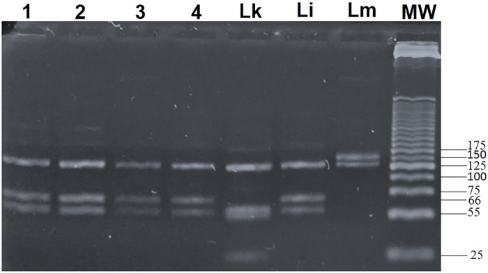
Parasite detection was carried out only in Lar. subgenus females, the suspected vector of L. infantum. A total of 1,029 females were collected and screened for Leishmania infection during the three years of the study. Among this population, 85 specimens were found infected with Leishmania spp. (8.26%). Ph. perniciosus (N = 32; 37.65%) was the most infected species followed by Ph. perfiliewi (N = 31; 36.47%) and Ph. longicuspis (N = 17; 20%). For some infected sand flies belonging to Phlebotomus Lar. subgenus (N = 5; 5.88%), the sand fly species identification was not possible. The one-way ANOVA test was applied to analyze the infection rates among the different sand flies species. Differences were statistically significant (p-value = 0.0452). Within the infected sand flies, 62 were unfed (72.94%), 22 were engorged (25.88%), and 1 was gravid (1.17%). Nested PCR for ITS1 was positive in 54 of the cases (63.52%) and negative in 31 cases (36.47%) (Ph. perniciosus, n = 7, Ph. perfiliewi, n = 13, Ph. longicuspis, n = 10 and Larroussius unidentifiable sand fly species, n = 1). The typing of Leishmania species using RFLP was done for 54 PCR-ITS1 positive samples (Fig 1). Among the infected Ph. perniciosus, Leishmania was identified as L. major (N = 6; 18.75%), L. infantum (N = 12; 37.50%) and L. killicki (N = 7; 21.87%). In Ph. perfiliewi infected sand flies, Leishmania was identified as L. infantum (N = 9; 29.03%), L. major (N = 7; 22.58%) and L. killicki (N = 2; 6.45%). The typing of Leishmania in Ph. longicuspis, revealed L. infantum (N = 4; 23.52%), L. major (N = 2; 11.76%) and L. killicki (N = 1; 5.88%). Furthermore, species identification was made for five unidentified Lar. species (5.88%) and revealed L. major (N = 3; 60%), and L. killicki (N = 1; 20%) (Table 3).
Fig 1. ITS1 nested PCR-RFLP results.
The Leishmania species typing using RFLP was done for the 54 PCR-ITS1 positive samples. The specific molecular band sizes are: 127, 55, 52, and 20 bp for L. killicki; 126, 66, and 55 bp for L. infantum; 145 and 126 bp for L. major. Lanes 1, 2, and 3: L. infantum identified from infected Ph. perfiliewi, Lane 4: L. infantum identified from infected Ph. perniciosus, Lk: L. killicki control, Li: L. infantum control, and Lm: L. major control. MW: Molecular Weight marker (25-bp DNA ladder).
Table 3. Leishmania typing DNA in infected sand flies.
| Leishmania typing | L. major | L. infantum | L. killicki | NI Leishmania. spp. | Total infected sand flies (%) |
P | ||||||||
|---|---|---|---|---|---|---|---|---|---|---|---|---|---|---|
| Abdomen stage | E | G | UN | E | G | UN | E | G | UN | E | G | UN | ||
| Sand fly species | ||||||||||||||
| Ph. perniciosus | 2 | 0 | 4 | 5 | 0 | 7 | 1 | 0 | 6 | 2 | 1 | 4 | 32 (37.64%) | P* = 0.0452 |
| Ph. perfiliewi | 4 | 0 | 3 | 2 | 0 | 7 | 0 | 0 | 2 | 3 | 0 | 10 | 31 (36.47%) | |
| Ph. longicuspis | 1 | 0 | 1 | 0 | 0 | 4 | 0 | 0 | 1 | 0 | 0 | 10 | 17 (20.00%) | |
|
Unidentified Ph. (Lar.) spp. |
2 | 0 | 1 | 0 | 0 | 0 | 0 | 0 | 1 | 0 | 0 | 1 | 5 (5.88%) | |
| Total (%) | 9 | 0 | 9 | 7 | 0 | 18 | 1 | 0 | 10 | 5 | 1 | 25 | 85 | |
| 18 (21.17%) |
25 (29.41%) |
11 (12.94%) |
31 (36.47%) |
|||||||||||
E: engorged, UN: unfed, G: gravid, and NI: non identified Leishmania species. P*: p-value using the one-way ANOVA test.
Leishmania DNA sequencing and phylogenetic analysis
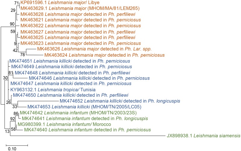
In order to confirm PCR-RFLP results, amplification products from the ITS1 nested PCR of seventeen infected sand flies and three reference strains of L. major (MK463629), L. killicki (MK474653), and L. infantum (MK474642) were sequenced. The obtained twenty partial ITS1 DNA sequences were compared to Leishmania spp. from GenBank. The phylogenetic analysis was performed to confirm the genetic relationship between strains isolated in human and those detected in sandflies. The ITS1 sequences exhibit a length range of 245–271 bp depending on the species. The topology of the phylogenetic tree showed a clear subdivision in three well-supported clades, corresponding to three Leishmania species (L. infantum, L. killicki, and L. major). The identified L. infantum strains in infected Ph. perniciosus and Ph. longicuspis were similar to L. infantum isolated from human VL (L. infantum MON-1) from Tunisia (MK474642) and Moroccan strain from human VL (MG980399). Leishmania major sequences identified from infected sand flies were clustered with L. major strain from patients with CL patients from Tunisia (MK463629) and L. major from Libya (KP691596). Leishmania killicki sequences identified in our study were similar to Tunisian L. killicki strain (MK474653) isolated from human CL and L. tropica from Psammomys vexillaris from Tunisia (KY963132). The phylogenetic analysis based on ITS1 sequences revealed that sequences identified in infected sand flies, sequences from human with CL and VL, and sequence from infected animal were clustered together (Fig 2).
Fig 2. Phylogenetic analysis by Maximum Likelihood method based on Leishmania ITS1 sequences.
The numbers above the branches indicate the bootstrap sampling percentages.
Blood meal analysis and sequencing
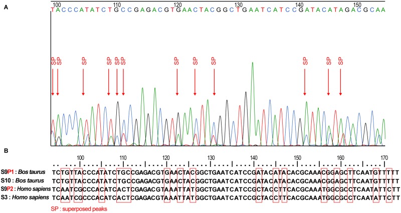
In order to identify the blood meal sources in the sand flies belonging to Lar. subgenus, DNA extracted from 177 engorged females sand flies were studied. DNA amplification targeting vertebrate cyt b was performed and a 359 bp PCR product was detected in 152 samples (85.87%). Furthermore, negative samples were analyzed using degenerated cyt b primers and gave positive results in 17 samples. Thus, a total of 169 positive PCR products (95.48%) were sequenced and compared to the GenBank database. The majority of sand flies were fed on cattle (Bos taurus; N = 66; 39.05%), followed by human (Homo sapiens; N = 42; 24.85%), sheep (Ovis aries; N = 10; 5.91%), chicken (Gallus gallus; N = 8; 4.73%), goat (Capra hircus; N = 7; 4.14%), donkey (Equus asinus africanus; N = 2; 1.18%), and turkey (Meleagris gallopavo; N = 1; 0.59%) (Table 4). In addition, the analysis of cyt b sequence revealed superposed peaks in the electropherogram at different positions suggesting mixed blood sources (Fig 3). Eight vertebrate host combinations were identified (cow/human; N = 3; 1.77%), (chicken/cow; N = 2; 1.18%), (chicken/sheep; N = 2; 1.18%), (sheep/human; N = 2; 1.18%), (goat/human; N = 1; 0.59%), (sheep/goat; N = 1; 0.59%), (human/rodent; N = 1; 0.59%), and (chicken/turkey; N = 1; 0.59%) (Table 4).
Table 4. Blood meal typing in Ph. (Lar.) species.
| Blood meal typing | Cow Bos taurus |
Human Homo sapiens |
Sheep Ovis aries |
Chicken Gallus gallus |
Goat Capra hircus |
Donkey Equus asinus africanus |
Turkey Meleagris gallopavo |
Human/ Cow Homo sapiens/ Bos taurus |
Chicken/ Cow Gallus gallus/ Bos taurus |
Chicken/ Sheep Gallus gallus/ Ovis aries |
Human/ Sheep Ovis aries/ Homo sapiens |
Goat/ Human Capra hircus/ Homo sapiens |
Chicken/ Guineafowl Gallus gallus/ Agelastes meleagrides |
Sheep/ Goat Ovis aries/ Capra hircus |
Human/ Black rat Homo sapiens/ Volemys musseri |
Not assigned sequences | Total |
|---|---|---|---|---|---|---|---|---|---|---|---|---|---|---|---|---|---|
| Sand flies species | |||||||||||||||||
| Ph. perniciosus | 18 | 19 | 4 | 2 | 6 | 0 | 0 | 0 | 1 | 1 | 2 | 1 | 1 | 1 | 0 | 13 | 69 |
| Ph. perfiliewi | 37 | 10 | 4 | 4 | 1 | 0 | 0 | 1 | 1 | 1 | 0 | 0 | 0 | 0 | 1 | 2 | 62 |
| Ph. longicuspis | 5 | 7 | 2 | 1 | 0 | 1 | 1 | 0 | 0 | 0 | 0 | 0 | 0 | 0 | 0 | 3 | 20 |
| Unidentified Lar. spp. | 6 | 6 | 0 | 1 | 0 | 1 | 0 | 2 | 0 | 0 | 0 | 0 | 0 | 0 | 0 | 2 | 18 |
| Total (%) | 66 (39.05%) | 42 (24.85%) | 10 (5.91%) | 8 (4.73%) | 7 (4.14%) | 2 (1.18%) | 1 (0.59%) | 3 (17.75%) | 2 (1.18%) | 2 (1.18%) | 2 (1.18%) | 1 (0.59%) | 1 (0.59%) | 1 (0.59%) | 1 (0.59%) | 20 (11.83%) | 169 |
Fig 3. Blood meal identification results.
A: electropherogram of part of the sequence S 9 showing superposed peaks, B: part of the alignment of four cytochrome b sequences showing superposed peaks for S9, S 10: cytochrome b sequence of Bos taurus, S 3: cytochrome b sequence of Homo sapiens, S 9: cytochrome b sequence of mixed blood meal (Bos taurus/Homo sapiens), two peaks were detected peak 1 (S9P1 corresponding to Bos taurus sequence) and peak 2 (S9P2 corresponding to Homo sapiens sequence).
Interestingly, 21 infected sand flies were engorged. The blood meal analysis in infected sand flies revealed a predominance of cattle (N = 11; 52.38%), followed by goat (N = 4; 19.04%), and chicken (N = 2; 9.52%). Even more, we detected mixed blood meals in several specimens: human/cow (N = 1; 4.76%), chicken/cow (N = 1; 4.76%), chicken/sheep (N = 1; 4.76%), and chicken/turkey (N = 1; 4.76%) (Table 5).
Table 5. The blood meal origins in infected Ph. (Lar.) species.
| Blood meal origin | Cow Bos taurus |
Chicken Gallus gallus |
Goat Capra hircus |
Chicken/ Cow Gallus gallus/ Bos taurus |
Chicken/ Sheep Gallus gallus/ Ovis aries |
Chicken/ Guineafowl Gallus gallus/ Agelastes meleagrides |
Human/ Cow Homo sapiens/ Bos taurus |
Total | |
|---|---|---|---|---|---|---|---|---|---|
| Sand flies and Leishmania species | |||||||||
| Ph. perniciosus | L. major | 1 | 0 | 1 | 0 | 0 | 0 | 0 | 2 |
| L. infantum | 1 | 0 | 3 | 0 | 0 | 0 | 1 | 5 | |
| L. killicki | 0 | 0 | 0 | 1 | 0 | 0 | 0 | 1 | |
| NI | 1 | 0 | 0 | 0 | 0 | 1 | 0 | 2 | |
| Ph. perfiliewi | L. major | 3 | 1 | 0 | 0 | 0 | 0 | 0 | 4 |
| L. infantum | 0 | 1 | 0 | 0 | 1 | 0 | 0 | 2 | |
| NI | 2 | 0 | 0 | 0 | 0 | 0 | 0 | 2 | |
| Ph. longicuspis | L. major | 1 | 0 | 0 | 0 | 0 | 0 | 0 | 1 |
| Unidentified Lar. spp. | L. major | 2 | 0 | 0 | 0 | 0 | 0 | 0 | 2 |
| Total (%) | 11 (52.38%) |
2 (9.52%) |
4 (19.04%) |
1 (4.76%) |
1 (4.76%) |
1 (4.76%) |
1 (4.76%) |
21 | |
NI: non identified Leishmania species.
Parasite load quantification
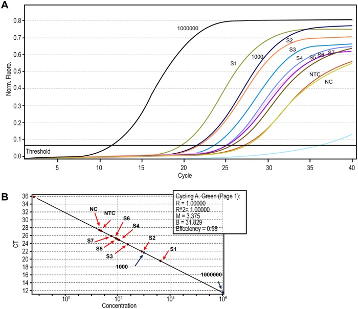
The parasite load was quantified using qPCR in 17 infected sand flies (6 Ph. perniciosus, 10 Ph. perfiliewi and 1 belonging to Lar. subgenus). Seven of them were engorged, and ten were unfed. Ct threshold values were calculated according to default parameters and NTC Ct values in each reaction (Fig 4). Infected flies were classified in four categories according to the parasite loads: very high loads (≥10,000 parasites/reaction), high loads (≥1,000 parasites/ reaction), moderate loads (> 10 parasites/ reaction), and low parasite loads (<10 parasites/ reaction). The highest parasite load was observed in unfed Ph. perfiliewi infected with L. infantum (10,000 parasites) and the lowest was observed in Ph. perfiliewi infected by L. infantum (19.9 promastigotes/ reaction) (Table 6). The mean parasite burden in unfed sand flies was 1,174 promastigotes/ reaction, while in engorged females was 90 promastigotes/ reaction. Statistical analysis through Fisher’s exact test showed significant difference between the parasite loads in fed and unfed sand flies (p<0.0001).
Fig 4. Real time PCR results.
A: Representative fluorescence acquisition graph showed comparison of each sand fly according to standards. NTC (non template control), NC (negative control corresponding to 30 ng of DNA from reared sand fly), and standards (106 and 103 promastigotes/ml). B: Graphic showing concentration results of each sample according to standards. Slope = -3.37, efficiency = 0.98. S1: Ph. perfiliewi infected with L. infantum, S2: Ph. perniciosus infected with L. major, S3: Ph. perniciosus infected with L. killicki, S4: Ph. perfiliewi infected with L. major, S5: Ph. (Lar.) spp. infected with L. major, S6: Ph. perniciosus infected with L. infantum, and S7: Ph. perniciosus infected with L. killicki.
Table 6. Parasite loads and blood meal analysis in infected sand flies collected in human visceral leishmaniasis focus.
| Species | Abdomen states/ blood feed sources | Leishmania spp. | Parasite loads estimation (NO. of parasites/ reaction) | Level of infection |
|---|---|---|---|---|
| Ph. perniciosus | E/Capra hircus | L. major | 100 | Moderate |
| Ph. perfiliewi | E/ Bos taurus | L. major | 100 | Moderate |
| Ph. perfiliewi | E/ Bos taurus | L. major | 199.5 | Moderate |
|
NI Ph. (Lar.) spp. |
E/ Bos taurus | L. major | 100 | Moderate |
| Ph. perniciosus | E/ (Gallus gallus/Bos taurus) | L. killicki | 50.1 | Moderate |
| Ph. perfiliewi | E/ Gallus gallus | L. infantum | 19.9 | Moderate |
| Ph. perfiliewi | E/ (Gallus gallus/Ovis aries) | L. infantum | 50.1 | Moderate |
| Ph. perfiliewi | UN | L. infantum | 10000 | Very high |
| Ph. perniciosus | UN | L. major | 100 | Moderate |
| Ph. perfiliewi | UN | L. infantum | 316.2 | Moderate |
| Ph. perfiliewi | UN | L. infantum | 3981 | High |
| Ph. perfiliewi | UN | L. infantum | 316.2 | Moderate |
| Ph. perfiliewi | UN | L. infantum | 1000 | High |
| Ph. perfiliewi | UN | L. infantum | 50.1 | Moderate |
| Ph. perniciosus | UN | L. major | 794.3 | Moderate |
| Ph. perniciosus | UN | L. infantum | 3981 | High |
| Ph. perniciosus | UN | L. killicki | 199.5 | Moderate |
E: engorged, UN: unfed, and NI: non identified species.
Discussion
This study describes the results of an epidemiological study carried out in the center of Tunisia to identify the life cycle of L. infantum and the potential vectors and reservoirs. The studied region is characterized by a high prevalence of CL and VL and the co-existence of three L. infantum zymodemes (MON-1, MON-24, and MON-80) [3]. Within the seven monitored sites of human VL, nine sand fly species were identified. Phlebotomus papatasi was the predominant (32.70%) followed by Ph. perniciosus (29.91%), Ph. perfiliewi (18.35%), Ph. longicuspis (11.11%), Ph. sergenti (0.54%), and Sergentomyia genus (6.78%). Phlebotomus papatasi, the vector of L. major in Tunisia, is known to be abundant in arid and Saharan regions. Aridity seems to be a limiting factor for its distribution and it was described especially in non-irrigated areas [18, 42–45]. However, the high number of Ph. papatasi described in our study (32.70%) is in opposition to previously cited studies and it seems that Ph. papatasi is also predominant in the irrigated area. In the Mediterranean basin, Ph. perniciosus is described in both humid and arid bioclimatic regions. In Algeria, it was described that the distribution of Ph. perniciosus spreads out to the Saharan region [46–48]. In Tunisia, Ph. perniciosus was known to be more frequent in the semi-arid bioclimatic region and much less abundant in sub-humid and arid areas [49, 50]. Recently, in a study conducted in the center of Tunisia, it was demonstrated that Ph. perniciosus was predominant in arid bioclimatic zones and irrigated areas [42, 51]. Our results corroborate such findings with a predominance of 29.91% for Ph. perniciosus. Furthermore, Ph. perfiliewi was known to be limited in the north part of Tunisia and aridity appeared to be a limiting factor for its distribution [49]. Recently, it was demonstrated that Ph. perfiliewi is the most abundant species in the irrigated area, and its geographical distribution is extending towards the center and the south of Tunisia [18]. Our findings align with the extension of Ph. perfiliewi (18.53%) towards the center and irrigated areas. Regarding to Phlebotomus longicuspis, it was described as no limited to any climatic zone in Tunisia, and appears to have the same distribution in Morocco since it has been found in all biogeographical areas including Saharan regions [51, 52]. In our study, this species was found in semi-arid and irrigated zones (11.11%). Thus, the increase in the trapping of sand flies belonging to subgenus Lar. (59.95%) is consistent with the hypothesis of the extension of VL in arid areas [18]. Our results highlight the extension of Lar. species to the center and their strong involvement in L. infantum transmission. Phlebotomus sergenti, the vector of L. killicki in Tunisia, was known as the dominant species in the south-east of the country [49]. The low number of Ph. sergenti observed in our study shows (0.54%) the expansion of this species towards the center. Thus, we demonstrate that the geographical dissemination and sand flies species abundance would be related to several factors associated to human activities and environmental changes (global warming and irrigation) as it was described before [53, 54].
In addition to sand flies species analysis, we screened for Leishmania infection in Lar. species. The overall rate of infection with Leishmania was 8.26%. In agreement with previous reports, our findings show Ph. perniciosus, Ph. longicuspis, and Ph. perfiliewi infected with L. infantum. Similar results were reported from the central part of Tunisia where Ph. perfiliewi and Ph. perniciosus were found infected with L. infantum [18]. In Algeria, L. infantum MON-24 was isolated from Ph. perfiliewi, proving its role in L. infantum transmission [55]. Such findings highlight the role of Ph. perfiliewi in L. infantum transmission in Tunisia and Mediterranean countries. Also, L. infantum infection was reported in Ph. longicuspis and Ph. perniciosus on the eastern coast of Tunisia [19]. Similar results were described in Ph. longicuspis from Morocco and Algeria [52, 56]. In the light of these findings, Ph. longicuspis is highly involved in L. infantum transmission.
Even more, we report for the first time the infection of Ph. (Lar.) species with L. major and L. killicki. Experimental studies described that Ph. perniciosus is a permissive vector for L. infantum (MON-1, MON-24, and MON-80), L. major, and L. tropica [57, 58]. Furthermore, we describe the incrimination of Ph. longicuspis and Ph. perfiliewi in L. major and L. tropica transmission.
In addition to molecular detection of Leishmania, we carried out a qPCR to quantify the parasite load in infected sand flies. In total, 17 sand flies were analyzed (10 unfed and 7 engorged). The average load was 1,174 parasites/reaction in unfed sand flies and 90 parasites/reaction in engorged sand flies. The highest parasite load was observed in unfed sand flies. Concerning to the unfed females, the highest parasite load detected was 10,000 parasites/reaction in a Ph. perfiliewi infected by L. infantum. Similar results were described before with wild caught Ph. perniciosus, Lu. longipalpis and Lu. migonei infected by L. infantum in Spain and Brazil, respectively [20, 27]. To the best of our knowledge, none of the previous studies has quantified parasite load in Ph. perfiliewi. Hence, we report the first quantification of L. infantum in Ph. perfiliewi. Investigations conducted by Roger et al. (2007) indicated that Leishmania parasites could manipulate sand fly feeding behavior. The high parasite loads in sand fly midguts are correlated with a persistent feeding pattern and lead to an increase in Leishmania transmission [21]. According to the criteria of Killick-Kendrick (1990), the incrimination of a sand fly species as a vector of leishmaniasis is based on a significant anthropophilic behavior, vectorial capacity, simultaneous presence of vector and disease, and the abundance of the vector [59]. In our study, criteria are already available for anthropophilic behavior, detection of L. infantum DNA in Ph. perfiliewi (29%), and the high parasite loads quantified in unfed sand flies demonstrate that Ph. perfiliewi is a potential vector for L. infantum in Tunisia. Furthermore, we found a moderate load of infection in Ph. perniciosus infected with L. major and L. killicki, which highlight its role as a permissive vector.
In the current study, the molecular identification of blood sources in engorged sand flies belonging to subgenus Lar. detected seven sources: cattle, human, sheep, chicken, goat, donkey, and turkey. Compared to similar studies, we report the most extensive range of hosts in mixed L. infantum focus [19, 30]. Interestingly, we identified mixed blood meals in twelve cases. Our results show that Lar. species have no host preferences and are opportunistic feeders while disturbed or challenged with less accessible capillary veins as it was demonstrated before for mosquitoes and phlebotomine sand flies behaviors [60, 61]. Although, dogs have been clearly defined as a proven reservoir of L. infantum in Tunisia and Mediterranean countries [13, 15, 40, 62, 63], in our study no dog blood was detected in engorged Ph. (Lar) subgenus. The presence of a broad host availability could probably explain this results in the vicinity of the traps confirming the opportunistic behavior of species of this subgenus. This finding was also reported in previous studies in Tunisian as well as Spanish leishmaniasis foci [19, 30, 40, 54, 64].
Leishmania detection in engorged sand flies showed females fed predominantly on cattle, followed by goat and chicken. Moreover, the analysis of superposed peaks of blood meals in infected sand flies revealed multiple blood origins: human/cow, chicken/cow, chicken/sheep and chicken/turkey. To our knowledge, only dogs were described as the reservoir of L. infantum MON-1 in Tunisia and a potential reservoir of L. infantum MON-24 and MON-80. In Mediterranean countries, the dog is the main reservoir of VL. However, many other hosts were suspected such as rabbit, cat, hare, jackal, fox, wild rodents, and horse [65–76]. This demonstrates the involvement of different mammals in L. infantum transmission. Moreover, our results of parasite typing and quantification in engorged sand flies showed an average load of 90 parasites/reaction. Our findings align with previous studies, which found that the highest parasite loads were observed in unfed sand flies [20, 77]. In addition, a moderate load was described for Ph. perfiliewi and Ph. perniciosus infected with L. major and fed on cow and goat, respectively. These findings could be explained by two hypotheses. The first suggests that cow and goat could be potential reservoirs, and the second hypothesis suggests that Ph. perfiliewi and Ph. perniciosus could be potential vectors of L. major. To the best of our knowledge, only rodents have been described as reservoirs of L. major in Tunisia [14]. Hence, the hypothesis that cow and goat could be a reservoir of L. major should be discarded. To conclude, our findings support the hypothesis that Ph. perfiliewi and Ph. perniciosus could be permissive vectors and the blood was only necessary for eggs maturation.
Unexpectedly, during the blood meal analysis of engorged infected sand flies, we identified mixed blood meals in two Ph. perniciosus and Ph. perfiliewi infected with L. killicki and L. infantum, respectively. The quantification of the parasite load revealed 50 parasites/reaction in both of them. It was described before using experimental infections that both L. infantum and Leishmania mexicana promote feeding on multiple hosts. Furthermore, it was demonstrated that Leishmania parasites could manipulate sand fly feeding behavior according to the presence of infective forms available for transmission [21]. Furthermore, it was recently proved that ingestion of a second blood by Leishmania infected sand flies triggers parasite dedifferentiation and amplification that greatly enhance disease transmission [78]. In the light of these findings, the moderate number of parasites observed in mixed blood meals could be explained by the necessity of a second blood meal for dedifferentiation and amplification of parasite as proven previously. In another hand, we observed a low parasite charge (19 parasites/reaction) in Ph. perfiliewi infected by L. infantum and fed on chicken. Indeed, it was previously described that chickens or birds are not susceptible to Leishmania infection due to some physiological characteristics such as their body temperature of 41°C and infected sand fly may eliminate Leishmania parasite during their second blood meal [79].
In light of this finding, the moderate and the low amount of parasite observed in our study demonstrate that chicken blood affects the presence of parasites in the sand fly gut and 50 parasites/reaction could be the quantity of parasite uptaken from the first host. So, the supposition that chicken could be a reservoir for L. infantum should be discarded. Further studies should be carried out to confirm the involvement of the described vertebrate in L. infantum life cycle such as experimental infection.
Conclusion
In the present study, we demonstrated the high involvement of Ph. longicuspis, Ph. perfiliewi, and Ph. perniciosus in L. infantum transmission. Moreover, we confirmed that Ph. perniciosus is a permissive vector in nature which strengthens its role in different Leishmania life cycles. Thereby, an efficient control strategy against these species and its distribution toward cutaneous and visceral leishmaniasis foci should be carried out in order to reduce Leishmania transmission in Tunisia. Moreover, Ph. perfiliewi could be a permissive vector for L. infantum and other Leishmania species. Thus, it would be preferable to combine sand fly dissection and Leishmania strain isolation from Ph. perfiliewi to determine the L. infantum zymodeme. The combination of the parasite detection and the blood meal analysis in infected flies revealed the possible incrimination of different mammals in L. infantum transmission. Such results await further exploration to a better understanding of L. infantum transmission.
Supporting information
(PDF)
Acknowledgments
We are very grateful to the team of the Center of Hygiene and Basic Health Care Kairouan and the Regional Commissariat for Agricultural Development of Kairouan, Tunisia, who helped us to accomplish this work. The authors are also very grateful for Oussama Babba and Salma Remadi for the graphical design.
Data Availability
All relevant data are within the manuscript and its Supporting Information files.
Funding Statement
This study was supported by a grant from the EMRO/TDR Small Grants Scheme for Operational Research in Tropical and Other Communicable Diseases (No. SGS14/23); by the Ministry of Higher Education and Scientific Research of Tunisia and was partially supported with the facilities of the ISCIII. The funders had no role in the study design, data collection and analysis, decision to publish, publication fees, or preparation of the manuscript.
References
- 1.Alvar J, Velez ID, Bern C, Herrero M, Desjeux P, Cano J, et al. Leishmaniasis worldwide and global estimates of its incidence. PloS one. 2012;7(5):e35671 Epub 2012/06/14. 10.1371/journal.pone.0035671 [DOI] [PMC free article] [PubMed] [Google Scholar]
- 2.Chaara D, Ravel C, Banuls A, Haouas N, Lami P, Talignani L, et al. Evolutionary history of Leishmania killicki (synonymous Leishmania tropica) and taxonomic implications. Parasites & vectors. 2015;8:198 10.1186/s13071-015-0821-6 [DOI] [PMC free article] [PubMed] [Google Scholar]
- 3.Haouas N, Chaker E, Chargui N, Gorcii M, Belhadj S, Kallel K, et al. Geographical distribution updating of Tunisian leishmaniasis foci: about the isoenzymatic analysis of 694 strains. Acta tropica. 2012;124(3):221–8. 10.1016/j.actatropica.2012.08.012 . [DOI] [PubMed] [Google Scholar]
- 4.Ben Ismail R, Gramiccia M, Gradoni L, Helal H, Ben Rachid MS. Isolation of Leishmania major from Phlebotomus papatasi in Tunisia. Transactions of the Royal Society of Tropical Medicine and Hygiene. 1987;81(5):749 Epub 1987/01/01. 10.1016/0035-9203(87)90018-6 . [DOI] [PubMed] [Google Scholar]
- 5.Ghawar W, Snoussi MA, Hamida NB, Boukthir A, Yazidi R, Chaabane S, et al. First report of natural infection of least weasel (Mustela nivalis Linnaeus, 1776) with Leishmania major in Tunisia. Vector borne and zoonotic diseases (Larchmont, NY). 2011;11(11):1507–9. Epub 2011/08/27. 10.1089/vbz.2011.0673 . [DOI] [PubMed] [Google Scholar]
- 6.Chemkhi J, Souguir H, Ali IB, Driss M, Guizani I, Guerbouj S. Natural infection of Algerian hedgehog, Atelerix algirus (Lereboullet 1842) with Leishmania parasites in Tunisia. Acta tropica. 2015;150:42–51. Epub 2015/06/19. 10.1016/j.actatropica.2015.06.009 . [DOI] [PubMed] [Google Scholar]
- 7.Souguir-Omrani H, Chemkhi J, Fathallah-Mili A, Saadi-BenAoun Y, BelHadjAli I, Guizani I, et al. Paraechinus aethiopicus (Ehrenberg 1832) and Atelerix algirus (Lereboullet 1842) hedgehogs: Possible reservoirs of endemic leishmaniases in Tunisia. Infection, genetics and evolution: journal of molecular epidemiology and evolutionary genetics in infectious diseases. 2018;63:219–30. Epub 2018/06/04. 10.1016/j.meegid.2018.05.029 . [DOI] [PubMed] [Google Scholar]
- 8.Jaouadi K, Haouas N, Chaara D, Gorcii M, Chargui N, Augot D, et al. First detection of Leishmania killicki (Kinetoplastida, Trypanosomatidae) in Ctenodactylus gundi (Rodentia, Ctenodactylidae), a possible reservoir of human cutaneous leishmaniasis in Tunisia. Parasites & vectors. 2011;4:159 10.1186/1756-3305-4-159 [DOI] [PMC free article] [PubMed] [Google Scholar]
- 9.Jaouadi K, Depaquit J, Haouas N, Chaara D, Gorcii M, Chargui N, et al. Twenty-four new human cases of cutaneous leishmaniasis due to Leishmania killicki in Metlaoui, southwestern Tunisia. Probable role of Phlebotomus sergenti in the transmission. Acta tropica. 2012;122(3):276–83. 10.1016/j.actatropica.2012.01.014 [DOI] [PubMed] [Google Scholar]
- 10.Bousslimi N, Ben-Ayed S, Ben-Abda I, Aoun K, Bouratbine A. Natural infection of North African gundi (Ctenodactylus gundi) by Leishmania tropica in the focus of cutaneous leishmaniasis, Southeast Tunisia. The American journal of tropical medicine and hygiene. 2012;86(6):962–5. Epub 2012/06/06. 10.4269/ajtmh.2012.11-0572 [DOI] [PMC free article] [PubMed] [Google Scholar]
- 11.Tabbabi A, Bousslimi N, Rhim A, Aoun K, Bouratbine A. First report on natural infection of Phlebotomus sergenti with Leishmania promastigotes in the cutaneous leishmaniasis focus in southeastern Tunisia. The American journal of tropical medicine and hygiene. 2011;85(4):646–7. Epub 2011/10/07. 10.4269/ajtmh.2011.10-0681 [DOI] [PMC free article] [PubMed] [Google Scholar]
- 12.Aoun K, Amri F, Chouihi E, Haouas N, Bedoui K, Benikhlef R, et al. [Epidemiology of Leishmania (L.) infantum, L. major and L. killicki in Tunisia: results and analysis of the identification of 226 human and canine isolates]. Bulletin de la Societe de pathologie exotique. 2008;101(4):323–8. 10.3185/pathexo3201 . [DOI] [PubMed] [Google Scholar]
- 13.Ben Ismail R. Incrimination de Phlebotomus perniciosus comme vecteur de Leishmania infantum. Arch Institut Pasteur Tunis. 1993;70:91–110. [Google Scholar]
- 14.Haouas N, Babba H. Leishmaniasis in Tunisia: History and New Insights into the Epidemiology of a Neglected Disease. 2017. 10.5772/65000 [DOI] [Google Scholar]
- 15.Benikhlef R, Harrat Z, Toudjine M, Djerbouh A, Bendali-Braham S, Belkaid M. [Detection of Leishmania infantum MON-24 in the dog]. Medecine tropicale: revue du Corps de sante colonial. 2004;64(4):381–3. Epub 2004/12/24. . [PubMed] [Google Scholar]
- 16.Benikhlef R, Aoun K, Bedoui K, Harrat Z, Bouratbine A. Premières identifications de Leishmania infantum MON-80 chez le chien en Algérie et en Tunisie. Revue de Médecine Vétérinaire. 2009;160(10):464–6. [Google Scholar]
- 17.Guerbouj S, Chemkhi J, Kaabi B, Rahali A, Ben Ismail R, Guizani I. Natural infection of Phlebotomus (Larroussius) langeroni (Diptera: Psychodidae) with Leishmania infantum in Tunisia. Transactions of the Royal Society of Tropical Medicine and Hygiene. 2007;101(4):372–7. Epub 2006/10/20. 10.1016/j.trstmh.2006.07.007 . [DOI] [PubMed] [Google Scholar]
- 18.Barhoumi W, Fares W, Cherni S, Derbali M, Dachraoui K, Chelbi I, et al. Changes of sand fly populations and Leishmania infantum infection rates in an irrigated village located in arid Central Tunisia. International journal of environmental research and public health. 2016;13(3):329. [DOI] [PMC free article] [PubMed] [Google Scholar]
- 19.Chargui N, Slama D, Haouas N, Rmadi L, Babba H. Transmission cycle analysis in a Leishmania infantum focus: Infection rates and blood meal origins in sand flies (Diptera: Psychodidae). Journal of vector ecology: journal of the Society for Vector Ecology. 2018;43(2):321–7. Epub 2018/11/09. 10.1111/jvec.12316 . [DOI] [PubMed] [Google Scholar]
- 20.Gonzalez E, Alvarez A, Ruiz S, Molina R, Jimenez M. Detection of high Leishmania infantum loads in Phlebotomus perniciosus captured in the leishmaniasis focus of southwestern Madrid region (Spain) by real time PCR. Acta tropica. 2017;171:68–73. Epub 2017/03/31. 10.1016/j.actatropica.2017.03.023 . [DOI] [PubMed] [Google Scholar]
- 21.Rogers ME, Bates PA. Leishmania manipulation of sand fly feeding behavior results in enhanced transmission. PLoS pathogens. 2007;3(6):e91 Epub 2007/07/03. 10.1371/journal.ppat.0030091 . [DOI] [PMC free article] [PubMed] [Google Scholar]
- 22.Dantas-Torres F, Tarallo VD, Latrofa MS, Falchi A, Lia RP, Otranto D. Ecology of phlebotomine sand flies and Leishmania infantum infection in a rural area of southern Italy. Acta tropica. 2014;137:67–73. Epub 2014/05/13. 10.1016/j.actatropica.2014.04.034 . [DOI] [PubMed] [Google Scholar]
- 23.Myskova J, Votypka J, Volf P. Leishmania in sand flies: comparison of quantitative polymerase chain reaction with other techniques to determine the intensity of infection. Journal of medical entomology. 2008;45(1):133–8. Epub 2008/02/21. 10.1603/0022-2585(2008)45[133:lisfco]2.0.co;2 . [DOI] [PubMed] [Google Scholar]
- 24.Bezerra-Vasconcelos DR, Melo LM, Albuquerque ES, Luciano MC, Bevilaqua CM. Real-time PCR to assess the Leishmania load in Lutzomyia longipalpis sand flies: screening of target genes and assessment of quantitative methods. Experimental parasitology. 2011;129(3):234–9. Epub 2011/08/26. 10.1016/j.exppara.2011.08.010 . [DOI] [PubMed] [Google Scholar]
- 25.Aghaei AA, Rassi Y, Sharifi I, Vatandoost H, Mollaie H, Oshaghi MA, et al. First report on natural Leishmania infection of Phlebotomus sergenti due Leishmania tropica by high resolution melting curve method in South-eastern Iran. Asian Pacific journal of tropical medicine. 2014;7(2):93–6. Epub 2014/01/28. 10.1016/S1995-7645(14)60002-X . [DOI] [PubMed] [Google Scholar]
- 26.Coleman RE, Hochberg LP, Swanson KI, Lee JS, McAvin JC, Moulton JK, et al. Impact of phlebotomine sand flies on U.S. military operations at Tallil Air Base, Iraq: 4. Detection and identification of leishmania parasites in sand flies. Journal of medical entomology. 2009;46(3):649–63. Epub 2009/06/06. 10.1603/033.046.0333 . [DOI] [PubMed] [Google Scholar]
- 27.Rodrigues AC, Melo LM, Magalhaes RD, de Moraes NB, de Souza Junior AD, Bevilaqua CM. Molecular identification of Lutzomyia migonei (Diptera: Psychodidae) as a potential vector for Leishmania infantum (Kinetoplastida: Trypanosomatidae). Veterinary parasitology. 2016;220:28–32. Epub 2016/03/21. 10.1016/j.vetpar.2016.02.018 . [DOI] [PubMed] [Google Scholar]
- 28.Cunha RC, Andreotti R, Cominetti MC, Silva EA. Detection of Leishmania infantum in Lutzomyia longipalpis captured in Campo Grande, MS. Revista brasileira de parasitologia veterinaria = Brazilian journal of veterinary parasitology: Orgao Oficial do Colegio Brasileiro de Parasitologia Veterinaria. 2014;23(2):269–73. Epub 2014/07/24. 10.1590/s1984-29612014049 . [DOI] [PubMed] [Google Scholar]
- 29.Benabid M, Ghrab J, Rhim A, Ben-Romdhane R, Aoun K, Bouratbine A. Temporal dynamics and Leishmania infantum infection prevalence of Phlebotomus perniciosus (Diptera, Phlebotominae) in highly endemic areas of visceral leishmaniasis in Tunisia. PloS one. 2017;12(9):e0184700 Epub 2017/09/22. 10.1371/journal.pone.0184700 [DOI] [PMC free article] [PubMed] [Google Scholar]
- 30.Jaouadi K, Bettaieb J, Bennour A, Salem S, Ghawar W, Rjeibi MR, et al. Blood Meal Analysis of Phlebotomine Sandflies (Diptera: Psychodidae: Phlebotominae) for Leishmania spp. Identification and Vertebrate Blood Origin, Central Tunisia, 2015–2016. The American journal of tropical medicine and hygiene. 2018;98(1):146–9. Epub 2017/11/23. 10.4269/ajtmh.17-0313 [DOI] [PMC free article] [PubMed] [Google Scholar]
- 31.Atlas du gouvernorat de Kairouan [Internet]. 2011. http://www.mehat.gov.tn/fileadmin/user_upload/Amenagement_Territoire/AtlasKairouanFrv2.pdf.
- 32.Ben Salah A, Ben Alaya Bouafif N, Chlif S, Gharbi A, Bel Haj Hamida N, Zaatour A, et al. [Risk factors of leishmanin-skin test positivity in transmission of Leishmania infantum in the center of Tunisia]. Archives de l’Institut Pasteur de Tunis. 2003;80(1–4):17–27. Epub 2005/06/09. . [PubMed] [Google Scholar]
- 33.Chalghaf B, Chlif S, Mayala B, Ghawar W, Bettaieb J, Harrabi M, et al. Ecological Niche Modeling for the Prediction of the Geographic Distribution of Cutaneous Leishmaniasis in Tunisia. The American journal of tropical medicine and hygiene. 2016;94(4):844–51. Epub 2016/02/10. 10.4269/ajtmh.15-0345 [DOI] [PMC free article] [PubMed] [Google Scholar]
- 34.Jimenez M, Gonzalez E, Iriso A, Marco E, Alegret A, Fuster F, et al. Detection of Leishmania infantum and identification of blood meals in Phlebotomus perniciosus from a focus of human leishmaniasis in Madrid, Spain. Parasitology research. 2013;112(7):2453–9. Epub 2013/03/29. 10.1007/s00436-013-3406-3 . [DOI] [PubMed] [Google Scholar]
- 35.Croset H, Rioux JA, Maistre M, Bayar N. [The phlebotomines of Tunisia (Diptera-Phlebotominae). A revision of the systematics, distribution and behaviour (author’s transl)]. Annales de parasitologie humaine et comparee. 1978;53(6):711–49. Epub 1978/11/01. . [PubMed] [Google Scholar]
- 36.Leger N, Pesson B, Madulo-Leblond G, Abonnenc E. [Differentiation of females of the subgenus Larroussius Nitzulescu 1931 (Diptera-Phlebotomidae) of the Mediterranean region]. Annales de parasitologie humaine et comparee. 1983;58(6):611–23. Epub 1983/01/01. . [PubMed] [Google Scholar]
- 37.Pesson B, Ready JS, Benabdennbi I, Martin-Sanchez J, Esseghir S, Cadi-Soussi M, et al. Sandflies of the Phlebotomus perniciosus complex: mitochondrial introgression and a new sibling species of P. longicuspis in the Moroccan Rif. Med Vet Entomol. 2004;18(1):25–37. Epub 2004/03/11. 10.1111/j.0269-283x.2004.0471.x . [DOI] [PubMed] [Google Scholar]
- 38.Spanakos G, Patsoula E, Kremastinou T, Saroglou G, Vakalis N. Development of a PCR-based method for diagnosis of Leishmania in blood samples. Molecular and cellular probes. 2002;16(6):415–20. Epub 2002/12/20. 10.1006/mcpr.2002.0436 . [DOI] [PubMed] [Google Scholar]
- 39.Cruz I, Millet A, Carrillo E, Chenik M, Salotra P, Verma S, et al. An approach for interlaboratory comparison of conventional and real-time PCR assays for diagnosis of human leishmaniasis. Experimental parasitology. 2013;134(3):281–9. Epub 2013/04/09. 10.1016/j.exppara.2013.03.026 . [DOI] [PubMed] [Google Scholar]
- 40.Gonzalez E, Gallego M, Molina R, Abras A, Alcover MM, Ballart C, et al. Identification of blood meals in field captured sand flies by a PCR-RFLP approach based on cytochrome b gene. Acta tropica. 2015;152:96–102. Epub 2015/09/08. 10.1016/j.actatropica.2015.08.020 . [DOI] [PubMed] [Google Scholar]
- 41.Nicolas L, Prina E, Lang T, Milon G. Real-time PCR for detection and quantitation of leishmania in mouse tissues. Journal of clinical microbiology. 2002;40(5):1666–9. Epub 2002/05/01. 10.1128/JCM.40.5.1666-1669.2002 [DOI] [PMC free article] [PubMed] [Google Scholar]
- 42.Barhoumi W, Qualls WA, Archer RS, Fuller DO, Chelbi I, Cherni S, et al. Irrigation in the arid regions of Tunisia impacts the abundance and apparent density of sand fly vectors of Leishmania infantum. Acta tropica. 2015;141(Pt A):73–8. Epub 2014/12/03. 10.1016/j.actatropica.2014.10.008 . [DOI] [PMC free article] [PubMed] [Google Scholar]
- 43.Zhioua E, Kaabi B, Chelbi I. Entomological investigations following the spread of visceral leishmaniasis in Tunisia. Journal of vector ecology: journal of the Society for Vector Ecology. 2007;32(2):371–4. Epub 2008/02/12. 10.3376/1081-1710(2007)32[371:eiftso]2.0.co;2 . [DOI] [PubMed] [Google Scholar]
- 44.Chelbi I, Kaabi B, Bejaoui M, Derbali M, Zhioua E. Spatial correlation between Phlebotomus papatasi Scopoli (Diptera: Psychodidae) and incidence of zoonotic cutaneous leishmaniasis in Tunisia. Journal of medical entomology. 2009;46(2):400–2. Epub 2009/04/09. 10.1603/033.046.0229 . [DOI] [PubMed] [Google Scholar]
- 45.Cross ER, Hyams KC. The potential effect of global warming on the geographic and seasonal distribution of Phlebotomus papatasi in southwest Asia. Environmental health perspectives. 1996;104(7):724–7. Epub 1996/07/01. 10.1289/ehp.96104724 [DOI] [PMC free article] [PubMed] [Google Scholar]
- 46.Benallal KE, Benikhlef R, Garni R, Gassen B, Dedet JP, Harrat Z. Presence of Phlebotomus perniciosus Atypical Form in Algeria. Journal of arthropod-borne diseases. 2017;11(1):139–46. Epub 2017/10/14. [PMC free article] [PubMed] [Google Scholar]
- 47.Rioux JA, Golvan YJ, Croset H, Houin R, Juminer B, Bain O, et al. [Ecology of leishmaniases in Southern France. 1. Phlebotomus. Sampling—ethology]. Annales de parasitologie humaine et comparee. 1967;42(6):561–603. Epub 1967/11/01. . [PubMed] [Google Scholar]
- 48.Berdjane-Brouk Z, Charrel RN, Hamrioui B, Izri A. First detection of Leishmania infantum DNA in Phlebotomus longicuspis Nitzulescu, 1930 from visceral leishmaniasis endemic focus in Algeria. Parasitology research. 2012;111(1):419–22. Epub 2012/02/22. 10.1007/s00436-012-2858-1 . [DOI] [PubMed] [Google Scholar]
- 49.Ghrab J, Rhim A, Bach-Hamba D, Chahed M, Aoun K, Nouira S, et al. Phlebotominae (Diptera: Psychodidae) of human leishmaniosis sites in Tunisia. Parasite. 2006;13(1):23–33. 10.1051/parasite/2006131023 [DOI] [PubMed] [Google Scholar]
- 50.Ben-Ahmed K, Aoun K, Jeddi F, Ghrab J, El-Aroui MA, Bouratbine A. Visceral leishmaniasis in Tunisia: spatial distribution and association with climatic factors. The American journal of tropical medicine and hygiene. 2009;81(1):40–5. Epub 2009/06/27. . [PubMed] [Google Scholar]
- 51.Barhoumi W, Fares W, Cherni S, Derbali M, Dachraoui K, Chelbi I, et al. Changes of Sand Fly Populations and Leishmania infantum Infection Rates in an Irrigated Village Located in Arid Central Tunisia. Int J Environ Res Public Health. 2016;13(3). Epub 2016/03/22. 10.3390/ijerph13030329 [DOI] [PMC free article] [PubMed] [Google Scholar]
- 52.Mhaidi I, El Kacem S, Ait Kbaich M, El Hamouchi A, Sarih M, Akarid K, et al. Molecular identification of Leishmania infection in the most relevant sand fly species and in patient skin samples from a cutaneous leishmaniasis focus, in Morocco. 2018;12(3):e0006315 10.1371/journal.pntd.0006315 . [DOI] [PMC free article] [PubMed] [Google Scholar]
- 53.Bennai K, Tahir D, Lafri I, Bendjaballah-Laliam A, Bitam I, Parola P. Molecular detection of Leishmania infantum DNA and host blood meal identification in Phlebotomus in a hypoendemic focus of human leishmaniasis in northern Algeria. 2018;12(6):e0006513 10.1371/journal.pntd.0006513 . [DOI] [PMC free article] [PubMed] [Google Scholar]
- 54.Gonzalez E, Jimenez M, Hernandez S, Martin-Martin I, Molina R. Phlebotomine sand fly survey in the focus of leishmaniasis in Madrid, Spain (2012–2014): seasonal dynamics, Leishmania infantum infection rates and blood meal preferences. Parasites & vectors. 2017;10(1):368 Epub 2017/08/03. 10.1186/s13071-017-2309-z [DOI] [PMC free article] [PubMed] [Google Scholar]
- 55.Izri MA, Belazzoug S. Phlebotomus (Larroussius) perfiliewi naturally infected with dermotropic Leishmania infantum at Tenes, Algeria. Transactions of the Royal Society of Tropical Medicine and Hygiene. 1993;87(4):399 Epub 1993/07/01. 10.1016/0035-9203(93)90011-e . [DOI] [PubMed] [Google Scholar]
- 56.Harrat Z, Belkaid M. [Leishmaniasis in Algiers: epidemiologic data]. Bulletin de la Societe de pathologie exotique. 2003;96(3):212–4. Epub 2003/10/30. . [PubMed] [Google Scholar]
- 57.Svarovska A, Ant TH, Seblova V, Jecna L, Beverley SM, Volf P. Leishmania major glycosylation mutants require phosphoglycans (lpg2-) but not lipophosphoglycan (lpg1-) for survival in permissive sand fly vectors. PLoS neglected tropical diseases. 2010;4(1):e580 Epub 2010/01/20. 10.1371/journal.pntd.0000580 [DOI] [PMC free article] [PubMed] [Google Scholar]
- 58.Remadi L, Jimenez M, Chargui N, Haouas N, Babba H, Molina R. The vector competence of Phlebotomus perniciosus for Leishmania infantum zymodemes of Tunisia. 2018;117(8):2499–506. 10.1007/s00436-018-5939-y . [DOI] [PubMed] [Google Scholar]
- 59.Killick-Kendrick R. Phlebotomine vectors of the leishmaniases: a review. Med Vet Entomol. 1990;4(1):1–24. Epub 1990/01/01. 10.1111/j.1365-2915.1990.tb00255.x . [DOI] [PubMed] [Google Scholar]
- 60.Rodrigues SC, Maruniak JE. Blood meal identification from mosquitoes collected at a commercial alligator farm. Journal of the American Mosquito Control Association. 2006;22(3):557–60. Epub 2006/10/28. 10.2987/8756-971X(2006)22[557:BMIFMC]2.0.CO;2 . [DOI] [PubMed] [Google Scholar]
- 61.Bongiorno G, Habluetzel A, Khoury C, Maroli M. Host preferences of phlebotomine sand flies at a hypoendemic focus of canine leishmaniasis in central Italy. Acta tropica. 2003;88(2):109–16. Epub 2003/10/01. 10.1016/s0001-706x(03)00190-6 . [DOI] [PubMed] [Google Scholar]
- 62.Guessous-Idrissi N, Hamdani A, Rhalem A, Riyad M, Sahibi H, Dehbi F, et al. Epidemiology of human visceral leishmaniasis in Taounate, a northern province of Morocco. Parasite. 1997;4(2):181–5. Epub 1997/06/01. 10.1051/parasite/1997042181 . [DOI] [PubMed] [Google Scholar]
- 63.Chargui N, Haouas N, Slama D, Gorcii M, Jaouadi K, Essabbah-Aguir N, et al. Transmission of visceral leishmaniasis in a previously non-endemic region of Tunisia: detection of Leishmania DNA in Phlebotomus perniciosus. Journal of vector ecology: journal of the Society for Vector Ecology. 2013;38(1):1–5. 10.1111/j.1948-7134.2013.12000.x . [DOI] [PubMed] [Google Scholar]
- 64.Haouas N, Pesson B, Boudabous R, Dedet JP, Babba H, Ravel C. Development of a molecular tool for the identification of Leishmania reservoir hosts by blood meal analysis in the insect vectors. The American journal of tropical medicine and hygiene. 2007;77(6):1054–9. . [PubMed] [Google Scholar]
- 65.Maia C, Campino L. Can domestic cats be considered reservoir hosts of zoonotic leishmaniasis? Trends Parasitol. 2011;27(8):341–4. Epub 2011/05/17. 10.1016/j.pt.2011.03.008 . [DOI] [PubMed] [Google Scholar]
- 66.Chitimia L, Munoz-Garcia CI, Sanchez-Velasco D, Lizana V, Del Rio L, Murcia L, et al. Cryptic Leishmaniosis by Leishmania infantum, a feature of canines only? A study of natural infection in wild rabbits, humans and dogs in southeastern Spain. Veterinary parasitology. 2011;181(1):12–6. Epub 2011/05/20. 10.1016/j.vetpar.2011.04.016 . [DOI] [PubMed] [Google Scholar]
- 67.Carrillo E, Moreno J, Cruz I. What is responsible for a large and unusual outbreak of leishmaniasis in Madrid? Trends Parasitol. 2013;29(12):579–80. Epub 2013/11/28. 10.1016/j.pt.2013.10.007 . [DOI] [PubMed] [Google Scholar]
- 68.Molina R, Jimenez MI, Cruz I, Iriso A, Martin-Martin I, Sevillano O, et al. The hare (Lepus granatensis) as potential sylvatic reservoir of Leishmania infantum in Spain. Veterinary parasitology. 2012;190(1–2):268–71. Epub 2012/06/09. 10.1016/j.vetpar.2012.05.006 . [DOI] [PubMed] [Google Scholar]
- 69.Chaara D, Haouas N, Dedet JP, Babba H, Pratlong F. Leishmaniases in Maghreb: an endemic neglected disease. Acta tropica. 2014;132:80–93. 10.1016/j.actatropica.2013.12.018 . [DOI] [PubMed] [Google Scholar]
- 70.Chatzis MK, Andreadou M, Leontides L, Kasabalis D, Mylonakis M, Koutinas AF, et al. Cytological and molecular detection of Leishmania infantum in different tissues of clinically normal and sick cats. Veterinary parasitology. 2014;202(3–4):217–25. Epub 2014/03/19. 10.1016/j.vetpar.2014.02.044 . [DOI] [PubMed] [Google Scholar]
- 71.Aharonson-Raz K, Baneth G, Lopes AP, Brancal H, Schallig H, Cardoso L, et al. Low Seroprevalence of Leishmania infantum and Toxoplasma gondii in the Horse Population in Israel. Vector borne and zoonotic diseases (Larchmont, NY). 2015;15(12):726–31. Epub 2015/11/19. 10.1089/vbz.2015.1826 . [DOI] [PubMed] [Google Scholar]
- 72.Can H, Doskaya M, Ozdemir HG, Sahar EA, Karakavuk M, Pektas B, et al. Seroprevalence of Leishmania infection and molecular detection of Leishmania tropica and Leishmania infantum in stray cats of Izmir, Turkey. Experimental parasitology. 2016;167:109–14. Epub 2016/06/05. 10.1016/j.exppara.2016.05.011 . [DOI] [PubMed] [Google Scholar]
- 73.Bessad A, Mouloua K, Kherrachi I, Benbetka S, Benikhlef R, Mezai G, et al. [Leishmania infantum MON-1 isolated from a golden jackal (Canis aureus) in Grande Kabylie (Algeria)]. Bulletin de la Societe de pathologie exotique. 2012;105(1):5–7. Epub 2011/08/30. 10.1007/s13149-011-0182-4 . [DOI] [PubMed] [Google Scholar]
- 74.Rioux JA, Albaret JL, Houin R, Dedet JP, Lanotte G. [Ecology of leishmaniasis in the south of France. 2. Selvatic reservoirs. Spontaneous infestation of the fox (Vulpes vulpes L.)]. Annales de parasitologie humaine et comparee. 1968;43(4):421–8. Epub 1968/07/01. . [PubMed] [Google Scholar]
- 75.Papadogiannakis E, Spanakos G, Kontos V, Menounos PG, Tegos N, Vakalis N. Molecular detection of Leishmania infantum in wild rodents (Rattus norvegicus) in Greece. Zoonoses and public health. 2010;57(7–8):e23–5. Epub 2009/11/17. 10.1111/j.1863-2378.2009.01264.x . [DOI] [PubMed] [Google Scholar]
- 76.Hervas J, Mendez A, Carrasco L, Gomez-Villamandos JC. Pathological study of visceral leishmaniasis in a jackal (Canis aureus). The Veterinary record. 1996;139(12):293–5. Epub 1996/09/21. 10.1136/vr.139.12.293 . [DOI] [PubMed] [Google Scholar]
- 77.Tiwary P, Kumar D, Mishra M, Singh RP, Rai M, Sundar S. Seasonal variation in the prevalence of sand flies infected with Leishmania donovani. PloS one. 2013;8(4):e61370 Epub 2013/04/16. 10.1371/journal.pone.0061370 [DOI] [PMC free article] [PubMed] [Google Scholar]
- 78.Serafim TD, Coutinho-Abreu IV, Oliveira F. Sequential blood meals promote Leishmania replication and reverse metacyclogenesis augmenting vector infectivity. 2018;3(5):548–55. 10.1038/s41564-018-0125-7 . [DOI] [PMC free article] [PubMed] [Google Scholar]
- 79.Ajaoud M, Es-Sette N, Charrel RN, Laamrani-Idrissi A, Nhammi H, Riyad M, et al. Phlebotomus sergenti in a cutaneous leishmaniasis focus in Azilal province (High Atlas, Morocco): molecular detection and genotyping of Leishmania tropica, and feeding behavior. PLoS neglected tropical diseases. 2015;9(3):e0003687 Epub 2015/04/01. 10.1371/journal.pntd.0003687 [DOI] [PMC free article] [PubMed] [Google Scholar]
Associated Data
This section collects any data citations, data availability statements, or supplementary materials included in this article.
Supplementary Materials
(PDF)
Data Availability Statement
All relevant data are within the manuscript and its Supporting Information files.


