Publisher's Note: There is a Blood Commentary on this article in this issue.
Key Points
Eculizumab is an effective therapeutic strategy for HSCT recipients with high-risk TA-TMA, with improved 1-year post-HSCT survival.
Subjects with a higher sC5b-9 level at the start of eculizumab therapy are less likely to respond to treatment and need more drug doses.
Abstract
Overactivated complement is a high-risk feature in hematopoietic stem cell transplant (HSCT) recipients with transplant-associated thrombotic microangiopathy (TA-TMA), and untreated patients have dismal outcomes. We present our experience with 64 pediatric HSCT recipients who had high-risk TA-TMA (hrTA-TMA) and multiorgan injury treated with the complement blocker eculizumab. We demonstrate significant improvement to 66% in 1-year post-HSCT survival in treated patients from our previously reported untreated cohort with same hrTA-TMA features that had 1-year post-HSCT survival of 16.7%. Responding patients benefited from a brief but intensive course of eculizumab using pharmacokinetic/pharmacodynamic–guided dosing, requiring a median of 11 doses of eculizumab (interquartile range [IQR] 7-20). Treatment was discontinued because TA-TMA resolved at a median of 66 days (IQR 41-110). Subjects with higher complement activation measured by elevated blood sC5b-9 at the start of treatment were less likely to respond (odds ratio, 0.15; P = .0014) and required more doses of eculizumab (r = 0.43; P = .0004). Patients with intestinal bleeding had the fastest eculizumab clearance, required the highest number of eculizumab doses (20 vs 9; P = .0015), and had lower 1-year survival (44% vs 78%; P = .01). Over 70% of survivors had proteinuria on long-term follow-up. The best glomerular filtration rate (GFR) recovery in survivors was a median 20% lower (IQR, 7.3%-40.3%) than their pre-HSCT GFR. In summary, complement blockade with eculizumab is an effective therapeutic strategy for hrTA-TMA, but some patients with severe disease lacked a complete response, prompting us to propose early intervention and search for additional targetable endothelial injury pathways.
Visual Abstract
Introduction
Transplant-associated thrombotic microangiopathy (TA-TMA) is now recognized as a severe complication of hematopoietic stem cell transplantation (HSCT), significantly affecting patients’ well-being and transplantation outcomes.1-4 Awareness of TA-TMA has increased in recent years, resulting in more centers implementing prospective screening for this condition. Our first prospective observational study of TA-TMA, reported in 2014, showed that both complement system overactivation, measured as elevated soluble terminal complement complex activity (sC5b-9) in the blood, and nephrotic range proteinuria were high-risk TA-TMA (hrTA-TMA) features, associated with multiorgan injury and dismal outcome. Untreated patients with hrTA-TMA defined by these criteria had 16.7% survival at 1 year after HSCT and 9% overall survival (OS).5 We identified the complement system as a targetable pathway of endothelial injury and studied the pharmacokinetics and pharmacodynamics (PK/PD) of the terminal complement blocker eculizumab in HSCT recipients. We showed that the degree of complement activation measured by sC5b-9 concentrations at the start of therapy, in addition to actual body weight, was a significant determinant of eculizumab clearance and disease response.6,7 This article summarizes our experience in treating hrTA-TMA patients with the terminal complement blocker eculizumab as a first-line treatment to aid in planning future prospective studies for this complex patient population.
Methods
TA-TMA diagnosis and risk stratification
TA-TMA was diagnosed in HSCT recipients with either histologic evidence of TMA on tissue sample or >4 diagnostic markers appearing at the same time: (1) lactate dehydrogenase above normal for age; (2) schistocytes on blood smear; (3) de novo thrombocytopenia or increased transfusion requirements; (4) de novo anemia or increased transfusion requirements; (5) hypertension >99% for age (<18 years) or >140/90 (≥18 years of age); (6) proteinuria ≥30 mg/dL or random urine protein/creatinine ratio (rUPCR) ≥2 mg/mg; and (7) elevated soluble terminal complement complex activity (plasma sC5b-9 above normal of <244 ng/mL).
Subjects were classified as hrTA-TMA if they had both a nephrotic range of proteinuria (rUPCR ≥2 mg/mg) and elevated sC5b-9 (≥244 ng/mL) at TA-TMA diagnosis, or 1 of these 2 high-risk laboratory features with clinical evidence of multiorgan dysfunction syndrome (MODS; supplemental Table 1, available on the Blood Web site),8-16 or tissue diagnosis of TA-TMA and received eculizumab. Subjects with TA-TMA but without the above-listed high-risk features did not receive any targeted TA-TMA therapy.
Study subjects
All HSCT recipients at our institution were prospectively monitored for TA-TMA starting with a baseline pretransplantation assessment and continuing until at least 100 days after HSCT or until TA-TMA resolution.
Prospective monitoring for TA-TMA included daily blood counts of hemoglobin, platelets, and schistocytes; renal panels; twice weekly measurement of the lactate dehydrogenase level; weekly urinalyses with rUPCR; and weekly cystatin C estimated glomerular filtration rate (cystC GFR), calculated by the Larsson formula, sC5b-9, and haptoglobin. We used standardized monitoring and intervention for hypertension. Cardiac echocardiography was performed as a screening test before transplantation, at days +7, +30, and +100 and on admission to the pediatric intensive care unit (PICU) and was reviewed by a cardiologist, looking specifically for pericardial effusion or pulmonary hypertension.2,17
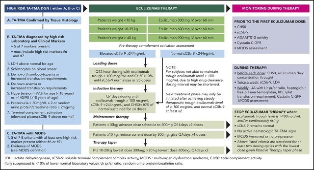
All patients with hrTA-TMA were offered eculizumab as a standard of care using uniform monitoring and drug dosing (Figure 1).2,7 Subjects without high-risk features received only supportive care without any targeted TA-TMA interventions.
Figure 1.
Eculizumab therapy for hrTA-TMA. LDH, lactate dehydrogenase.
Informed consent was obtained from all study subjects participating in the HSCT data and tissue repository. The institutional review board approved retrospective data analysis from electronic medical records and HSCT databases.
Eculizumab treatment and monitoring
Patients with hrTA-TMA were offered eculizumab as a first-line treatment. Blood total complement activity (CH50), sC5b-9, ADAMTS13 activity, and MODS assessment were performed before starting eculizumab. The dosage of eculizumab was determined using PK/PD monitoring, as displayed in Figure 1.7 In brief, the first eculizumab dose for patients <10 kg was 300 mg, patients 10 to 39 kg were given 600 mg, and those ≥40 kg received 900 mg. For patients with elevated pretherapy sC5b-9 (≥244 ng/mL), eculizumab loading doses were given at 72-hour intervals until sC5b-9 normalized and then advanced to weekly (induction) dosing, while maintaining therapeutic eculizumab trough concentration in the serum (eculizumab level) of ≥100 µg/mL and fully suppressed CH50 (<10% of lower normal laboratory value). Patients with a normal pretherapy sC5b-9 value were started on eculizumab induction at 7-day intervals for at least 4 doses or until active TA-TMA was controlled. Eculizumab level and CH50 were obtained before each eculizumab dose, and sC5b-9 was monitored twice a week. Eculizumab dosing frequency was shortened if needed to achieve and maintain a therapeutic trough level and fully suppressed trough CH50. If a therapeutic eculizumab level was not achieved with shortening the dosing interval to 48 hours, then the eculizumab dose was increased by 300 mg/dose. Maintenance and taper schedules were only considered after sustaining therapeutic eculizumab level and CH50 <10% for at least 2 consecutive doses after normalization of sC5b-9. Eculizumab was discontinued after resolution of hematologic TA-TMA and stabilization or improvement in MODS, when therapeutic eculizumab level and normal sC5b-9 were documented for 2 consecutive doses after starting the taper schedule. Discontinuation of the drug was considered successful when there was no evidence of active TA-TMA after eculizumab was cleared from the blood (<25 µg/mL), and CH50 activity was restored to normal without elevation of sC5b-9. All patients received antibacterial prophylaxis against Neisseria meningitidis and antifungal prophylaxis during eculizumab treatment and until CH50 normalized after full clearance of eculizumab.
Treatment response assessment
TA-TMA and MODS response were evaluated after the last eculizumab dose, and survival was evaluated at 6 months from TA-TMA diagnosis and 1-year after HSCT. Patients were classified as achieving complete response (CR) to TA-TMA targeted therapy if they showed resolution of high-risk markers (normalized sC5b-9 and resolved proteinuria <2 mg/mg), were not receiving red blood cell and platelet transfusions, and showed resolution from MODS, as listed in supplemental Table 1. Patients were classified as having no response (NR) when they did not achieve resolution of high markers, remained dependent on red blood cell and/or platelet transfusions, did not recover vital organ function after last eculizumab dose, or died with active TA-TMA. Partial response (PR) was assigned to those who did not meet CR or NR criteria.
Assessment of the incidence of bloodstream infections
The incidence of bacterial blood stream infections (BSIs) was evaluated in study subjects from HSCT (day 0) through the start of eculizumab, as well as from the start of eculizumab through the last eculizumab dose. In addition, we calculated the rate of bacterial BSIs during the 2 time periods (before eculizumab and during the eculizumab regimen) as the number of bacterial BSIs divided by the cumulative number of days at risk (rate = number of bacterial BSIs/days × 1000). The incidence of fungal and mold BSIs in study subjects was assessed from the start of eculizumab until the last follow-up or death.
Statistical analyses
Continuous and categorical data were described by median (interquartile range [IQR]) and frequency (percentage), respectively. Response defined as CR or PR and associations with predictor variables were reported as the odds ratio (OR) from a logistic regression model. OS was defined as the number of days from transplantation to death. Surviving patients were censored at the time of last follow-up. The Kaplan-Meier method was used to compute OS curves and percentages for categorical predictors. The hazard ratio (HR) describes the OS for continuous variables. The log-rank test and Cox proportional hazards regression were used to determine the significance of categorical and continuous variables, respectively. Significance was set at the .05 level. All tests were 2-sided, and all computations were carried out in R (version 3.5.1; Vienna, Austria).
Results
Study subjects
A total of 566 subjects underwent HSCT during the study period of 1 March 2012 to 1 October 2018. TA-TMA was diagnosed in 177 subjects (31%). High-risk TA-TMA was diagnosed in 64 subjects (11% [64/566] of all HSCT recipients and 36% [64/177]of patients with TA-TMA). Only HSCT recipients with hrTA-TMA (n = 64) received treatment with eculizumab and were included in further analyses described in this article. Eighteen of these patients were reported in prior publications describing eculizumab PK/PD in an HSCT population.7 Patient demographics are displayed in Table 1. All patients were children less than 18 years of age, the majority (79.7%) were Caucasian, and 62.5% were male. Most of the patients had transplantation performed for malignancy or immune deficiency, with the main stem cell source being bone marrow from related and unrelated donors, reflecting our usual clinical practice. Calcineurin inhibitors were used as the main drug for graft-versus-host disease (GVHD) prophylaxis, with the exception of a few patients who received T-depleted peripheral blood stem cell (PBSC) grafts.
Table 1.
Demographics
| Variable | Total subjects |
|---|---|
| Male sex, n (%) | 40 (62.5) |
| Age at transplant (range), y | 5.5 (2.7-11.7) |
| Race, n (%) | |
| Caucasian | 51 (79.7) |
| African American | 7 (10.9) |
| Asian | 3 (4.7) |
| Mixed | 3 (4.7) |
| Diagnostic group, n (%) | |
| Malignancy | 18 (28.1) |
| Immune deficiency | 28 (43.8) |
| Marrow failure | 8 (12.5) |
| Benign hematology | 7 (10.9) |
| Genetic/metabolic | 3 (4.7) |
| Stem cell donor type, n (%) | |
| Related (MRD, MMRD, haplo) | 30 (46.9) |
| Unrelated (MUD, MMUD) | 26 (40.6) |
| Autologous | 8 (12.5) |
| Stem cell source, n (%) | |
| Bone marrow (BM) | 41 (64) |
| Peripheral blood (PBSC) | 19 (29.7) |
| Cord blood (Cord) | 4 (6.3) |
| HLA match (allo-HSCT only), n (%) | |
| Fully matched (MRD, MUD) | 30 (53.6) |
| Mismatched (MMR, MMUD, haplo) | 26 (46.4) |
| Conditioning regimen type, n (%) | |
| Myeloablative | 35 (54.7) |
| Reduced intensity | 29 (45.3) |
| GVHD prophylaxis (allo-HSCT only), n (%) | |
| Calcineurin inhibitors | 49 (87.5) |
| T-cell depletion | 7 (12.5) |
| Pretransplant cystatin C GFR (IQR), mL/min | 129 (110-143) |
| Pretransplant rUPCR (range), mg/mg | 0.4 (0.2-0.8) |
MRD, matched related; MMRD, mismatched related; haplo: haploidentical; MUD, matched unrelated; MMUD, mismatched unrelated; BM, bone marrow.
TA-TMA was diagnosed during the first 100 days after transplantation in 59 of 64 (92%) subjects, at a median of 23 days (IQR, 3-48) after HSCT. Five patients had late TA-TMA between 118 and 221 days after transplantation. Four of those patients were allogeneic HSCT recipients who received prolonged therapy for GVHD (skin, lung, or bowel involvement) and 1 autologous HSCT recipient with neuroblastoma who developed late TA-TMA triggered by planned post–autologous transplantation radiation therapy.
TA-TMA response to eculizumab
Thirty-six of 64 treated patients (56%) achieved CR, 5 (8%) achieved PR, and 23 (36%) were classified as NR by the end of eculizumab treatment. The median number of eculizumab doses was 11 (IQR, 7-20; range, 2-50), and overall median duration of treatment was 66 days (IQR, 41-110). Eculizumab was well tolerated without any significant side effects attributed to the drug.
Fifty-two of the 64 patients (81%) had elevated sC5b9 at the start of eculizumab, with a median level of 398 ng/mL (IQR, 282-544). sC5b-9 normalized after eculizumab initiation in responders at a median of 15 days (IQR, 7-43). Higher sC5b-9 value (max sC5b-9) before starting eculizumab was associated with a longer time to normalize sC5b-9 (log[max sC5b-9]: HR, 2.0; P = .001), and showed significant positive correlation with the number of eculizumab doses required [r = 0.43; P = .0004].
Subjects with a higher sC5b-9 at TA-TMA diagnosis and with higher maximum sC5b-9 (sC5b-9 Max) elevation at the time of initiation of eculizumab were less likely to respond to treatment (OR, 0.28; P = .026 and OR, 0.15; P = .0014, respectively; Table 2).
Table 2.
Response to eculizumab therapy
| Variable | OR | P |
|---|---|---|
| Log (sC5b-9) at TA-TMA diagnosis | 0.28 | .026 |
| Log (sC5b-9 max) before starting eculizumab | 0.15 | .0014 |
| rUPCR at TA-TMA diagnosis | 0.76 | .65 |
| aGVHD diagnosed before TA-TMA* | 0.96 | .95 |
| aGVHD diagnosed after TA-TMA*,† | 0.3 | .16 |
The OR is from a univariate logistic regression model with response defined as a CR or a PR. An OR > 1 indicates that the variable corresponds to a decrease in the odds of a response.
aGVHD, acute GVHD.
Analysis excludes autologous HSCT recipients.
Analysis excludes those with aGVHD before TA-TMA.
Eculizumab was successfully discontinued in all survivors after TA-TMA resolution. The median time to clear eculizumab was 31 days (IQR, 25-40) and total blood complement activity (normalization of CH50) was recovered a median of 38 days (IQR 28-77) after stopping eculizumab. One patient with primary immunodeficiency and mixed-donor chimerism of 38% had recurrence of TMA during severe illness with disseminated histoplasmosis 5 years after initial hrTA-TMA diagnosis. This patient received 23 doses of eculizumab and is now doing well, having been off the drug for 14 months. There were no TA-TMA relapses in other patients at last follow-up at a median of 450 days (IQR, 268-1023) after HSCT.
Clinical outcomes
MODS and survival
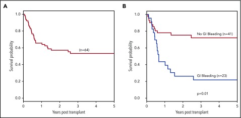
Fifty-four of 64 patients (84.4%) had MODS during hrTA-TMA therapy. Forty-eight subjects (75%) had at least 1 PICU admission. Seventy-seven percent of cases survived 6 months from TA-TMA diagnosis and 66% at 1 year after HSCT (Figure 2). OS 3 years after HSCT was 53.2%. Of the 29 subjects who died, 2 had resolution of TA-TMA before death, and all others had active laboratory signs of TMA at death. Autopsies of 6 of 7 patients showed histologic evidence of TMA in at least 1 organ. Deaths that occurred within 1 year after HSCT (n = 22) were related to multiorgan failure, with 9 patients having acute GVHD and 5 having Candida infections as the contributing cause. There were 7 late deaths (>1 year after HSCT). Causes of death of these patients was chronic GVHD/bronchiolitis obliterans (n = 2), intestinal GVHD with gastrointestinal bleeding (n = 3; 1 with Candida infection), cytomegalovirus (CMV) infection with multiorgan failure (n = 1), and neuroblastoma relapse (n = 1).
Figure 2.
Outcomes in patients with hrTA-TMA treated with eculizumab. Survival in HSCT recipients with hrTA-TMA treated with the terminal complement blocker eculizumab was calculated using Kaplan–Meier and log-rank tests starting at the beginning of HSCT (day 0, stem cell infusion day). (A) One-year post-HSCT survival in all treated patients was 66%. (B) One-year post-HSCT survival in patients with GI bleeding and without was 44% vs 78% (P = .01).
Renal outcomes
The median pre-HSCT (baseline) cystC GFR for this cohort was 129 mL/min (IQR, 110-143). The median baseline rUPCR was 0.4 mg/mg (range, 0.2-0.8) in 60 subjects with proteinuria; 4 patients had no proteinuria on baseline urinalyses.
Two patients were undergoing renal replacement therapy (RRT) at the start of eculizumab, and 15 of 64 (23%) subjects required RRT at some point during TA-TMA treatment. The median maximum rUPCR during active TA-TMA was 10.6 mg/mg (IQR, 4.9-23.9). The cystC GFR was reduced in all cases, with the median lowest GFR during treatment of patients who did not need RRT of 43 mL/min (IQR, 32-61). All patients had severe hypertension requiring at least 2 antihypertensive medications, and 16 required continuous infusion of antihypertension medication while in PICU.
Thirty of 35 survivors resolved nephrotic range proteinuria (rUPCR <2 mg/mg) a median of 82.5 days (IQR, 44-157) after starting eculizumab. The last renal function follow-up was performed at a median of 450 days (IQR, 268-1023) after HSCT. Median rUPCR was 0.45 mg/mg (IQR, 0.33-1.2). Five patients still had nephrotic range proteinuria at this time. The median cystC GFR was 98 mL/min (IQR, 84-114), and cystC GFR at last follow-up was a median of 20% lower (IQR 7.3% to 40.4% lower) than the pretransplant baseline. Two long-term survivors who had required RRT for TA-TMA had cystC GFRs of 84 and 86 mL/min and rUPCR of 0.35 and 0.2 mg/mg 8 and 5 years after HSCT.
Cardiopulmonary outcomes
No patients had pulmonary hypertension (PH) or pericardial effusions on pre-HSCT echocardiography screening. PH was diagnosed in 11 of 64 (17%) patients after TA-TMA diagnosis and was managed by a cardiologist. All patients with PH received inhaled nitric oxide during acute illness. One patient underwent emergency septostomy and has survived without PH for 4 years after HSCT. Two of 5 survivors with PH received maintenance sildenafil but discontinued the treatment by 6 months after TA-TMA diagnosis.
Twelve of 64 (18.8%) patients underwent pericardiocentesis for cardiac tamponade related to pericardial effusion at a median of 31 days (range, 2-98) after TA-TMA diagnosis and a median of 28 days (range, 0-74) from the start of eculizumab. Six of these patients underwent more than one pericardiocentesis. A pericardial drain was needed for a median of 5 days (range, 2-23). Two patients underwent pleurocentesis.
Five of 26 patients needing invasive mechanical ventilation were successfully extubated. Seven patients had pulmonary bleeding; 1 survived.
GI outcomes
Twenty-three of 64 (36%) patients with hrTA-TMA had lower GI bleeding, including 1 autologous HSCT recipient. Histologic tissue analysis was available in 18 allogeneic HSCT recipients at 1 to 6 different time points. At the time of the first GI tissue biopsy, patients clinically were classified as stage 4 (n = 16) and stage 3 (n = 2) gut GVHD by the Glucksberg criteria. Histologic evaluation of tissue according to the Lerner criteria documented no evidence of GVHD in 8 patients, grade 1 intestinal GVHD in 8 patients, and grade 2 GVHD in 2 patients. Fifteen of those 18 patients had documented histologic features compatible with intestinal TMA, and the remaining 3 had no intestinal vasculature on the biopsy for assessment. Later in the disease course, histologic tissue reports on patients with multiple evaluations progressed to describe mucosal ulceration or focal denudation, mucosal replacement by granulation tissue, mucosal hemorrhages, and changes representing ischemic injury. These changes were interpreted as “compatible with severe GVHD.” Available autopsies from 5 patients with GI hemorrhage showed massive amounts of blood in the bowel, GI ulcerations or sloughed mucosa, and various degrees of vascular injury. Three patients had small-bowel strictures: 2 identified by radiologic studies and surgically resected and 1 diagnosed on autopsy.
Patients with GI bleeding required more doses of eculizumab than those without bleeding (20 [IQR, 12-31] vs 9 [IQR, 7-13]; P = .0015). Response to treatment was also significantly worse (35% vs 80%; P = .0004) and the rate of 1-year post-HSCT survival was lower (44% vs 78%, P = .01; Figure 2).
Central nervous system symptomatology
Central nervous system (CNS) symptoms were documented in 21 of 64 (32.8%) patients with hrTA-TMA. Twelve patients had seizures, 6 of them associated with posterior reversible encephalopathy syndrome and 4 with CNS bleeding. One patient had a subdural hematoma requiring draining. Mental status changes presenting as encephalopathy were documented in 8 patients. One patient was diagnosed with vancomycin-resistant enterococcal meningitis in association with vancomycin-resistant enterococcus bacteremia and fully recovered. No meningococcal infections were documented.
Infections
Twenty-seven bacterial BSIs were diagnosed in 20 of the 64 patients (31%) from HSCT (day 0) through the first dose of eculizumab (combined 4150 days of risk for BSI). The bacterial BSI rate before the first dose of eculizumab was 6.5 bacterial BSIs per 1000 days. Twenty-six bacterial BSIs were diagnosed in 16 of the 64 patients (25%) who were receiving eculizumab (5136 days of risk for BSI). The bacterial BSI rate while receiving eculizumab was 5.1 bacterial BSI per 1000 days. None of the BSIs were recorded as a cause of death.
No patients developed invasive mold infections. Six patients developed a yeast BSI (all Candida species) after starting eculizumab for TA-TMA. Median time to develop candidemia after the first eculizumab dose was 83 days (range, 17-132 days), and patients had received a median of 17 doses (range, 8-35) before developing fungal BSI. Four of 6 patients developed the first Candida BSI during the complement blockade period. Eculizumab was discontinued in all patients after a positive fungal BSI was found. Five of the 6 patients had steroid refractory GVHD in addition to TA-TMA and were on multiple immunosuppressants for a prolonged time before developing candidemia, and 1 patient developed a disseminated Candida infection 33 days after stem cell infusion and 17 days after starting eculizumab for TA-TMA. None of those patients survived. Three of the 6 patients had autopsies confirming yeast infection.
All patients had viral screening for blood CMV, Epstein-Barr virus, adenovirus, and BK polymerase chain reactions 1 to 2 times per weeks, and 48 of 64 subjects (75%) had at least 1 type of viremia documented during the study period. Only 1 patient’s death was attributed to CMV infection.
GVHD and TA-TMA
Thirty of 64 patients (47%) had a diagnosis of acute GVHD. Half of the patients were diagnosed with GVHD before and half after TA-TMA diagnosis. Response to TA-TMA treatment was more likely in those with lower grades of GVHD (grade 2 or lower [85%] vs higher than grade 2 [43%]; P = .09). Generally, calcineurin prophylaxis was continued after the diagnosis of TA-TMA, organ function permitting, in patients who were at high risk for GVHD or had active GVHD. Calcineurin inhibitors were discontinued in patients requiring RRT or with uncontrolled hypertension resulting in posterior reversible encephalopathy syndrome or CNS bleeding.
Veno-occlusive disease
Five patients were diagnosed with veno-occlusive disease of the liver, and 4 of them were treated with defibrotide before and 1 of them after TA-TMA diagnosis. Two were TA-TMA responders and 3 were TA-TMA nonresponders.
Complement gene testing
A total of 30 of 64 subjects with hrTA-TMA had recipient DNA collected before transplantation and were tested for a clinically available hemolytic atypical uremic syndrome (aHUS) genetic susceptibility panel that included sequencing of 10 genes (C3, CFB, CFH, CFHR1, CFHR3, CFHR5, CFI, DGKE, MCP, and THBD) and multiplex ligation probe amplification testing for CFHR3/CFHR1 deletion analysis. Of the 30 subjects tested, 21 had at least 1 complement gene variant identified (11 are alive, 10 are deceased), and 9 had no variants identified (5 are alive and 4 are deceased; supplemental Table 2).
Discussion
We report our experience of the largest cohort to date of patients given the terminal complement blocker eculizumab as a first-line treatment for hrTA-TMA. We recorded a response rate of 64% and survival of 66% 1 year after HSCT. Although these outcomes are not ideal, it shows significant improvement from our previously reported untreated historical observational cohort with the same high-risk features in which 16.7% of cases survived 1 year after HSCT.5
In our current cohort, subjects with higher pretherapy sC5b-9 (higher systemic complement activation) were less likely to respond to treatment, took longer to control complement activation, required more doses of eculizumab, and had worse outcomes.6 Although sC5b-9 monitoring aids in identifying high-risk patients and guiding eculizumab dosage, the test is currently only available for clinical use in several specialized laboratories. Harder et al18 recently reported that overwhelming complement activation, as could be seen in HSCT recipients with TA-TMA presenting with elevated blood sC5b-9, may escape blockade, even with adequate eculizumab dosing, and a combination of complement blockers or new-generation blockers may assist in overcoming this challenge in the future.
We observed 2 main types of eculizumab NRs: those who were started on eculizumab therapy in the terminal stages of their illness (in the early years of our experience with the drug), with very advanced organ injury and were not able to get the benefit of the therapy, and those who had severe intestinal bleeding. Patients with intestinal bleeding were a unique group that had very high eculizumab clearance, required more eculizumab doses, and had significantly worse outcomes. Some also received multiple immunosuppressive agents for clinical diagnosis of therapy refractory GI GVHD and started eculizumab for TA-TMA with significant delay (also in the early years of our experience with the drug).
Review of available GI tissue from our cohort demonstrated a significant discrepancy between clinical symptomatology (bleeding) that was attributable to GVHD and histologic evidence of GVHD at symptom presentation, suggesting that a TA-TMA component of the bowel injury may have been overlooked. An early intervention preserving vascular endothelial health and controlling complement activation, along with effective GVHD prophylaxis or therapy, may prevent progression to severe mucosal injury and improve outcomes, but we have yet to learn the best strategies in such patients.9,10,19,20 Our experience indicates that appropriate intensification of eculizumab treatment based on PK/PD end points is essential in these patients with GI bleeding.
We offered eculizumab only to patients with hrTA-TMA features, so the majority of persons in our cohort had multiorgan injury at the start of the treatment. Renal, cardiac, and GI complications were common in this cohort. Pulmonary hypertension requiring intervention was identified by targeted examination of patients with hypoxemia using echocardiography. We observed that serositis often develops late during the treatment course, making high awareness and a low threshold for echocardiography and radiological examination valuable. Patients requiring pericardiocentesis need careful assessment before pericardial drains are removed, as effusions tend to reaccumulate. In our clinical experience, eculizumab treatment, if already documented to be therapeutic, does not have to be further intensified just for serositis.
Our patients with hrTA-TMA had significant renal injury, and such patients must be monitored by a nephrologist for long-term effects, as over 70% of survivors still had some proteinuria on long-term follow-up, and best cystC GFR after recovering from TA-TMA remained below the pretransplantation baseline value.14,21 Subjects with intestinal bleeding were the sickest group of patients, and the occurrence of profuse GI bleeding after transplantation should trigger consideration of TA-TMA as well as GVHD, as both can be present at the same time.
Patients in our cohort who responded to therapy benefited from a brief but intensive course of eculizumab, a very different strategy from that reported in aHUS.22-24 Eculizumab clearance was very high in HSCT recipients with highly activated complement and a great deal of ligand to bind drug, and frequent (occasionally every 48 hours) eculizumab loading doses were necessary at the beginning of the therapy to achieve control of activated complement. Despite intensive upfront dosing, the amount of eculizumab used for HSCT recipients with TA-TMA was comparable to similar sized patients with aHUS receiving eculizumab administered according to the drug packet insert in the first 3 months of therapy. The difference is that subjects with TA-TMA receive most of the drug in the beginning of the therapy right after diagnosis, followed by rapid taper and discontinuation, and do not require indefinite therapy, as recommended in some aHUS cases.
A recent report from Cassol et al25 suggests that eculizumab can remain incorporated within arterioles for a significant time after cessation of therapy. This may explain the prolonged time to normalize CH50 in some of our patients after stopping eculizumab therapy. The role of eculizumab in TA-TMA is to control damaging complement overactivation caused by the transplant process and to restore normal complement activity. As the toxicities of the TA-TMA-inciting transplantation process resolve, it is then possible and advisable to stop eculizumab as soon as possible, to reduce additional risk of infections.
We previously observed a high overall incidence of BSIs in HSCT recipients with untreated TA-TMA, probably because of bacterial translocation with severe mucosal compromise, especially in patients with intestinal vascular endothelial injury.26 Although there is a concern that eculizumab may increase infection risk, we did not see a significant increase in bacterial BSIs after initiation of eculizumab treatment. Antibacterial prophylaxis was provided to all our treated patients until CH50 normalization was documented after cessation of eculizumab, given that antimeningococcal vaccines are not effective in this setting.27,28
Eculizumab has been associated with increased rates of invasive molds in some cases. However, we did not see any invasive molds in our treated patients, although we did see candidemia. Blocking terminal complement using eculizumab is not expected to affect the function of C3, which potentially mediates opsonization and immune clearance of fungi, but the exact role of the complement in the control of fungal pathogens still should be studied in transplantation settings, because HSCT recipients receive other immunosuppressive agents elevating risk of fungemia.29 HSCT recipients receiving complement-blocking agents should be provided with antifungal prophylaxis against yeasts and molds.
Multiple factors, such as chemotherapy, viral infections, calcineurin inhibitors, and GVHD, have been implicated as TA-TMA triggers. Additional studies are necessary to determine how to manage calcineurin inhibitors in HSCT recipients with a TA-TMA diagnosis.30-34 Abrupt discontinuation of GVHD prophylaxis may trigger a flare of GVHD that will make TA-TMA management challenging, as both conditions trigger tissue inflammation. On the other hand, unnecessary escalation in immunosuppression for presumed GVHD in patients with intestinal bleeding may lead to life-threatening infections that can further activate complement and endothelial damage.
In summary, hrTA-TMA is a significant contributor to acute morbidity and mortality, and we are only just beginning to understand the effect of hrTA-TMA on long-term organ function in transplant survivors. Our experience is limited to a pediatric population mainly with immune deficiencies, thus representing a unique group. Adult HSCT-recipients also have been reported to have complement-mediated TA-TMA that benefits from targeted complement blocker therapies.30,35,36 We anticipate that there are likely differences between TA-TMA observed in children and adults that must be learned. Although at this time studies of complement inhibitors are very limited in the HSCT population, narsoplimab (OMS721), a MASP-2 inhibitor (lectin pathway), received a Breakthrough Therapy Designation from the US Food and Drug Administration for HSCT-TMA, based on improved survival compared with historical controls in a phase 2 study with a phase 3 program in progress. Ravulizumab, a long-acting anti-C5 monoclonal antibody engineered from eculizumab, is currently being investigated for the treatment of aHUS and may be of interest in patients with TA-TMA with very high eculizumab clearance. Coversin, a small protein complement C5 inhibitor, may benefit patients who are resistant to eculizumab because of complement C5 polymorphisms.37 Well-structured prospective studies are needed to improve our understanding of complement blockade in HSCT, especially as novel agents are introduced into clinical practice.38 In addition, lack of adequate response to complement blockade in some patients prompts us to search for additional targetable endothelial injury pathways, such as interferons, that can induce complement system activation, IL-8 release from injured endothelial cells that can activate neutrophil extracellular traps, and others. These novel observations will provide us with the opportunity to consider combination treatment for TA-TMA to further improve outcomes.39-42
Supplementary Material
The online version of this article contains a data supplement.
Acknowledgments
The authors acknowledge the inspirational work of Javier El-Bietar in intestinal TA-TMA and thank Mary Block and the Hematology Laboratory at CCHMC, Bradley Dixon, Thelma Kathman, and the Nephrology laboratory at CCHMC for collaboration in establishing and validating TA-TMA clinical tests and the CCHMC Bone Marrow Tissue Repository for outstanding ongoing technical assistance.
This study was performed using Division of Bone Marrow Transplantation and Immune Deficiency divisional funds. Part of the research reported in this publication was supported by the Eunice Kennedy Shriver National Institute of Child Health and Human Development of the National Institutes of Health (NIH) under award number R01HD093773 (S.J.).
Footnotes
Supporting data may be obtained by e-mail from the corresponding author.
The publication costs of this article were defrayed in part by page charge payment. Therefore, and solely to indicate this fact, this article is hereby marked “advertisement” in accordance with 18 USC section 1734.
Authorship
Contribution: S.J. and S.M.D. monitored eculizumab therapy for all patients, collected and analyzed the data, and wrote the manuscript; C.E.D. provided vital contributions in study planning and multidisciplinary team integration in prospective TA-TMA screening and collected and analyzed the clinical data; A.T.-C. monitored eculizumab administration and summarized the pharmacology data; A.L. performed the statistical analyses and prepared the data tables and figures; K.C.M., G.W., A.N., and J.B. performed patient monitoring and participated in study planning and data analysis; B.L.L. and S.B. managed renal complications and prepared the kidney analyses; R.S.C. oversaw care of the patients and data collection in the pediatric intensive care unit. R.H. and T.D.R. performed cardiac monitoring of the study patients and prepared cardiac data and analysis; K.M. performed eculizumab PK/PD testing; M.W. performed histologic tissue review and prepared the pathology photographs; and all authors edited the manuscript.
Conflict-of-interest disclosure: S.J., S.M.D., B.L.L., and K.M. have a US Provisional Patent Application pending. The remaining authors declare no competing financial interests.
Correspondence: Sonata Jodele, Division of Bone Marrow Transplantation and Immune Deficiency, Cincinnati Children’s Hospital Medical Center, 3333 Burnet Ave, MLC 11027, Cincinnati, OH 45229; e-mail: sonata.jodele@cchmc.org.
REFERENCES
- 1.Dvorak CC, Higham C, Shimano KA. Transplant-Associated Thrombotic Microangiopathy in Pediatric Hematopoietic Cell Transplant Recipients: A Practical Approach to Diagnosis and Management. Front Pediatr. 2019;7:133. [DOI] [PMC free article] [PubMed] [Google Scholar]
- 2.Jodele S, Dandoy CE, Myers KC, et al. . New approaches in the diagnosis, pathophysiology, and treatment of pediatric hematopoietic stem cell transplantation-associated thrombotic microangiopathy. Transfus Apheresis Sci. 2016;54(2):181-190. [DOI] [PMC free article] [PubMed] [Google Scholar]
- 3.Moiseev IS, Tsvetkova T, Aljurf M, et al. . Clinical and morphological practices in the diagnosis of transplant-associated microangiopathy: a study on behalf of Transplant Complications Working Party of the EBMT. Bone Marrow Transplant. 2019;54(7):1022-1028. [DOI] [PubMed] [Google Scholar]
- 4.Gavriilaki E, Sakellari I, Batsis I, et al. . Transplant-associated thrombotic microangiopathy: Incidence, prognostic factors, morbidity, and mortality in allogeneic hematopoietic cell transplantation. Clin Transplant. 2018;32(9):e13371. [DOI] [PubMed] [Google Scholar]
- 5.Jodele S, Davies SM, Lane A, et al. . Diagnostic and risk criteria for HSCT-associated thrombotic microangiopathy: a study in children and young adults. Blood. 2014;124(4):645-653. [DOI] [PMC free article] [PubMed] [Google Scholar]
- 6.Jodele S. Complement in Pathophysiology and Treatment of Transplant-Associated Thrombotic Microangiopathies. Semin Hematol. 2018;55(3):159-166. [DOI] [PubMed] [Google Scholar]
- 7.Jodele S, Fukuda T, Mizuno K, et al. . Variable Eculizumab Clearance Requires Pharmacodynamic Monitoring to Optimize Therapy for Thrombotic Microangiopathy after Hematopoietic Stem Cell Transplantation. Biol Blood Marrow Transplant. 2016;22(2):307-315. [DOI] [PMC free article] [PubMed] [Google Scholar]
- 8.Leteurtre S, Duhamel A, Salleron J, Grandbastien B, Lacroix J, Leclerc F; Groupe Francophone de Réanimation et d’Urgences Pédiatriques (GFRUP) . PELOD-2: an update of the PEdiatric logistic organ dysfunction score. Crit Care Med. 2013;41(7):1761-1773. [DOI] [PubMed] [Google Scholar]
- 9.El-Bietar J, Warren M, Dandoy C, et al. . Histologic Features of Intestinal Thrombotic Microangiopathy in Pediatric and Young Adult Patients after Hematopoietic Stem Cell Transplantation. Biol Blood Marrow Transplant. 2015;21(11):1994-2001. [DOI] [PMC free article] [PubMed] [Google Scholar]
- 10.Warren M, Jodele S, Dandoy C, et al. . A Complete Histologic Approach to Gastrointestinal Biopsy From Hematopoietic Stem Cell Transplant Patients With Evidence of Transplant-Associated Gastrointestinal Thrombotic Microangiopathy. Arch Pathol Lab Med. 2017;141(11):1558-1566. [DOI] [PubMed] [Google Scholar]
- 11.Dandoy CE, Davies SM, Flesch L, et al. . A team-based approach to reducing cardiac monitor alarms. Pediatrics. 2014;134(6):e1686-e1694. [DOI] [PubMed] [Google Scholar]
- 12.Rotz SJ, Ryan TD, Jodele S, et al. . The injured heart: early cardiac effects of hematopoietic stem cell transplantation in children and young adults. Bone Marrow Transplant. 2017;52(8):1171-1179. [DOI] [PubMed] [Google Scholar]
- 13.Dandoy CE, Davies SM, Hirsch R, et al. . Abnormal echocardiography 7 days after stem cell transplantation may be an early indicator of thrombotic microangiopathy. Biol Blood Marrow Transplant. 2015;21(1):113-118. [DOI] [PMC free article] [PubMed] [Google Scholar]
- 14.Benoit SW, Dixon BP, Goldstein SL, et al. . A novel strategy for identifying early acute kidney injury in pediatric hematopoietic stem cell transplantation. Bone Marrow Transplant. 2019;54(9):1453-1461. [DOI] [PubMed] [Google Scholar]
- 15.Laskin BL, Nehus E, Goebel J, Furth S, Davies SM, Jodele S. Estimated versus measured glomerular filtration rate in children before hematopoietic cell transplantation. Biol Blood Marrow Transplant. 2014;20(12):2056-2061. [DOI] [PMC free article] [PubMed] [Google Scholar]
- 16.Laskin BL, Nehus E, Goebel J, Khoury JC, Davies SM, Jodele S. Cystatin C-estimated glomerular filtration rate in pediatric autologous hematopoietic stem cell transplantation. Biol Blood Marrow Transplant. 2012;18(11):1745-1752. [DOI] [PubMed] [Google Scholar]
- 17.Dandoy CE, Davies SM, Hirsch R, et al. . Abnormal echocardiography 7 days after stem cell transplantation may be an early indicator of thrombotic microangiopathy. Biol Blood Marrow Transplant. 2015;21(1):113-118. [DOI] [PMC free article] [PubMed] [Google Scholar]
- 18.Harder MJ, Kuhn N, Schrezenmeier H, et al. . Incomplete inhibition by eculizumab: mechanistic evidence for residual C5 activity during strong complement activation. Blood. 2017;129(8):970-980. [DOI] [PMC free article] [PubMed] [Google Scholar]
- 19.Gavriilaki E, Sakellari I, Karafoulidou I, et al. . Intestinal thrombotic microangiopathy: a distinct entity in the spectrum of graft-versus-host disease. Int J Hematol. 2019;110(5):529-532. [DOI] [PubMed] [Google Scholar]
- 20.Yamada R, Nemoto T, Ohashi K, et al. . Distribution of Transplantation-Associated Thrombotic Microangiopathy (TA-TMA) and Comparison between Renal TA-TMA and Intestinal TA-TMA: Autopsy Study. Biol Blood Marrow Transplant. 2020;26:178-188. [DOI] [PubMed] [Google Scholar]
- 21.Hingorani S, Pao E, Stevenson P, et al. . Changes in Glomerular Filtration Rate and Impact on Long-Term Survival among Adults after Hematopoietic Cell Transplantation: A Prospective Cohort Study. Clin J Am Soc Nephrol. 2018;13(6):866-873. [DOI] [PMC free article] [PubMed] [Google Scholar]
- 22.Raina R, Grewal MK, Radhakrishnan Y, et al. . Optimal management of atypical hemolytic uremic disease: challenges and solutions. Int J Nephrol Renovasc Dis. 2019;12:183-204. [DOI] [PMC free article] [PubMed] [Google Scholar]
- 23.Menne J, Delmas Y, Fakhouri F, et al. . Outcomes in patients with atypical hemolytic uremic syndrome treated with eculizumab in a long-term observational study. BMC Nephrol. 2019;20(1):125. [DOI] [PMC free article] [PubMed] [Google Scholar]
- 24.Olson SR, Lu E, Sulpizio E, Shatzel JJ, Rueda JF, DeLoughery TG. When to Stop Eculizumab in Complement-Mediated Thrombotic Microangiopathies. Am J Nephrol. 2018;48(2):96-107. [DOI] [PubMed] [Google Scholar]
- 25.Cassol CA, Brodsky SV, Satoskar AA, Blissett AR, Cataland S, Nadasdy T. Eculizumab deposits in vessel walls in thrombotic microangiopathy. Kidney Int. 2019;96(3):761-768. [DOI] [PubMed] [Google Scholar]
- 26.Grossmann L, Alonso PB, Nelson A, et al. . Multiple bloodstream infections in pediatric stem cell transplant recipients: A case series. Pediatr Blood Cancer. 2018;65(12):e27388. [DOI] [PubMed] [Google Scholar]
- 27.Konar M, Granoff DM. Eculizumab treatment and impaired opsonophagocytic killing of meningococci by whole blood from immunized adults. Blood. 2017;130(7):891-899. [DOI] [PMC free article] [PubMed] [Google Scholar]
- 28.Davies SM. Accept the complement (blockade). Blood. 2017;130(7):842-843. [DOI] [PubMed] [Google Scholar]
- 29.Tsoni SV, Kerrigan AM, Marakalala MJ, et al. . Complement C3 plays an essential role in the control of opportunistic fungal infections. Infect Immun. 2009;77(9):3679-3685. [DOI] [PMC free article] [PubMed] [Google Scholar]
- 30.Jan AS, Hosing C, Aung F, Yeh J. Approaching treatment of transplant-associated thrombotic Microangiopathy from two directions with Eculizumab and transitioning from Tacrolimus to Sirolimus. Transfusion. 2019;59(11):3519-3524. [DOI] [PubMed] [Google Scholar]
- 31.Wall SA, Zhao Q, Yearsley M, et al. . Complement-mediated thrombotic microangiopathy as a link between endothelial damage and steroid-refractory GVHD. Blood Adv. 2018;2(20):2619-2628. [DOI] [PMC free article] [PubMed] [Google Scholar]
- 32.Li A, Wu Q, Davis C, et al. . Transplant-Associated Thrombotic Microangiopathy Is a Multifactorial Disease Unresponsive to Immunosuppressant Withdrawal. Biol Blood Marrow Transplant. 2019;25(3):570-576. [DOI] [PMC free article] [PubMed] [Google Scholar]
- 33.Kraft S, Bollinger N, Bodenmann B, et al. . High mortality in hematopoietic stem cell transplant-associated thrombotic microangiopathy with and without concomitant acute graft-versus-host disease. Bone Marrow Transplant. 2019;54(4):540-548. [DOI] [PubMed] [Google Scholar]
- 34.Zeisbrich M, Becker N, Benner A, et al. . Transplant-associated thrombotic microangiopathy is an endothelial complication associated with refractoriness of acute GvHD. Bone Marrow Transplant. 2017;52(10):1399-1405. [DOI] [PubMed] [Google Scholar]
- 35.Rudoni J, Jan A, Hosing C, Aung F, Yeh J. Eculizumab for transplant-associated thrombotic microangiopathy in adult allogeneic stem cell transplant recipients. Eur J Haematol. 2018;101(3):389-398. [DOI] [PubMed] [Google Scholar]
- 36.Dhakal P, Giri S, Pathak R, Bhatt VR. Eculizumab in Transplant-Associated Thrombotic Microangiopathy. Clin Appl Thromb Hemost. 2017;23(2):175-180. [DOI] [PubMed] [Google Scholar]
- 37.Goodship THJ, Pinto F, Weston-Davies WH, et al. . Use of the complement inhibitor Coversin to treat HSCT-associated TMA. Blood Adv. 2017;1(16):1254-1258. [DOI] [PMC free article] [PubMed] [Google Scholar]
- 38.Patriquin CJ, Kuo KHM. Eculizumab and Beyond: The Past, Present, and Future of Complement Therapeutics. Transfus Med Rev. 2019;33:256-265. [DOI] [PubMed] [Google Scholar]
- 39.Gloude NJ, Khandelwal P, Luebbering N, et al. . Circulating dsDNA, endothelial injury, and complement activation in thrombotic microangiopathy and GVHD. Blood. 2017;130(10):1259-1266. [DOI] [PMC free article] [PubMed] [Google Scholar]
- 40.Garcia-Romo GS, Caielli S, Vega B, et al. . Netting neutrophils are major inducers of type I IFN production in pediatric systemic lupus erythematosus. Sci Transl Med. 2011;3(73):73ra20. [DOI] [PMC free article] [PubMed] [Google Scholar]
- 41.An P, Sáenz Robles MT, Duray AM, Cantalupo PG, Pipas JM. Human polyomavirus BKV infection of endothelial cells results in interferon pathway induction and persistence. PLoS Pathog. 2019;15(1):e1007505. [DOI] [PMC free article] [PubMed] [Google Scholar]
- 42.Allinovi M, Cirami CL, Caroti L, et al. . Thrombotic microangiopathy induced by interferon beta in patients with multiple sclerosis: three cases treated with eculizumab. Clin Kidney J. 2017;10(5):625-631. [DOI] [PMC free article] [PubMed] [Google Scholar]
Associated Data
This section collects any data citations, data availability statements, or supplementary materials included in this article.


