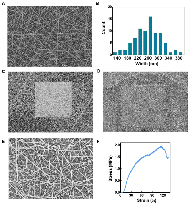
Figure 7. Electronics-biomaterial scaffold hybrid.
A. Scanning electron micrograph of the electrospun PCL-gelatin nanofibers. B. Histogram showing the distribution of nanofiber diameter. C. Scanning electron micrograph of the electrospun nanofibers on the small recording electrode. D. Scanning electron micrograph of the electrospun nanofibers covering the drug-loaded polypyrrole layer, which is deposited on the large drug release electrode. E. Zoom-in on the electrospun nanofibers covering the polypyrrole layer. F. Typical stress-strain curve resulting from the tensile test performed on the hybrid device.

