Abstract
Purpose of Review
This review highlights some of the recent concerning emerging infectious diseases, a number of them specifically that the World Health Organization has categorized as priorities for research.
Recent Findings
Emerging and reemerging infectious diseases account for significant losses in not only human life, but also financially. There are a number of contributing factors, most commonly surrounding human behavior, that lead to disease emergence. Zoonoses are the most common type of infection, specifically from viral pathogens. The most recent emerging diseases in the USA are Emergomyces canadensis, the Heartland virus, and the Bourbon virus.
Summary
In addition to the aforementioned pathogens, the Severe Acute Respiratory Syndrome, Middle East Respiratory Syndrome, Nipah virus, New Delhi metallo-ß-lactamase-1 Enterobacteriaceae, Rift Valley Fever virus, and Crimean-Congo Hemorrhagic Fever virus are reviewed. These pathogens are very concerning with a high risk for potential epidemic, ultimately causing both significant mortality and financial costs. Research should be focused on monitoring, prevention, and treatment of these diseases.
Keywords: Emerging infectious diseases, Reemerging infections, Viral pathogens
Introduction
In 1992, an expert committee that produced the Institute of Medicine report on emerging infections defined them as “new, reemerging, or drug-resistant infections whose incidence in humans has increased within the past two decades or whose incidence threatens to increase in the near future.” Additionally, six major contributors to these diseases were presented and included changes in human demographics and behavior, advances in technology and changes in industry practices, economic development and changes in land-use patterns, dramatic increases in volume and speed of international travel and commerce, microbial adaptation and change, and breakdown of public health capacity [1]. A common theme to recent emerging infectious diseases is that the majority are of animal origin [2]. In fact, some vector-borne pathogens that are projected to be introduced into new regions include Rift Valley Fever and Japanese Encephalitis viruses in the Americas and Crimean-Congo Hemorrhagic Fever virus in Eurasia [3]. Additionally, the majority of them have been viral. In addition to the cost of human life, emerging or reemerging infectious diseases can be very costly financially.
The recent Zika outbreak is estimated to have cost $3.5 billion in economic losses in 2016 [4]. The World Health Organization has prioritized a number of infectious diseases as requiring urgent need for research and development given the concern for potential of severe outbreaks. This article reviews the majority of emerging infections on this list, a few others that have emerged in the USA as well, and those that have not recently emerged but that the WHO has prioritized as having a high likelihood of causing outbreaks in the future.
Severe Acute Respiratory Syndrome
In 2002, an outbreak in southern China of atypical pneumonia of undetermined etiology spread to neighboring countries and eventually across the world. The coronavirus that caused the severe acute respiratory syndrome (SARS) was then discovered [5]. Hong Kong and Beijing were the most severely affected cities with estimated costs in the Far East of approximately $30 billion by May 2003 alone [6]. Over 8000 probable cases were reported in 29 countries with a mortality rate of about 10% by the end of the epidemic in July 2003. SARS reemerged in small scales from the end of 2003 to 2004. It is a positive-sense RNA coronavirus whose mean incubation period is 5 days [7]. The clinical presentation varied by patient age: children developed typical mild upper respiratory tract infection while teenagers and adults developed a more severe but predictable course [5, 7]. Phase I of the course was associated with increasing viral load with flu-like symptoms. Phase II was characterized by recurrence of fever with hypoxemia and progression of pneumonia. About 50% would require supplementary oxygen and approximately 20% would require intubation due to the development of acute respiratory distress syndrome (ARDS). Watery diarrhea was a prominent extrapulmonary manifestation. Additionally, hepatitis was a common complication and patients occasionally developed neurologic manifestations including status epilepticus. The clinical worsening in phase II is thought to be immune-mediated. Laboratory findings include lymphopenia, elevated aminotransferases, elevated lactate dehydrogenase (LDH), and creatine kinase (CK), with results consistent with disseminated intravascular coagulopathy. Progression from unilateral opacities to bilateral involvement was found on imaging. SARS is detected in respiratory secretions, urine, and feces [7]. However, the most notable finding is diffuse alveolar damage [5]. Advanced age, severe hepatitis, high initial LDH, high neutrophilia on presentation, diabetes mellitus, low CD4 and CD8 counts, and high initial viral load are all prognostic factors for severe disease. Some survivors have shown persistent lung function abnormalities. It is spread by close contact via droplet transmission, though there is some evidence that it might follow airborne transmission [7]. The virus has a high rate of infecting healthcare workers, accounting for approximately 20% of cases [8]. The original outbreak was thought to have started from the handling of wild animals, particularly the civet cat and raccoon dog [7, 9].
If suspected, a patient should be placed in respiratory isolation. A single test result, either positive or negative, is insufficient to make conclusions about a patient’s diagnosis as both false positives and false negatives occur. It is initially tested for via serology or reverse-transcriptase-polymerase chain reaction (RT-PCR). However, two specimens obtained from either different sources or from the same source on different occasions confirm the diagnosis. Specimens can be obtained from a nasopharyngeal swab or stool. Additionally, seroconversion is another diagnostic method. It is vital to identify patients with SARS, as isolation within 3 days of illness significantly reduces secondary transmission [10]. Although ribavirin was used in the initial outbreak, it has not been shown to have in vitro activity against the virus. Corticosteroids should be used only in the late-phase for rescue purposes as it may prolong viremia if given early [7]. Most of the other agents that have shown promise, including monoclonal antibodies, nitric oxide, protease inhibitors, and interferons have not yet shown activity in humans and there have been very few animal studies [7, 11]. Part of the concern about the potential for future epidemics lies in the fact that the family of viruses from which SARS comes is notorious for frequent mutations [6]. Additionally, there has been decreased worldwide attention, leading to decreased funding for research, as there have been no reported cases of SARS since 2005 [11].
Middle East Respiratory Syndrome
The Middle East respiratory syndrome coronavirus (MERS-CoV) was first identified in 2012. It is a ß coronavirus that is enveloped with a positive-sense RNA genome. The clinical features may include flu-like symptoms, gastrointestinal symptoms, severe pneumonia with acute respiratory distress syndrome, septic shock, disseminated intravascular coagulopathy, and multi-organ failure [12]. Lab findings may include lymphopenia, thrombocytopenia, elevated LDH levels, and elevated aminotransferases [13, 14]. However, approximately 20% of cases had no or mild symptoms while almost 50% had severe symptoms [15••]. The incubation period is 2–14 days. Children and younger adults seem to be less susceptible [14]. A large proportion of the confirmed pediatric cases have been asymptomatic and have been acquired via household contacts [16]. Primary infection results from contact with a dromedary camel or its products, though human-to-human transmission is possible. Cases acquired via human transmission tend to be more mild and the number of infected contacts tends to be limited. In fact, the majority of secondary cases have been associated with healthcare settings, where the majority of outbreaks have occurred and about 18% of cases have been healthcare workers [14]. With the exception of South Korea, outbreaks have been limited to the Middle East where dromedary camels are found [12]. In the South Korean outbreak, there were 186 cases and 38 deaths [15••]. Cases have been imported to Europe, Asia, Africa, and the USA [Table 1]. The cases in the USA were detected promptly, so no secondary cases occurred. Both cases occurred in May 2014 and were healthcare workers in Saudi Arabia [14]. As of July 2017, there have been 2040 laboratory-confirmed cases, with the majority of these in Saudi Arabia. The reproduction number is less than 1 with significant heterogeneity [15••, 17]. In the South Korean outbreak, “spreaders” had significantly higher rates of underlying pulmonary disease. Most transmissions occurred early in the disease course, during days 4–7 after symptom onset and were associated with patients going to multiple healthcare facilities before diagnosis [17]. Human-to-human transmission seems to require close contact, though has not been sustained. Most cases requiring hospitalization are those with chronic comorbidities, such as obesity, diabetes, end-stage renal disease, or chronic lung disease [13]. The mortality rate is about 35% [15••]. Among those who were critically ill, patients frequently had underlying chronic disease, and extrapulmonary manifestations were common [18].
Table 1.
Number of confirmed cases reported by countries as of July 2017
| Algeria | 2 |
| Austria | 2 |
| Bahrain | 1 |
| China | 1 |
| Egypt | 1 |
| France | 2 |
| Germany | 3 |
| Greece | 1 |
| Iran | 6 |
| Italy | 1 |
| Jordan | 28 |
| Kuwait | 4 |
| Lebanon | 2 |
| Malaysia | 1 |
| Netherlands | 2 |
| Oman | 8 |
| Philippines | 2 |
| Qatar | 19 |
| Republic of Korea | 185 |
| Saudi Arabia | 1672 |
| Thailand | 3 |
| Tunisia | 3 |
| Turkey | 1 |
| UK | 4 |
| United Arab Emirates | 83 |
| USA | 2 |
| Yemen | 1 |
| Total | 2040 |
MERS can be detected in respiratory tract secretions with bronchoalveolar lavage specimens providing a better yield than nasopharyngeal swabs; however, serum samples may also be obtained [13]. The Centers for Disease Control and Prevention (CDC) recommends evaluation for patients with respiratory symptoms and travel to an affected country within 14 days or for those who are in a cluster of patients with severe acute respiratory disease in which MERS-CoV is being evaluated [14]. Testing should not be performed before 24 h of exposure [19]. Healthcare workers with exposure during aerosol-generating procedures are at high risk for transmission; because of this, the CDC recommends airborne and contact precautions for suspected or known cases [14]. Patients suspected to have MERS should be admitted if they have shortness of breath, pneumonia, or hypoxemia. Otherwise, they may be cared for at home in isolation; however, these cases should be carefully selected for potential of spread [19]. Treatment is primarily supportive with mechanical ventilation and renal replacement therapy as needed as there are no specific antivirals that have been developed [13]. A number of studies have been performed with a variety of medications, including interferons (IFN), ribavirin, protease inhibitors, mycophenolic acid, cyclosporin A, chloroquine, nitazoxanide, antibiotics, fusion inhibitors, and steroids. Perhaps the most promising human studies have involved a combination of ribavirin and interferons, though overall results have been mixed. Some have shown no benefit, but have been critiqued that treatment was too late. Other studies have shown improved 14-day survival but not at 28 days, and others have shown improved survival at both [12, 20, 21]. A small retrospective study had all eight patients who received combination therapy with IFN-ß and mycophenolic acid survive, though they had lower Acute Physiology and Chronic Health Evaluation II scores compared to patients who received other agents [21, 22].
Heartland and Bourbon Viruses
Two new viruses that have recently been discovered in the USA are the Heartland and Bourbon viruses. The Heartland virus is a member of the Bunyaviridae family in the genus Phlebovirus with single-stranded, negative-sense RNA [23]. The Lone Star tick (Amblyomma americanum) transmits the disease [24]. It was first discovered in 2009 from a farmer in Missouri. Clinical features include flu-like symptoms, leukopenia, thrombocytopenia, and mild transaminitis and develop within 14 days of tick bite. Six additional cases were identified during 2012–2013; four of these patients were hospitalized and one patient with multiple underlying diseases died. Five patients were in Missouri and one was in Tennessee [23, 24]. The deceased patient had developed hypoxemic respiratory failure with severe thrombocytopenia, acute kidney injury, and upper gastrointestinal bleeding and his family ultimately opted to transition to comfort measures. His clinical course was consistent with severe infection from Ehrlichia chaffeensis except that he did not improve with doxycycline [25]. Diagnosis should be suspected when a patient does not improve with doxycycline therapy and can be made with detection of viral RNA by RT-PCR [26•]. The Bourbon virus is in the genus Thogotovirus within the family Orthomyxoviridae and is pleomorphic with an RNA genome. The case patient was from Bourbon County, Kansas, in 2014. Within several days of tick bite, he developed flu-like symptoms. Later findings included leukopenia, thrombocytopenia, hyponatremia, and mild transaminitis; the case patient then developed congestive heart failure and later died after withdrawal of care [27]. A study of tick populations in Missouri revealed that Amblyomma americanum ticks were carriers of the virus, though transmission from these ticks is not certain [28]. Little is known otherwise about the disease as confirmed cases have been scarce.
Nipah Virus
Nipah encephalitis was first noted in an outbreak of fatal encephalitis starting in 1998 in Malaysia. It is an RNA virus from the family Paramyxoviridae, genus Henipavirus. The outbreak initially involved pig-farming villages but spread to other areas [29]. Since discovery, outbreaks have been recognized almost yearly in Bangladesh and occasionally in India [30]. It is characterized by a high attack rate with neurologic features at presentation. The strain in Malaysia rarely displays respiratory symptoms, though the more concerning Bangladeshi strain often has a respiratory component [29, 30]. Prodromal symptoms consisting of sore throat, myalgias, fever, headache, and vomiting are common with the ultimate development of altered mental status and potentially seizures and coma. The majority of patients show brainstem dysfunction with pinpoint pupils, dysautonomia, and abnormal vestibulo-ocular reflex. Myoclonus may also be present and characteristically involves the diaphragm and anterior neck muscles. Patients commonly develop thrombocytopenia and abnormal liver function tests. Cerebral spinal fluid analysis yields normal glucose with elevated white cell counts and protein typical of viral infections. Diagnosis can be made by RT-PCR. Most patients with encephalitis had abnormal electroencephalograms and magnetic resonance imaging studies (MRI). MRIs commonly show small discrete hyperintense lesions in the subcortical and deep white matter [29]. Pathophysiologically, Nipah virus causes a widespread vasculitis most commonly involving the brain and lungs [30].
Mortality rates have been quoted to be between 39 and 70%. The majority of people infected develop severe disease. Neuropsychiatric sequelae are not uncommon (one third of survivors) with persistent cognitive impairment, cerebellar signs, and peripheral nerve lesions [29, 30]. Relapse and late-onset encephalitis may also occur months or years after the acute illness, though rates are rare (about 5%). Additionally, relapse or late-onset disease has a lower mortality rate of about 18% with minimal brainstem involvement.
Transmission can consist of consumption of food contaminated by bat saliva (raw sap is common), contact with infected animals, or human-to-human spread [29, 30]. Sustained human-to-human transmission beyond 5 generations has not been recognized and the reproductive number has averaged 0.48. Those that are at greatest risk of infection are providers of fatally infected patients with prominent respiratory secretions. However, most patients do not transmit infection to anyone [30]. Bats of the family Pteropodidae are the natural reservoirs, though numerous mammals can become infected. Treatment consists of supportive care. Treatment with ribavirin was tried during a Malaysian outbreak with 36% reduction in mortality; however, this was not a controlled clinical trial and did not reach statistical significance [29]. The concern about a potential for pandemic arises from the fact that it is an RNA virus and the Bangladeshi strains have high genetic variability with potential for an increase in the reproductive number [30].
An experimental human monoclonal antibody (m102.4) has shown promise in nonhuman studies [31].
Emergomyces canadensis
Another emerging infectious disease is the dimorphic fungus, Emergomyces canadensis. Although other Emergomyces species have been discovered throughout Europe and Africa, E. canadensis was only recently discovered in North America [32]. Emergomyces was previously named “Emmonsia-like” pathogens. There are numerous species of Emergomyces, with the most common being E. africanus that causes infections in those with HIV in South Africa. No animal infections have been documented [33]. There have been four cases noted so far in immunocompromised patients that developed systemic disease with fungemia. Patients commonly had pneumonia as well as skin lesions. It is suspected that inhalational infection occurs and that geographic range involves central and western North America [32]. The diagnosis is made via histopathology or culture of affected tissue [33]. Limited susceptibility testing has shown susceptibility to itraconazole and amphotericin B. Treatment should follow typical guidelines for dimorphic fungal infections in immunocompromised hosts with amphotericin B for 1–2 weeks followed by itraconazole for 12 months [32].
NDM-1
One of the most concerning emerging infectious diseases is the New Delhi metallo-ß-lactamase-1 (NDM-1) Enterobacteriaceae. These “superbugs” seemed to have originated in the Indian subcontinent and cases were first reported in 2008, though retrospective analysis shows its presence as early as 2006. The NDM-1 gene is carried on a plasmid, blaNDM-1, that is easily spread among bacteria [34]. Although carbapenamases are not new, the level of promiscuity of NDM-1 is higher than others [35]. Additionally, studies have shown both a high level of inter-lineage and inter-species gene transfer though the dominant mechanism seems to be the latter. It is most commonly found among Escherichia coli and Klebsiella pneumoniae, but has also been found within all species of Enterobacteriaceae as well as Acinetobacter baumanii and Pseudomonas species [34]. Although Gram-negative bacteria have typically been treated with ß-lactams, fluoroquinolones, and aminoglycosides, NDM-positive pathogens contain up to 14 antibiotic resistance genes and are only susceptible to tigecycline and colistin, with the exception of a few strains sensitive to aztreonam and certain aminoglycosides [36]. Multiple variants have been reported (i.e., NDM-2). NDM-1 is widely found throughout both hospital strains as well as community strains in India, which is consistent with the rates of extended-spectrum ß-lactamase (ESBL) of 79% in both the community and hospital setting [34, 35]. It is likely that the overuse of antibiotics led to excretion into the waste water systems, ultimately resulting in selection-pressure for multidrug-resistant organisms [36, 37]. In fact, some estimate that at least 100 million Indian residents have gut flora consisting of NDM-1-containing bacteria [37]. NDM-1 has spread to numerous other countries; it is believed that the medical tourism industry propagated its dissemination. In fact, seven out of eight tourists to India were colonized with ESBL-positive bacteria upon return, which they did not have upon departure [35].
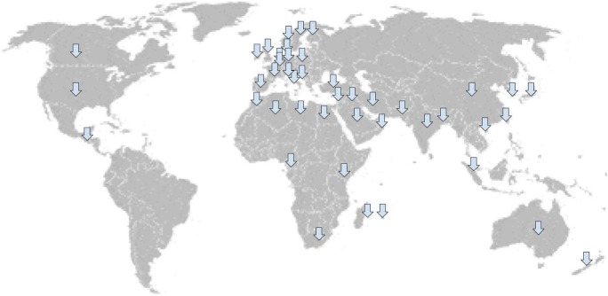
The Balkans have been another area with high rates of NDM-positive bacteria, which has now spread to all continents except South America and Antarctica [Fig. 1 from reference 34]]. NDM-positive bacteria have even now been found in companion animals in the USA [34]. Adding to the concern is that there are few antimicrobials in the research pipeline with activity against Gram-negative pathogens.
Fig. 1.
Countries where NDM-positive bacteria have been reported
Rift Valley Fever Virus
Rift Valley fever virus (RVFV) is a single-stranded RNA virus of the genus Phlebovirus, family Bunyaviridae. The majority of infected individuals are asymptomatic. Most patients who are symptomatic have a self-limiting febrile illness; however, a small minority develop severe disease with acute hepatitis, renal failure, and hemorrhagic manifestations. Those who survive this phase often develop neurologic symptoms consisting of vision loss and encephalitis. Long-term sequelae may develop with blindness, hemiparesis, quadriparesis, incontinence, hallucinations, or coma. The fatality rate is 1–3%, though patients with hemorrhagic disease have a fatality rate as high as 50%. Diagnosis is made by detection of viral RNA and immunoglobulin serology, particularly during the acute febrile stage [38]. It is often transmitted by mosquitoes, typically of the Aedes genus, though exposure to infected animal tissue or consumption of their products can also cause disease [39]. RVFV is endemic in sub-Saharan Africa with periodic outbreaks after heavy rainfall and flooding. It has spread to the Middle East and the French island of Mayotte. Outbreaks often have significant effects on both humans and livestock. Characteristics of an RVFV outbreak include the sudden increase in abortions and mortality of young animals with the appearance of disease in humans. Models have been developed with successful predictions of outbreaks using satellite imagery of sea surface temperatures, rainfall, and vegetation. Given this spread out of Africa, there is concern about it reaching the Mediterranean basin and Europe. A number of competent vectors for RVFV have been identified in Europe. It has additionally been isolated from other hematophagous arthropods including ticks and sand flies, though their significance is not currently understood. There is currently no specific treatment, though a vaccine, TSI-GSD-200, has provided protection following primary inoculation and single boost schedule [38].
Crimean-Congo Hemorrhagic Fever
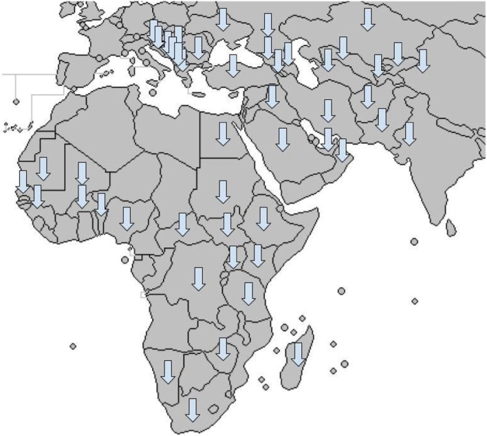
Crimean-Congo hemorrhagic fever virus (CCHFV) produced its first major known outbreak from 1944 to 1945 in the Crimean Peninsula, though it has likely been causing outbreaks for centuries. It was not identified until 1968. As such, it has not recently emerged but is listed as a priority agent for research and development by WHO due to its potential for spreading and causing more severe outbreaks. CCHFV is a member of the Bunyaviridae family, genus Nairovirus. It has many aliases, including Asian Ebola, Karakhalak (black death), Khunymuny (nose bleeding), and Khungribta (blood taking). CCHFV derives its name from outbreaks both in Crimea and the Belgian Congo and has been reported throughout many parts of Africa, the Middle East, Europe, and Asia [Fig. 2 from reference 40]. Crimean-Congo hemorrhagic fever (CCHF) is the most widespread tick-borne viral infection of humans [41]. It is a negative-sense RNA virus transmitted by numerous tick species; because of this, agricultural workers are most commonly affected [40, 41]. Additionally, human-to-human transmission may occur via contact with skin, mucous membranes, or body fluids of infected patients [40]. Standard barrier precautions are recommended for patients with suspected disease [41].
Fig. 2.
Distribution of Crimean-Congo Hemorrhagic Fever Virus
Domestic livestock are the main reservoirs. Outbreaks have been increasing in recent years. Likely targets of the virus are endothelial cells, hepatic cells, and Kupffer cells. The clinical course of CCHF follows 4 phases consisting of incubation, prehemorrhagic, hemorrhagic, and convalescence [40]. There is a spectrum of severity from a mild, febrile illness to hemorrhagic shock with multiorgan failure [41]. The incubation period varies by method of acquisition; it typically takes 1–3 days by tick bite and 5–6 days by exposure to infected patients. The prehemorrhagic stage is nonspecific with flu-like symptoms. The hemorrhagic stage follows and develops within 3–6 days of symptoms in severe disease [40]. One differentiating feature is that severely ill patients often develop a pattern of large ecchymosis not seen in other hemorrhagic fevers [41]. Laboratory findings include leukopenia, thrombocytopenia, elevated aminotransferases, elevated CK and LDH, and prolonged prothrombin time and activated partial thromboplastin time. Convalescence usually occurs 15–20 days after onset of illness [40].
Diagnosis is commonly made via detection of viral RNA with RT-PCR or serology. There has traditionally been no specific treatment for CCHF, though ribavirin has demonstrated both in vitro and in vivo activity [40]. Furthermore, both the CDC and WHO recommend use of ribavirin [42•]. A report from Turkey claimed that a combination of methylprednisolone, intravenous immunoglobulin (IVIG), and fresh-frozen plasma was beneficial, but there was no control group for comparison [41]. IVIG and methylprednisolone may be beneficial by improving thrombocytopenia [42•, 43]. Two vaccines have been developed, though they have not been through randomized clinical trials [40]. CCHF has never been demonstrated in vaccinated individuals and the Bulgarian Ministry of Health has reported a fourfold decrease in cases since vaccine implementation [41, 43]. Ribavirin might be beneficial for post-exposure prophylaxis [43]. Mortality rates have varied between 10 and 50%, with worsening prognosis if the disease is acquired nosocomially, with higher levels of aminotransferases, higher viremia, or with coagulopathy [40, 41]. Long-term sequelae have been documented but are rarely permanent and include impaired vision and other neurologic symptoms [42•].
CCHF has epidemic potential with high rates of mortality, nosocomial infection, and difficulty with prevention and treatment [40]. Additionally, it is the most genetically diverse of the arboviruses [41]. Attempts to control CCHF via eradication of tick vectors have proven inefficient and domestic animals are asymptomatic even when highly viremic [42•].
Conclusions
Infectious diseases promise to be one of the biggest challenges in the coming decades, with new ones emerging unpredictably. Successful clinical outcomes require a proactive approach with research and development, as human behavior contributes to new infections emerging, particularly with zoonoses. Although this strategy would prove costly, this article has demonstrated a few examples of how much an emerging infectious disease can cost during an outbreak. This article has reviewed a number of emerging infections that the WHO has categorized as priorities for research and development due to their potential for epidemics or even a pandemic. Additionally, it has reviewed a concerning drug-resistance mechanism that threatens to provide bacteria with resistance to all antibiotics. Finally, a few infectious diseases that have emerged in the USA have been covered, though little is known about them, with a paucity of cases to date.
Compliance with Ethical Standards
Conflict of Interest
The author declares that he has no conflict of interest.
Human and Animal Rights and Informed Consent
This article does not contain any studies with human or animal subjects performed by any of the authors.
Footnotes
This article is part of the Topical Collection on Infectious Disease
References
Papers of particular interest, published recently, have been highlighted as: • Of importance •• Of major Importance
- 1.Hughes JM. Emerging infectious diseases: a CDC perspective. Emerg Infect Dis. 2001;7(3 Suppl):494–496. doi: 10.3201/eid0707.017702. [DOI] [PMC free article] [PubMed] [Google Scholar]
- 2.Morens DM, Fauci AS. Emerging infectious diseases in 2012: 20 years after the Institute of Medicine Report. MBio. 2012;3(6):e00494–e00412. doi: 10.1128/mBio.00494-12. [DOI] [PMC free article] [PubMed] [Google Scholar]
- 3.Kilpatrick AM, Randolph SE. Drivers, dynamics, and control of emerging vector-borne zoonotic diseases. Lancet. 2012;380(9857):1946–1955. doi: 10.1016/S0140-6736(12)61151-9. [DOI] [PMC free article] [PubMed] [Google Scholar]
- 4.Bloom DE, Black S, Rappuoli R. Emerging infectious disease: a proactive approach. Proc Natl Acad Sci U S A. 2017;114(16):4055–4059. doi: 10.1073/pnas.1701410114. [DOI] [PMC free article] [PubMed] [Google Scholar]
- 5.Ksiazek TG, Erdman D, Goldsmith CS, Zaki SR, Peret T, Emery S, Tong S, Urbani C, Comer JA, Lim W, Rollin PE, Dowell SF, Ling AE, Humphrey CD, Shieh WJ, Guarner J, Paddock CD, Rota P, Fields B, DeRisi J, Yang JY, Cox N, Hughes JM, LeDuc J, Bellini WJ, Anderson LJ, SARS Working Group A novel coronavirus associated with severe acute respiratory syndrome. N Engl J Med. 2003;348(20):1953–1966. doi: 10.1056/NEJMoa030781. [DOI] [PubMed] [Google Scholar]
- 6.Demmler GJ, Ligon BL. Severe acute respiratory syndrome (SARS): a review of the history, epidemiology, prevention, and concerns for the future. Semin Pediatr Infect Dis. 2003;14(3):240–244. doi: 10.1016/S1045-1870(03)00056-6. [DOI] [PMC free article] [PubMed] [Google Scholar]
- 7.Hui DS, Chan PK. Severe acute respiratory syndrome and coronavirus. Infect Dis Clin N Am. 2010;24(3):619–638. doi: 10.1016/j.idc.2010.04.009. [DOI] [PMC free article] [PubMed] [Google Scholar]
- 8.Peiris JS. Severe acute respiratory syndrome (SARS) J Clin Virol. 2003;28(3):245–247. doi: 10.1016/j.jcv.2003.08.005. [DOI] [PMC free article] [PubMed] [Google Scholar]
- 9.Shortridge KF. Severe acute respiratory syndrome and influenza: virus incursions from southern China. Am J Respir Crit Care Med. 2003;168(12):1416–1420. doi: 10.1164/rccm.2310005. [DOI] [PubMed] [Google Scholar]
- 10.World Health Organization (2004) WHO guidelines for the global surveillance of severe acute respiratory syndrome (SARS). http://www.who.int/csr/resources/publications/WHO_CDS_CSR_ARO_2004_1/en/.
- 11.Kumar V, Jung YS, Liang PH. Anti-SARS coronavirus agents: a patent review (2008-present) Expert Opin Ther Pat. 2013;23(10):1337–1348. doi: 10.1517/13543776.2013.823159. [DOI] [PubMed] [Google Scholar]
- 12.Shehata MM, Gomaa MR, Ali MA, Kayali G. Middle East respiratory syndrome coronavirus: a comprehensive review. Front Med. 2016;10(2):120–136. doi: 10.1007/s11684-016-0430-6. [DOI] [PMC free article] [PubMed] [Google Scholar]
- 13.Sampathkumar P. Middle East respiratory syndrome: what clinicians need to know. Mayo Clin Proc. 2014;89(8):1153–8. doi: 10.1016/j.mayocp.2014.06.008. [DOI] [PMC free article] [PubMed] [Google Scholar]
- 14.Rasmussen SA, Gerber SI, Swerdlow DL. Middle East respiratory syndrome coronavirus: update for clinicians. Clin Infect Dis. 2015;60(11):1686–1689. doi: 10.1093/cid/civ118. [DOI] [PMC free article] [PubMed] [Google Scholar]
- 15.•• World Health Organization (2017) WHO MERS-CoV Global Summary and Assessment of Risk. http://www.who.int/emergencies/mers-cov/risk-assessment-july-2017.pdf. This article gives the majority of a summary of perhaps most significant recent emerging infectious disease in terms of both mortality and number of cases. It describes not only the cases noted in the Middle East, but additionally the South Korean outbreak as well as cases imported to nations globally.
- 16.Al-Tawfiq JA, Kattan RF, Memish ZA. Middle East respiratory syndrome coronavirus disease is rare in children: an update from Saudi Arabia. World J Clin Pediatr. 2016;5(4):391–396. doi: 10.5409/wjcp.v5.i4.391. [DOI] [PMC free article] [PubMed] [Google Scholar]
- 17.Kim SW, Park JW, Jung HD, Yang JS, Park YS, Lee C, Kim KM, Lee KJ, Kwon D, Hur YJ, Choi BY, Ki M. Risk factors for transmission of Middle East respiratory syndrome coronavirus infection during the 2015 outbreak in South Korea. Clin Infect Dis. 2017;64(5):551–557. doi: 10.1093/cid/ciw768.. [DOI] [PMC free article] [PubMed] [Google Scholar]
- 18.Arabi YM, Arifi AA, Balkhy HH, Najm H, Aldawood AS, Ghabashi A, Hawa H, Alothman A, Khaldi A, al Raiy B. Clinical course and outcomes of critically ill patients with Middle East respiratory syndrome coronavirus infection. Ann Intern Med. 2014;160(6):189–197. doi: 10.7326/M13-2486. [DOI] [PubMed] [Google Scholar]
- 19.Madani TA et al (2015) Infection Prevention and Control Guidelines for Middle East Respiratory Syndrome Coronavirus (MERS-CoV) Infection, 3rd Ed. https://www.moh.gov.sa/endepts/Infection/Documents/Guidelines-for-MERS-CoV.PDF. [PubMed]
- 20.Chan JF, et al. Broad-spectrum antivirals for the emerging Middle East respiratory syndrome coronavirus. J Inf Secur. 2013;67(6):606–616. doi: 10.1016/j.jinf.2013.09.029. [DOI] [PMC free article] [PubMed] [Google Scholar]
- 21.Mo Y, Fisher D. A review of treatment modalities for Middle East respiratory syndrome. J Antimicrob Chemother. 2016;71(12):3340–3350. doi: 10.1093/jac/dkw338. [DOI] [PMC free article] [PubMed] [Google Scholar]
- 22.Al Ghamdi M, et al. Treatment outcomes for patients with middle eastern respiratory syndrome coronavirus (MERS CoV) infection at a coronavirus referral center in the Kingdom of Saudi Arabia. BMC Infect Dis. 2016;16:174. doi: 10.1186/s12879-016-1492-4. [DOI] [PMC free article] [PubMed] [Google Scholar]
- 23.McMullan LK, et al. A new Phlebovirus associated with severe febrile illness in Missouri. N Engl J Med. 2012;367(9):834–841. doi: 10.1056/NEJMoa1203378. [DOI] [PubMed] [Google Scholar]
- 24.Pastula DM, et al. Notes from the field: heartland virus disease—United States, 2012-2013. MMWR Morb Mortal Wkly Rep. 2014;63(12):270–271. [PMC free article] [PubMed] [Google Scholar]
- 25.Muehlenbachs A, Fata CR, Lambert AJ, Paddock CD, Velez JO, Blau DM, Staples JE, Karlekar MB, Bhatnagar J, Nasci RS, Zaki SR. Heartland virus associated death in Tennessee. Clin Infect Dis. 2014;59(6):845–850. doi: 10.1093/cid/ciu434. [DOI] [PMC free article] [PubMed] [Google Scholar]
- 26.Wormser GP, Pritt B. Update and commentary on four emerging tick-borne infections. Infect Dis Clin N Am. 2015;29(2):371–381. doi: 10.1016/j.idc.2015.02.009. [DOI] [PubMed] [Google Scholar]
- 27.Kosoy OI, Lambert AJ, Hawkinson DJ, Pastula DM, Goldsmith CS, Hunt DC, Staples JE. Novel Thogotovirus associated with febrile illness and death, United States, 2014. Emerg Infect Dis. 2015;21(5):760–764. doi: 10.3201/eid2105.150150. [DOI] [PMC free article] [PubMed] [Google Scholar]
- 28.Savage HM, Burkhalter KL, Godsey MS, Jr, Panella NA, Ashley DC, Nicholson WL, Lambert AJ. Bourbon virus in field-collected ticks, Missouri, USA. Emerg Infect Dis. 2017;23(12):2017–2022. doi: 10.3201/eid2312.170532. [DOI] [PMC free article] [PubMed] [Google Scholar]
- 29.Sherrini BA, Chong TT. Nipah encephalitis—an update. Med J Malaysia. 2014;69(Supplement A):103–111. [PubMed] [Google Scholar]
- 30.Luby SP. The pandemic potential of Nipah virus. Antivir Res. 2013;100(1):38–43. doi: 10.1016/j.antiviral.2013.07.011. [DOI] [PubMed] [Google Scholar]
- 31.Geisbert TW, Mire CE, Geisbert JB, Chan YP, Agans KN, Feldmann F, Fenton KA, Zhu Z, Dimitrov DS, Scott DP, Bossart KN, Feldmann H, Broder CC. Therapeutic treatment of Nipah virus infection in nonhuman primates with a neutralizing human monoclonal antibody. Sci Transl Med. 2014;6(242):242ra82. doi: 10.1126/scitranslmed.3008929. [DOI] [PMC free article] [PubMed] [Google Scholar]
- 32.Schwartz IS, Sanche S, Wiederhold NP, Patterson TF, Sigler L. Emergomyces canadensis, a dimorphic fungus causing fatal systemic human disease in North America. Emerg Infect Dis. 2018;24(4):758–761. doi: 10.3201/eid2404.171765. [DOI] [PMC free article] [PubMed] [Google Scholar]
- 33.Schwartz IS, Maphanga TG, Govender NP. Emergomyces: a new genus of dimorphic fungal pathogens causing disseminated disease among immunocompromised persons globally. Curr Fungal Infect Rep. 2018;12(1):44–50. doi: 10.1007/s12281-018-0308-y. [DOI] [Google Scholar]
- 34.Johnson AP, Woodford N. Global spread of antibiotic resistance: the example of New Delhi metallo-ß-lactamase (NDM)-mediated carbapenem resistance. J Med Microbiol. 2013;62(Pt. 4):499–513. doi: 10.1099/jmm.0.052555-0. [DOI] [PubMed] [Google Scholar]
- 35.Walsh TR, Toleman MA. The new medical challenge: why NDM-1? Why Indian? Expert Rev Anti-Infect Ther. 2011;9(2):137–141. doi: 10.1586/eri.10.159. [DOI] [PubMed] [Google Scholar]
- 36.Kumarasamy KK, Toleman MA, Walsh TR, Bagaria J, Butt F, Balakrishnan R, Chaudhary U, Doumith M, Giske CG, Irfan S, Krishnan P, Kumar AV, Maharjan S, Mushtaq S, Noorie T, Paterson DL, Pearson A, Perry C, Pike R, Rao B, Ray U, Sarma JB, Sharma M, Sheridan E, Thirunarayan MA, Turton J, Upadhyay S, Warner M, Welfare W, Livermore DM, Woodford N. Emergence of a new antibiotic resistance mechanism in India, Pakistan, and the UK: a molecular, biological, and epidemiological study. Lancet Infect Dis. 2010;10(9):597–602. doi: 10.1016/S1473-3099(10)70143-2. [DOI] [PMC free article] [PubMed] [Google Scholar]
- 37.Walsh TR, Toleman MA. The emergence of pan-resistant Gram-negative pathogens merits a rapid global political response. J Antimicrob Chemother. 2012;67(1):1–3. doi: 10.1093/jac/dkr378. [DOI] [PubMed] [Google Scholar]
- 38.Mansfield KL, Banyard AC, McElhinney L, Johnson N, Horton DL, Hernández-Triana LM, Fooks AR. Rift Valley fever virus: a review of diagnosis and vaccination, and implications for emergence in Europe. Vaccine. 2015;33(42):5520–5531. doi: 10.1016/j.vaccine.2015.08.020. [DOI] [PubMed] [Google Scholar]
- 39.Nicholas DE, Jacobsen KH, Waters NM. Risk factors associated with human Rift Valley fever virus infection: systematic review and meta-analysis. Tropical Med Int Health. 2014;19(12):1420–1429. doi: 10.1111/tmi.12385. [DOI] [PubMed] [Google Scholar]
- 40.Shayan S, Bokaean M, Shahrivar MR, Chinikar S. Crimean-Congo hemorrhagic fever. Lab Med. 2015;46(3):180–189. doi: 10.1309/LMN1P2FRZ7BKZSCO. [DOI] [PubMed] [Google Scholar]
- 41.Bente DA, Forrester NL, Watts DM, McAuley AJ, Whitehouse CA, Bray M. Crimean-Congo hemorrhagic fever: history, epidemiology, pathogenesis, clinical syndrome and genetic diversity. Antivir Res. 2013;100(1):159–189. doi: 10.1016/j.antiviral.2013.07.006. [DOI] [PubMed] [Google Scholar]
- 42.• Mendoza EJ et al. (2018) Crimean-Congo haemorrhagic fever virus: past, present and future insights for animal modelling and medical countermeasures. Zoonoses Public Health 1–16. 10.1111/zph.12469. This article highlights potential management strategies of the most widespread tick-borne viral disease globally. [DOI] [PMC free article] [PubMed]
- 43.Papa A, Mirazimi A, Köksal I, Estrada-Pena A, Feldmann H. Recent advances in research on Crimean-Congo hemorrhagic fever. J Clin Virol. 2015;64:137–143. doi: 10.1016/j.jcv.2014.08.029. [DOI] [PMC free article] [PubMed] [Google Scholar]


