Abstract
We performed a systematic review and meta-analysis of studies performing sclerotherapy for treatment of venous malformations (VMs) of the face, head and neck. It is our hope that data from this study could be used to better inform providers and patients regarding the benefits and risks of percutaneous sclerotherapy for treatment of face, head and neck VMs. We searched PubMed, MEDLINE, and EMBASE from 2000–2018 for studies evaluating the safety and efficacy of percutaneous sclerotherapy of neck, face and head VMs. Two independent reviewers selected studies and abstracted data. The primary outcomes were complete and partial resolution of the VM. Data were analyzed using random-effects meta-analysis. Thirty-seven studies reporting on 2,067 patients were included. The overall rate of complete cure following percutaneous sclerotherapy with any agent was 64.7% (95% confidence interval [CI], 57.4–72.0%). Sodium tetradecyl sulfate had the lowest complete cure rate at 55.5% (95% CI, 36.1–74.9%) while pingyangmycin had the highest cure rate at 82.9% (95% CI, 71.1–94.7%). Overall patient satisfaction rates were 91.0% (95% CI, 86.1–95.9%). Overall quality of life improvement was 78.9% (95% CI, 67.0–90.8%). Overall permanent morbidity/mortality was 0.8% (95% CI, 0.3–1.3%) with no cases of mortality. Our systematic review and meta-analysis of 37 studies and over 2,000 patients found that percutaneous sclerotherapy is a very safe and effective treatment modality for treatment of VMs of the head, neck and face.
Keywords: Venous malformations, Venous, Head and neck, Sclerotherapy
INTRODUCTION
Venous malformations (VMs) are slow flow developmental anomalies of the veins which do not proliferate and normally do not involute [1-37]. These lesions can develop anywhere in the body, including structures of the face, head and neck. Due to the delicate interplay between aesthetics, function, and anatomy in this region, management of these lesions with any treatment modality can be challenging.
Over the past several decades, sclerotherapy with various agents has been demonstrated to be effective for face, head and neck VMs [1,6,13,38-40]. However, the evidence for treatment of these lesions is often based off of smaller case series which makes generalizing results to the greater population difficult. Furthermore, comparative studies examining the efficacy and safety of various sclerotherapy agents are few and far between. The goals of this systematic review and meta-analysis were to 1) understand the overall safety and efficacy rates of percutaneous sclerotherapy for treatment of VMs in the face, head and neck and 2) compare safety and efficacy rates of commonly used sclerotherapy agents. It is our hope that data from this study could be used to better inform providers and patients regarding the benefits and risks of percutaneous sclerotherapy for treatment of face, head and neck VMs.
MATERIALS AND METHODS
Literature search
The systematic review is reported according to the Preferred Reporting Items for Systematic Reviews and Meta-Analysis guidelines [37]. A comprehensive literature search of the databases PubMed, Ovid MEDLINE, and Ovid EMBASE was designed and conducted by an experienced librarian with input from the authors. The search duration was 2 months. The key words “sclerotherapy,” “vascular malformations,” “venous malformations,” “arteriovenous malformation,” “hemangioma,” “lymphatic malformation, “head,” “neck,” “facial,” “oropharyngeal,” and “orbital” were used in “AND” and “OR” combinations. The search was limited to articles published from 2000 to 2018. Inclusion criteria were the following: 1) English or Italian language, 2) case series reporting greater than 5 patients, 3) studies reporting image guided percutaneous sclerotherapy, 4) studies reporting exclusively face, head and neck VMs or subdividing outcomes and complications by anatomical region, and 5) studies classifying VMs appropriately using the International Society for the Study of Vascular Anomalies. Exclusion criteria were: 1) case series reporting fewer than 5 patients, 2) case reports, 3) vascular malformation not of the head and/or neck region (e.g., sclerotherapy for varicose veins in legs), and 4) studies not classifying lesions according to the International Society for the Study of Vascular Anomalies criteria. International Society for the Study of Vascular Anomalies criteria on imaging and clinical exam for VMs on imaging include 1) septated lobulated T2 hyperintense and T1 hypointense mass without mass effect, 2) phleboliths which are characteristically hypointense on T1/T2, 3) presence of fluid-fluid levels, 4) no flow voids on spin echo sequences, 5) the lesion infiltrates tissue planes, 6) no arterial or early venous enhancement, and 7) diffuse enhancement on delayed images.41 On clinical exam, VMs appear as faint blue, soft and easily compressible non-pulsatile masses. The lesions characteristically enlarged with Valsalva maneuver and in dependent positions and decompress with local compression.
In studies with overlapping patient populations written by the same author/institution, we only included the largest or most complete dataset. In cases where outcomes were separated out by the type of sclerotherapy agent used, we abstracted outcomes separately for each agent in order to perform our subgroup analyses. Two authors determined inclusion and exclusion criteria for the studies in the literature search with differences resolved by the senior author.
Outcomes and data extraction
For each study, we extracted the following baseline information: number of patients, mean or median age and gender, number of malformations treated, location of malformations, sclerosing agent and its mean volume used, mean number of treatment sessions, and mean length of radiographic and clinical follow-up. The primary outcome of this study is the efficacy of sclerotherapy which includes complete cure of the vascular malformation (resolution of the VM on physical exam), partial cure of the vascular malformation (partial decrease in VM size), lack of benefit following sclerotherapy, improvement in quality of life (QoL), and patient satisfaction. Secondary outcomes are adverse events after sclerotherapy, including respiratory complications, skin necrosis/scars, any permanent morbidity/mortality, local temporary complications). Permanent morbidity and mortality were defined as mortality or any permanent neurological deficit. Local temporary complications included erythema, swelling, and pain.
For our subgroup analysis by sclerotherapy agent, we separated outcomes by agent. We were able to abstract data for the following individual agents: bleomycin, ethanol, sodium tetradecyl sulfate (STS), ethanolamine and pingamycin.
Study risk of bias
We modified the Newcastle-Ottawa Quality Assessment Scale to assess the methodologic quality of the studies included in this meta-analysis. This tool is designed for use in comparative studies; however, because the studies did not include a control group, we assessed study risk of bias based on selected items from the tool, focusing on the following questions: 1) Did the study include all patients or consecutive patients versus a selected sample?, 2) Was the study retrospective or prospective?, 3) Was clinical follow-up satisfactory, thus allowing ascertainment of all outcomes?, 4) Were outcomes clearly reported?, and 5) Were there clearly defined inclusion and exclusion criteria?
Statistical analysis
We estimated from each cohort the cumulative prevalence and 95% confidence interval (CI) for each outcome. Event rates were pooled across studies with a random-effects meta-analysis. Heterogeneity across studies was evaluated using the I2 statistic. An I2 value of >50% suggests substantial heterogeneity. We also extracted a 2×2 table to calculate P values for the comparisons among the results. For the purpose of statistical comparisons we chose bleomycin sclerotherapy as the reference group, since it is the sclerosing agent most commonly used in the USA. Meta-regression was not used in this study. Statistical analyses were performed using OpenMeta[Analyst] (http://www.cebm.brown.edu/openmeta/; Biostat, Englewood, NJ, USA).
RESULTS
Literature search
The initial literature search yielded 1,211 articles. On review of the abstracts and titles, we excluded 1,126 articles. Eightyfive articles were selected for full-text screening, of which 37 met inclusion criteria [1-37]. The remaining 48 articles were excluded for reasons including 1) failure to separate outcomes by anatomic location (9 articles), 2) inclusion of lymphatic or venolymphatic malformations rather than pure VMs (14 articles), 3) use of confusing or unclear terminology making it difficult to ascertain whether lesions were VMs or hemangiomas (12 articles), and 4) mixture of VMs, AVMs and lymphatic malformations (13 articles). All studies included in the analysis had at least one or more outcome measure available for one or more of the patients groups analyzed. Fig. 1 shows the flow chart according to the PRISMA statement [37].
Fig. 1.
PRISMA flow diagram. VMs, venous malformations; PRISMA, Preferred Reporting Items for Systematic Reviews and Meta-Analysis.
These 37 studies included a total of 2,067 patients. The smallest study included 10 patients and the largest included 358 patients. Mean age was 24.9 years. There was a female predilection (1:1.2). The mean number of malformations per patient was 1.08 and they were all located in the head and/or neck region. The highest number of treated malformations per study was 358, while the least was 10. The mean number of treatment sessions per patient was 2.4. The mean length of radiographic and clinical follow-up from the time of the first treatment was 16.61 months and 18.04 months respectively.
Most included studies used a single sclerosing agent for each vascular malformation, while the remainder used a combination of them. Five studies reported outcomes of bleomycin sclerotherapy, 10 studies reported outcomes of ethanol sclerotherapy, 7 studies reported outcomes of STS sclerotherapy, 4 studies reported outcomes of ethanolamine sclerotherapy, and 4 studies reported outcomes of pingamycin sclerotherapy. In 11 studies either multiple agents were used and we could not separate outcomes by agent or other sclerosing agent including OK-432 were used. A summary of included studies is provided in Table 1.
Table 1.
| No. | Study | No. of patients | Mean/median age (years) | M:F | No. of malformations treated | Location of malformations (if specific) | Sclerosing agent | Mean volume used (mL) | Mean No. of treatment sessions | Mean length of radiographic follow-up (months) | Mean length of clinical follow-up (months) |
|---|---|---|---|---|---|---|---|---|---|---|---|
| 1 | Akan et al. [1] (2017) | 13 | 30 | 1:12 | 66 | Oropharynx | Ethanol | 6 | 1.8 | NR | 11 |
| 2 | Alakailly et al. [2] (2015) | 13 | 18.2 | 1:3.3 | 14 | NR | STS | NR | 1.4 | 9 | 9 |
| 3 | Alexander et al. [3] (2016) | 37 | 21.7 | 14:23 | 37 | NR | STS, ethanolamine | NR | 1.5 | 20.8 | 20.8 |
| 4 | Baek et al. [4] (2011) | 22 | 30.3 | 1:1.2 | NR | NR | Ethanol | 8.5 | 1.7 | NR | 13 |
| 5 | Bajpai and Bajpai [5] (2012) | 8 | 17 | 1:1 | NR | NR | Bleomycin | NR | 1.1 | 12 | 30 |
| 6 | Bajpai and Bajpai [5] (2012) | 8 | 15 | 1:1 | NR | NR | STS | NR | 5 | NR | 30 |
| 7 | Bourgouin et al. [6] (2018) | 33 | 43 | 1:1 | 46 | Oropharynx | STS | NR | 2.9 | 9 | 3 |
| 8 | Castren et al. [7] (2016) | 75 | 35.9 | 1:1.2 | 75 | NR | STS, polidocanol, bleomycin, ethanol doxycyclin | NR | 2 | NR | 18 |
| 9 | Chen et al. [8] (2015) | 11 | 36 | 1:1 | NR | NR | NR | 21 | 4 | 6 | |
| 10 | Chen et al. [9] (2010) | 18 | 11.8 | 1:1.25 | NR | Face, neck | OK-432 , PYM | NR | NR | NR | 8.4 |
| 11 | Chen et al. [10] (2008) | 19 | 15 | 1:1 | NR | Orbital | PYM | NR | NR | 23 | 23 |
| 12 | Choi et al. [11] (2002) | 29 | 22 | 10:19 | 29 | Cheek, lip, submandibular, temporalis muscle, scalp, parotid, orbit, oropharynx | Ethanolamine | NR | 2 | 8.5 | 8.5 |
| 13 | Colletti et al. [12] (2017) | 69 | 34 | NR | 69 | NR | STS | 7.75 | 2.1 | NR | NR |
| 14 | Costa et al. [13] (2011) | 53 | 51.5 | 1:1.2 | 66 | Oral, maxillofacial | Ethanolamine | NR | 1 | ||
| 15 | Jia et al. [14] (2014) | 33 | 23.8 | 15:18 | 33 | Orbital, periorbital | PYM | NR | 2 | 7.9 | 7.9 |
| 16 | Kim et al. [15] (2004) | 10 | 5.4 | 1:1.5 | NR | NR | Bleomycin | NR | 4.3 | NR | NR |
| 17 | Kim et al. [15] (2004) | 35 | 21 | 1:1 | NR | NR | OK-432 | 8.5 | 1.8 | NR | 11 |
| 18 | Kim et al. [15] (2004) | 29 | 22 | 1:1.9 | NR | NR | Ethanolamine | NR | NR | NR | NR |
| 19 | Kishi et al. [16] (2014) | 23 | 32 | 1:1.5 | 35 | NR | Ethanol | NR | NR | ||
| 20 | Lamba et al. [17] (2012) | 15 | 25 | 10:5 | 15 | Face, neck | Ethanol | 21 | 1.2 | 7.6 | 7.6 |
| 21 | Lee et al. [18] (2009) | 87 | 17.5 | 40:47 | NR | NR | Ethanol | 38 | 3.5 | 35 | 35 |
| 22 | Li et al. [19] (2010) | 20 | 17.14 | 10:10 | 21 | Maxillofacial | PYM, Ethanol | 18,9 (PYM), 12.5 (ethanol) | 2.4 | 13.5 | 13.5 |
| 23 | Liu et al. [20] (2009) | 23 | 21 | 12:11 | 23 | Cheek, parotid, infraorbital, scalp, chin, submandibular, oral | PYM, Ethanol | 22 (PYM), 2.8 (ethanol) | 2.5 | 2.5 | 20.3 |
| 24 | Meng et al. [21] (2014) | 43 | 20.5 | 26:17 | 43 | Tongue, pharynx, parotid | PYM, Ethanol | NR | NR | 24 | 24 |
| 25 | Orlando et al. [22] (2014) | 51 | 23 | 14:37 | 51 | Face, tongue, neck, lip | Ethanol | 2.5 | 7 | 18 | 18 |
| 26 | Ribeiro et al. [23] (2018) | 17 | 41.6 | 6:11 | 34 | NR | Ethanolamine | NR | 2.8 | 6 | 6 |
| 27 | Rosbe et al. [24] (2010) | 10 | 20.3 | 4:06 | 12 | Masseter | STS | NR | 1 | 28 | 28 |
| 28 | Sachin et al. [25] (2013) | 358 | NR | NR | 358 | NR | Butyl cyanoacrylate, polyvinyl alcohol, STS, bleomycine | NR | NR | 4.7 | 56.4 |
| 29 | Shigematsu et al. [26] (2019) | 18 | 34.3 | 6:12 | 18 | Eyes | Bleomycin | 34.5 | 3 | 43 | 43 |
| 30 | Siniluoto et al. [27] (1997) | 34 | 28 | 14.24 | 34 | NR | STS | 11.8 | 2.2 | NR | nr |
| 31 | Songsaeng et al. [28] (2015) | 33 | 25.1 | 6:27 | 43 | NR | Bleomycin, ethanol | NR | 3 | 14.7 | 14.7 |
| 32 | Spence et al. [29] (2011) | 17 | 40 | 7:09 | 17 | Face, tongue, parotid | Ethanol | 8.1 | 1.7 | 6 | 6 |
| 33 | Spence et al. [29] (2011) | 17 | 32.4 | 5:12 | 17 | Face, tongue, parotid | Bleomicyn | NR | 3.4 | 6 | 6 |
| 34 | Spence et al. [30] (2010) | 31 | 34.4 | 14:17 | 32 | Face, orbital, parotid, oropharinx | Bleomicyn | NR | 3.5 | 5.2 | 5.2 |
| 35 | Stimpson et al. [31] (2012) | 12 | 7 | 7:05 | 12 | Oropharynx | STS | NR | 3 | 28 | 28 |
| 36 | Su et al. [32] (2010) | 60 | 22.6 | 26:34 | 60 | Face, neck | Ethanol | 11.2 | 1.8 | 8 | 8 |
| 37 | Wang et al. [33] (2017) | 21 | 25.4 | 12:09 | 72 | Oropharynx, head, neck | Ethanol | 79.6 | 1.5 | 9.1 | 9.1 |
| 38 | Wang et al. [34] (2010) | 23 | 19 | 13:10 | 23 | Parotid, oral, neck, face | Ethanol | 73 | 1.5 | 25 | 25 |
| 39 | Zhao et al. [35] (2004) | 260 | NR | 131:159 | 260 | Oral, face | PYM, sodium morrhuate | NR | 1.7 | NR | NR |
| 40 | Zheng et al. [36] (2009) | 297 | 20.6 | 159:138 | 297 | Oral, maxillofacial, head, neck | PYM | NR | 3.5 | 36 | 36 |
| 41 | Zhi et al. [37] (2008) | 82 | 12 | 26:56 | 82 | Maxillofacial | PYM | NR | 2.9 | 24 | 24 |
No., number; M, male; F, female; NR, not reported; STS, sodium tetradecyl sulfate; PYM, pingyangmycin.
Efficacy outcomes
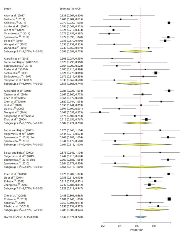
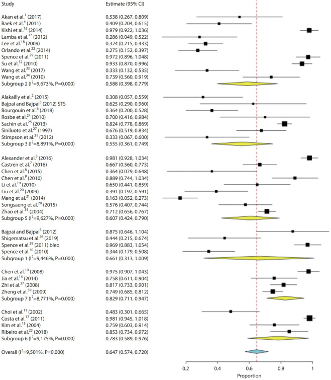
Overall complete cure rates were reported in 1,736 patients. The overall rate of complete cure following percutaneous sclerotherapy with any agent was 64.7% (95% CI, 57.4–72.0%). STS had the lowest complete cure rate at 55.5% (95% CI, 36.1–74.9%) while pingyangmycin had the highest cure rate at 82.9% (95% CI, 71.1–94.7%). Fig. 2 shows the complete cure rates forest plot. Overall partial cure rates were reported in 1,703 patients. The overall rate of partial cure following percutaneous sclerotherapy with any agent was 28.0% (95% CI, 22.1–34.0%). Partial cure rates ranged from 16.2% (95% CI, 4.7–27.6%) for pingyangmycin to 35.3% (95% CI, 19.1–51.5%) with ethanol. The overall rate of no benefit following percutaneous sclerotherapy was reported in 1,736 patients and was 4.5% (95% CI, 3.0–6.1%). STS sclerotherapy had the highest rate of no benefit (14.9%; 95% CI, 4.3–25.4%) while pingyangmycin had the lowest rate of no benefit (0.6%; 95% CI, 0.0–1.6%). Patient satisfaction was reported in 315 patients. Overall patient satisfaction rates were 91.0% (95% CI, 86.1–95.9%). Patient satisfaction rates ranged from 72.8% (95% CI, 63.6–81.9%) with STS to 96.0% (95% CI, 92.5–99.6%) with ethanol. Improvement in QoL was reported in 243 patients. Overall QoL improvement was 78.9% (95% CI, 67.0–90.8%) ranging from 46.7% (95% CI, 22.4–71.0%) for STS to 98.1% (95% CI, 94.5–100%) with ethanolamine.
Fig. 2.
Forest plot: complete cure rates. Subgroup 1, bleomycin; subgroup 2, ethanol; subgroup 3, sotradecol; subgroup 5, mixed/other; subgroup 6, ethanolamine; subgroup 7, pingyangmycin. CI, confidence interval.
Safety outcomes
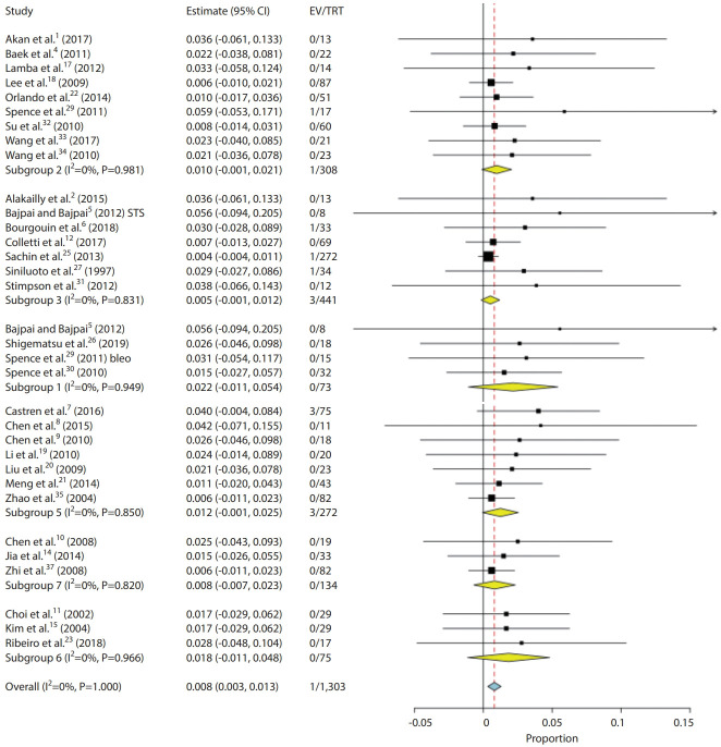
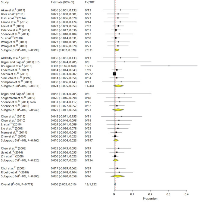
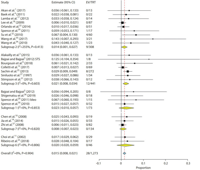
Overall permanent morbidity and mortality rates were reported in 1,303 patients. The overall rate of permanent morbidity and mortality was 0.8% (95% CI, 0.3–1.3%). The lowest rate was reported with STS, and pingamycin (0.5%, 0.8%) and the highest rate reported was with bleomycin (2.2%; 95% CI, 0.0–5.4%). Fig. 3 shows the permanent morbidity and mortality rates forest plot. Local temporary complication rates were reported in 1,312 patients. The overall rate of local temporary complications was 41.8% (95% CI, 27.0–56.5%). Local temporary complications were highest with ethanolamine (51.0%; 95% CI, 24.7–26.6%) and were lowest with bleomycin (27.0%; 95% CI, 5.4–59.4%). Pulmonary complication rates were reported in 1,222 patients. The overall rate was 0.6% (95% CI, 0.2–1.0%). Pulmonary complication rate was lowest for pingamycin (0.8%; 95% CI, 0.0–2.3%), and was highest for STS (2.4%; 95% CI, 0.5–5.3%). Fig. 4 shows the pulmonary complication rates forest plot. Skin necrosis and scarring rates were reported in 1,273 patients and the overall rate was 1.5% (95% CI, 0.8–2.1%). Rates were highest with bleomycin (2.3%; 95% CI, 1.0–5.7%) and lowest with pingamycin (0.8%; 95% CI, 0.0–2.3%). Fig. 5 shows the skin necrosis and scarring rates forest plot. The efficacy and safety outcomes are summarized in Tables 2 and 3.
Fig. 3.
Forest plot: permanent morbidity and mortality rates. Subgroup 1, bleomycin; subgroup 2, ethanol; subgroup 3, sotradecol; subgroup 5, mixed/other; subgroup 6, ethanolamine; subgroup 7, pingyangmycin. CI, confidence intervall; EV, number of events; TRT, number of treated patients.
Fig. 4.
Forest plot: pulmonary complication rates. Subgroup 1, bleomycin; subgroup 2, ethanol; subgroup 3, sotradecol; subgroup 5, mixed/other; subgroup 6, ethanolamine; subgroup 7, pingyangmycin. CI, confidence interval; EV, number of events; TRT, number of treated patients.
Fig. 5.
Forest plot: skin necrosis and scarring rates. Subgroup 1, bleomycin; subgroup 2, ethanol; subgroup 3, sotradecol; subgroup 5, mixed/other; subgroup 6, ethanolamine; subgroup 7, pingyangmycin. CI, confidence interval; EV, number of events; TRT, number of treated patients.
Table 2.
Summary of overall outcomes
| Overall (%) (95% CI) | |
|---|---|
| Complete cure | 64.7 (57.4–72.0) |
| Partial cure | 28.0 (22.1–34.0) |
| No benefit | 4.5 (3.0–6.1) |
| Improvement in QoL | 78.9 (67.0–90.8) |
| Patient satisfaction | 91.0 (86.1–95.9) |
| Pulmonary complication | 0.6 (0.2–1.0) |
| Skin necrosis/scar | 1.5 (0.8–2.1) |
| Any permanent morbidity/mortality | 0.8 (0.3–1.3) |
| Local temporary complication | 41.8 (27.0–56.5) |
QoL, quality of life; CI, confidence interval.
Table 3.
Summary of outcomes by agents
| Bleomycin |
Ethanol |
Sodium tetradecyl sulfate |
Ethanolamine |
Pingamycin |
||||||
|---|---|---|---|---|---|---|---|---|---|---|
| % (95% CI) | P-value vs. bleomycin | % (95% CI) | P-value vs. bleomycin | % (95% CI) | P-value vs. bleomycin | % (95% CI) | P-value vs. bleomycin | % (95% CI) | P-value vs. bleomycin | |
| Complete cure | 66.1 (31.3–100.9) | Ref. | 58.8 (39.8–77.9) | 0.004 | 55.5 (36.1–74.9 ) | 0.005 | 78.3 (58.9–97.6) | <0.001 | 85.9 (72.7–99.2) | <0.001 |
| Partial cure | 24.5 (1.7–47.3) | Ref. | 35.3 (19.1–51.5) | 0.18 | 25.9 (13.1–38.6) | 0.08 | 19.5 (2.1–37.0) | 0.05 | 12.5 (1.0–24.0) | 0.03 |
| No benefit | 12.9 (0.7–26.6) | Ref. | 4.0 (1.2–6.8) | 0.003 | 14.9 (4.3–25.4) | 0.02 | 2.1 (0.2–4.4) | <0.001 | 2.6 (0.1–5.2) | <0.001 |
| Improvement in QoL | Ref. | 89.8 (76.4–3.2) | 46.7 (22.4–71.0) | 98.1 (94.5–100) | ||||||
| Patient satisfaction | 94.3 (88.3–100.3) | Ref. | 96.0 (92.5–99.6) | 0.72 | 72.8 (63.6–81.9) | <0.001 | 76.5 (62.2–90.7) | 0.02 | ||
| Pulmonary complication | 2.2 (0.0–5.4) | Ref. | 1.5 (0.2–2.8) | 1.00 | 2.4 (0.5–5.3) | 0.38 | 2.0 (0.0–5.9) | 1.00 | 0.8 (0.0–2.3) | 1.00 |
| Skin necrosis/scar | 2.3 (1.0–5.7) | Ref. | 1.4 (0.1–2.7) | 0.69 | 2.1 (0.8–3.4) | 0.71 | 2.0 (0.0–5.9) | 1.00 | 0.8 (0.0–2.3) | 0.35 |
| Any permanent morbidity/mortality | 2.2 (0.0–5.4) | Ref. | 1.0 (0.1–2.1) | 1.00 | 0.5 (0.1–1.2) | 1.00 | 1.8 (0.0–4.8) | 1.00 | 0.8 (0.0–2.3) | 1.00 |
| Local temporary complication | 27.0 (5.4–59.4) | Ref. | 30.0 (3.3–56.7) | 0.61 | 44.0 (10.6–77.4) | <0.001 | 51.0 (24.7–26.6) | <0.001 | 36.2 (19.7–92.1) | 0.45 |
CI, confidence interval; Ref., reference; QoL, quality of life.
Study heterogeneity
I2 values were >50% indicating substantial heterogeneity for the following outcomes: lack of benefit, improvement in QoL, local temporary complication, overall cure, partial cure, patient satisfaction and systemic side effects. I2 values were <50% indicating lack of substantial heterogeneity for the following outcomes: permanent morbidity and mortality, pulmonary complications and skin necrosis/scarring.
DISCUSSION
Our systematic review and meta-analysis of percutaneous image guided sclerotherapy for treatment of VMs of the head face and neck found that the overall efficacy rate of sclerotherapy was high with complete and partial cure rates of over 90%. Over 90% of patients reported satisfaction with the results of their sclerotherapy treatments and over 70% reported improvements in QoL. Temporary local complication rates were high, however rates of permanent morbidity and mortality as well as permanent scarring were low. When performing subgroup analyses by agent, we found that agents such as ethanol, pingamycin and ethanolamine were generally associated with the highest rates of cure and patient satisfaction, however these agents were also associated with higher rates of local temporary complications. These findings are important as they provide important information regarding the risks and benefits of percutaneous sclerotherapy for VMs of the head, face and neck.
The efficacy of sclerotherapy for VMs is dependent on both the angioarchitecture of the venous malformation, the sclerosing agent used and the dwell time of the sclerosant within the venous malformation [42]. Angioarchitecture can be easily assessed on contrast venography following percutaneous injection of the lesion. According to Gemmette et al, the 4 morphological subtypes of VMs are as follows: type I, isolated malformation without discernible venous drainage; type II, lesions draining into normal veins; type III, lesions draining into dysplastic veins; type IV, lesion consists primarily of venous ectasia [42]. Types I and II are thought to respond best to sclerotherapy. None of the studies included in our analysis examined outcomes by lesion angioarchitecture, however this undoubtedly is an important factor in determining outcomes.
A summary of the different sclerosing agents is provided in Table 4. Based on our study, it is clear that sclerosants such as ethanol, ethanolamine oleate and pingyangmycin are generally more effective at inducing a complete or partial cure of the VM. However, this comes at the cost of increased risk of permanent morbidity as well as local side effects. Medications such as ethanol, ethanolamine and pingyangmycin act by directly inducing endothelial injury and thrombosis of the VM [43]. Meanwhile, less potent sclerosing agents such as STS and bleomycin work by inducing a nonspecific inflammatory reaction within the VM [43]. Adverse side effects of sclerosing agents also differ by agent and are summarized in Table 4. In brief, adverse side effects are generally most severe with ethanol and include nerve injury, skin necrosis, and cardiopulmonary collapse [43]. Side effects are least severe with bleomycin and include mucositis and skin pigmentation.
Table 4.
Summary of commonly used agents
| Agent | Mechanism of action | Standard dose | Adverse effects |
|---|---|---|---|
| Ethanol | Endothelial denudation and thrombosis | 1 mL/kg, most studies have max of 10–20 mL | Skin necrosis, tachycardia, DVT, PE, pain |
| Bleomycin | DNA damage. Lipid peroxidation, non-specific inflammatory reaction | 1 mg/kg up to 15 mg | Skin pigmentation, fever, mucositis, pulmonary toxicity (rare) |
| Sodium tetradecyl sulfate | Detergent resulting in lipid damage in vessel wall, inflammatory reaction | 0.5–2 mL of 3% solution | Pain, edema, ecchymosis, nerve injury |
| Ethanolamine oleate | Fatty acid emulsion inducing endothelial damage and thrombosis | 2 mL of 50 mg/mL solution | Skin ulceration and necrosis |
| Pingyangmycin | DNA damage. Lipid peroxidation, non-specific inflammatory reaction | 8 mg in a 2 mg/mL solution | Atrophy of subcutaneous tissues, fever, swelling, anaphylactic shock |
| OK-432 | Natural killer cell activator resulting in inflammation and endothelial permeability | 0.1 mg dried cocci in 10 mL of normal saline | Fever, pain, edema, inflammation |
| Doxycycline | Inhibition of matrix metalloproteins and VEGF > decreased angiogenesis | 10–20 mg/kg | Severe pain on injection |
DVT, deep vein thrombosis; PE, pulmonary embolism; VEGF, vascular endothelial growth factor.
It is important to point out that a variety of factors should go into the decision regarding which sclerotherapeutic agent to use. Choice of agent should be based on the depth of the venous malformation from the skin surface as well as the ability to limit non-target sclerotherapy and venous thrombosis. Alcohol is generally best used for deeper VMs due to the risk of skin necrosis from extravasation [4,28,29]. Superficial VMs are likely best treated with milder agents such as bleomycin or STS [5,26,28,29].
The results from our study compare favorably to other modalities of treatment including medical therapy, surgical resection and laser therapy. Conservative management techniques for VMs are centered around pain control, prevention of phlebitis or thrombosis and prevention of bleeding [43]. Pain can generally be managed with anti-inflammatory medications and analgesics. Low dose aspirin can provide pain relief in patients with venous malformation thrombosis [24,31]. Recently the mammalian target of rapamycin inhibitor sirolismus has been trialed for treatment of VMs and has been found to result in symptom improvement in approximately 80% of patients [44]. Surgical excision of VMs is usually part of staged multimodal treatment of VMs [45]. However, surgical excision alone is often considered when lesions are smaller and are not adjacent to vital structures. Surgical excision of large invasive VMs is often very challenging due to the presence of poorly defined tissue planes and proximity to important structures. In such cases, intralesional sclerotherapy can be considered as a useful adjunct to surgery as part of an interdisciplinary approach [46]. Small case series have reported various levels of efficacy with surface and endovenous/interstitial laser therapy for treatment of VMs, most series report efficacy rates ranging from 70–90%. Combined laser and sclerotherapy treatment has also been shown to be effective as part of an interdisciplinary approach [46].
Limitations
Our study has limitations. This meta-analysis was based primarily off of single-center case series and thus has limitations inherent to single center retrospective studies. There was high risk of bias in 21 series. While we were able to perform subgroup analyses based on sclerotherpeutic agent used, we were unable to perform more granular analyses stratifying outcomes by lesion location. Follow-up in the included studies is limited. Many of our outcomes are subjective and volumetric analyses documenting lesion improvement were not available for a vast majority of the included studies. Nonetheless, our study provides helpful information for both patients and providers who are considering percutaneous sclerotherapy for treatment of VMs and provides guidance for future areas of investigation.
CONCLUSIONS
Our systematic review and meta-analysis of 37 studies and over 2,000 patients found that percutaneous sclerotherapy is a very safe and effective treatment modality for treatment of VMs of the head, neck and face. As expected, mild sclerosing agents such as bleomycin and STS were associated with lower efficacy rates but generally superior safety profiles than stronger sclerosing agents such as ethanol, ethanolamine and pingyangmycin. Further prospective studies are needed to validate our results and determine the optimal treatment strategies for patients with VMs of the head, face and neck.
Footnotes
Fund
None.
Ethics Statement
For this type of study formal consent is not required.
Conflicts of Interest
The authors have no conflicts to disclose.
REFERENCES
- 1.Akan H, Uzunkaya F, Soylu AI. Percutaneous ethanol sclerotherapy of venous malformations of the oral cavity and the oropharynx. Erciyes Med. 2017;39:183–188. [Google Scholar]
- 2.Alakailly X, Kummoona R, Quereshy FA, Baur DA, González AE. The use of sodium tetradecyl sulphate for the treatment of venous malformations of the head and neck. J Maxillofac Oral Surg. 2015;14:332–338. doi: 10.1007/s12663-014-0623-y. [DOI] [PMC free article] [PubMed] [Google Scholar]
- 3.Alexander MD, McTaggart RA, Choudhri OA, Pandit RP, Wu A, Ross M, et al. Quantitative volumetric analysis of head and neck venous and lymphatic malformations to assess response to percutaneous sclerotherapy. Acta Radiol. 2016;57:205–209. doi: 10.1177/0284185115575779. [DOI] [PubMed] [Google Scholar]
- 4.Baek HJ, Hong JP, Choi JW, Suh DC. Direct percutaneous alcohol sclerotherapy for venous malformations of head and neck region without fluoroscopic guidance: technical consideration and outcome. Neurointervention. 2011;6:84–88. doi: 10.5469/neuroint.2011.6.2.84. [DOI] [PMC free article] [PubMed] [Google Scholar]
- 5.Bajpai H, Bajpai S. Comparative analysis of intralesional sclerotherapy with sodium tetradecyl sulfate versus bleomycin in the management of low flow craniofacial soft tissue vascular lesions. J Maxillofac Oral Surg. 2012;11:13–20. doi: 10.1007/s12663-011-0325-7. [DOI] [PMC free article] [PubMed] [Google Scholar]
- 6.Bourgouin P, Thomas-Chaussé F, Gilbert P, Giroux MF, Périgny S, Guertin L, et al. Effectiveness and safety of sclerotherapy for treatment of low-flow vascular malformations of the oropharyngeal region. J Vasc Interv Radiol. 2018;29:809–815. doi: 10.1016/j.jvir.2017.12.028. [DOI] [PubMed] [Google Scholar]
- 7.Castren E, Aronniemi J, Klockars T, Pekkola J, Lappalainen K, Vuola P, et al. Complications of sclerotherapy for 75 head and neck venous malformations. Eur Arch Otorhinolaryngol. 2016;273:1027–1036. doi: 10.1007/s00405-015-3577-x. [DOI] [PubMed] [Google Scholar]
- 8.Chen AW, Liu YR, Li K, Zhang K, Wang T, Liu SH. Efficacy of sclerotherapy with radio-opaque foam guided by digital subtraction angiography for the treatment of complex venous malformations of the head and neck. Br J Oral Maxillofac Surg. 2015;53:809–813. doi: 10.1016/j.bjoms.2015.05.024. [DOI] [PubMed] [Google Scholar]
- 9.Chen WL, Huang ZQ, Zhang DM, Chai Q. Percutaneous sclerotherapy of massive venous malformations of the face and neck using fibrin glue combined with ok-432 and pingyangmycin. Head Neck. 2010;32:467–472. doi: 10.1002/hed.21206. [DOI] [PubMed] [Google Scholar]
- 10.Chen Y, Li Y, Zhu Q, Zeng Q, Zhao J, He X, et al. Fluoroscopic intralesional injection with pingyangmycin lipiodol emulsion for the treatment of orbital venous malformations. AJR Am J Roentgenol. 2008;190:966–971. doi: 10.2214/AJR.07.2851. [DOI] [PubMed] [Google Scholar]
- 11.Choi YH, Han MH, O-Ki K, Cha SH, Chang KH. Craniofacial cavernous venous malformations: percutaneous sclerotherapy with use of ethanolamine oleate. J Vasc Interv Radiol. 2002;13:475–482. doi: 10.1016/s1051-0443(07)61527-9. [DOI] [PubMed] [Google Scholar]
- 12.Colletti G, Deganello A, Bardazzi A, Mattassi R, Dalmonte P, Gazzabin L, et al. Complications after treatment of head and neck venous malformations with sodium tetradecyl sulfate foam. J Craniofac Surg. 2017;28:e388. doi: 10.1097/SCS.0000000000003723. [DOI] [PubMed] [Google Scholar]
- 13.Costa JR, Torriani MA, Hosni ES, D’Avila OP, de Figueiredo PJ. Sclerotherapy for vascular malformations in the oral and maxillofacial region: treatment and follow-up of 66 lesions. J Oral Maxillofac Surg. 2011;69:e88. doi: 10.1016/j.joms.2010.06.204. [DOI] [PubMed] [Google Scholar]
- 14.Jia R, Xu S, Huang X, Song X, Pan H, Zhang L, et al. Pingyangmycin as first-line treatment for low-flow orbital or periorbital venous malformations: evaluation of 33 consecutive patients. JAMA Ophthalmol. 2014;132:942–948. doi: 10.1001/jamaophthalmol.2013.8229. [DOI] [PubMed] [Google Scholar]
- 15.Kim KH, Sung MW, Roh JL, Han MH. Sclerotherapy for congenital lesions in the head and neck. Otolaryngol Head Neck Surg. 2004;131:307–316. doi: 10.1016/j.otohns.2004.02.018. [DOI] [PubMed] [Google Scholar]
- 16.Kishi K, Morita N, Terada T, Sato M. Dose-saving isolation procedure in percutaneous ethanol sclerotherapy for venous malformations. Phlebology. 2014;29:276–286. doi: 10.1177/0268355513475604. [DOI] [PubMed] [Google Scholar]
- 17.Lamba S, Keshava SKN, Moses V, Surendrababu N, Gupta AK. Ethanol sclerotherapy for treatment of venous malformations of face and neck—a single centre experience. Eur J Plast Surg. 2012;35:345–350. [Google Scholar]
- 18.Lee IH, Kim KH, Jeon P, Byun HS, Kim HJ, Kim ST, et al. Ethanol sclerotherapy for the management of craniofacial venous malformations: the interim results. Korean J Radiol. 2009;10:269–276. doi: 10.3348/kjr.2009.10.3.269. [DOI] [PMC free article] [PubMed] [Google Scholar]
- 19.Li J, Chen J, Zheng G, Liao G, Fu Z, Li J, et al. Digital subtraction angiography-guided percutaneous sclerotherapy of venous malformations with pingyangmycin and/or absolute ethanol in the maxillofacial region. J Oral Maxillofac Surg. 2010;68:2258–2266. doi: 10.1016/j.joms.2009.06.024. [DOI] [PubMed] [Google Scholar]
- 20.Liu Y, Liu D, Wang Y, Zhang W, Zhao F. Clinical study of sclerotherapy of maxillofacial venous malformation using absolute ethanol and pingyangmycin. J Oral Maxillofac Surg. 2009;67:98–104. doi: 10.1016/j.joms.2008.06.053. [DOI] [PubMed] [Google Scholar]
- 21.Meng J, Zhuang QW, Gu QP, Zhang J, Li ZP, Si YM. Digital subtraction angiography (DSA) guided sequential sclerotherapy for maxillofacial vein malformation. Eur Rev Med Pharmacol Sci. 2014;18:1709–1712. [PubMed] [Google Scholar]
- 22.Orlando JL, Caldas JG, Campos HG, Nishinari K, Krutman M, Wolosker N. Ethanol sclerotherapy of head and neck venous malformations. Einstein (Sao Paulo) 2014;12:181–186. doi: 10.1590/S1679-45082014AO2844. [DOI] [PMC free article] [PubMed] [Google Scholar]
- 23.Ribeiro MC, de Mattos Camargo Grossmann S, do Amaral MBF, de Castro WH, Navarro TP, Procopio RJ, et al. Effectiveness and safety of foam sclerotherapy with 5% ethanolamine oleate in the treatment of low-flow venous malformations in the head and neck region: a case series. Int J Oral Maxillofac Surg. 2018;47:900–907. doi: 10.1016/j.ijom.2017.12.004. [DOI] [PubMed] [Google Scholar]
- 24.Rosbe KW, Hess CP, Dowd CF, Frieden IJ. Masseteric venous malformations: diagnosis, treatment, and outcomes. Otolaryngol Head Neck Surg. 2010;143:779–783. doi: 10.1016/j.otohns.2010.08.053. [DOI] [PubMed] [Google Scholar]
- 25.Sachin K, Rashmi S, Manish S, Siddhartha W, Uday L. Haemangiomas and venous malformations of the head and neck: a retrospective analysis of endovascular management in 358 patients. Indian J Plast Surg. 2013;46:109–116. doi: 10.4103/0970-0358.113727. [DOI] [PMC free article] [PubMed] [Google Scholar]
- 26.Shigematsu T, Sorscher M, Dier EC, Berenstein A. Bleomycin sclerotherapy for eyelid venous malformations as an alternative to surgery or laser therapy. J Neurointerv Surg. 2019;11:57–61. doi: 10.1136/neurintsurg-2018-013813. [DOI] [PubMed] [Google Scholar]
- 27.Siniluoto TM, Svendsen PA, Wikholm GM, Fogdestam I, Edström S. Percutaneous sclerotherapy of venous malformations of the head and neck using sodium tetradecyl sulphate (sotradecol) Scand J Plast Reconstr Surg Hand Surg. 1997;31:145–150. doi: 10.3109/02844319709085481. [DOI] [PubMed] [Google Scholar]
- 28.Songsaeng D, Churojana A, Khumthong R, Mahiwan L. Comparative outcomes for sclerotherapy of head and neck venous vascular malformation between alcohol and bleomycin. J Med Assoc Thai. 2015;98:408–413. [PubMed] [Google Scholar]
- 29.Spence J, Krings T, TerBrugge KG, Agid R. Percutaneous treatment of facial venous malformations: a matched comparison of alcohol and bleomycin sclerotherapy. Head Neck. 2011;33:125–130. doi: 10.1002/hed.21410. [DOI] [PubMed] [Google Scholar]
- 30.Spence J, Krings T, terBrugge KG, da Costa LB, Agid R. Percutaneous sclerotherapy for facial venous malformations: subjective clinical and objective MR imaging follow-up results. AJNR Am J Neuroradiol. 2010;31:955–960. doi: 10.3174/ajnr.A1940. [DOI] [PMC free article] [PubMed] [Google Scholar]
- 31.Stimpson P, Hewitt R, Barnacle A, Roebuck DJ, Hartley B. Sodium tetradecyl sulphate sclerotherapy for treating venous malformations of the oral and pharyngeal regions in children. Int J Pediatr Otorhinolaryngol. 2012;76:569–573. doi: 10.1016/j.ijporl.2012.01.019. [DOI] [PubMed] [Google Scholar]
- 32.Su L, Fan X, Zheng L, Zheng J. Absolute ethanol sclerotherapy for venous malformations in the face and neck. J Oral Maxillofac Surg. 2010;68:1622–1627. doi: 10.1016/j.joms.2009.07.094. [DOI] [PubMed] [Google Scholar]
- 33.Wang D, Su L, Han Y, Wang Z, Zheng L, Li J, et al. Direct intralesional ethanol sclerotherapy of extensive venous malformations with oropharyngeal involvement after a temporary tracheotomy in the head and neck: initial results. Head Neck. 2017;39:288–296. doi: 10.1002/hed.24588. [DOI] [PubMed] [Google Scholar]
- 34.Wang YA, Zheng JW, Zhu HG, Ye WM, He Y, Zhang ZY. Sclerotherapy of voluminous venous malformation in head and neck with absolute ethanol under digital subtraction angiography guidance. Phlebology. 2010;25:138–144. doi: 10.1258/phleb.2009.009019. [DOI] [PubMed] [Google Scholar]
- 35.Zhao JH, Zhang WF, Zhao YF. Sclerotherapy of oral and facial venous malformations with use of pingyangmycin and/or sodium morrhuate. Int J Oral Maxillofac Surg. 2004;33:463–466. doi: 10.1016/j.ijom.2003.10.003. [DOI] [PubMed] [Google Scholar]
- 36.Zheng JW, Yang XJ, Wang YA, He Y, Ye WM, Zhang ZY. Intralesional injection of pingyangmycin for vascular malformations in oral and maxillofacial regions: an evaluation of 297 consecutive patients. Oral Oncol. 2009;45:872–876. doi: 10.1016/j.oraloncology.2009.02.011. [DOI] [PubMed] [Google Scholar]
- 37.Zhi K, Wen Y, Li L, Ren W. The role of intralesional Pingyangmycin in the treatment of venous malformation of facial and maxillary region. Int J Pediatr Otorhinolaryngol. 2008;72:593–597. doi: 10.1016/j.ijporl.2008.01.007. [DOI] [PubMed] [Google Scholar]
- 38.Luo QF, Gan YH. Pingyangmycin with triamcinolone acetonide effective for treatment of lymphatic malformations in the oral and maxillofacial region. J Craniomaxillofac Surg. 2013;41:345–349. doi: 10.1016/j.jcms.2012.10.022. [DOI] [PubMed] [Google Scholar]
- 39.Shiels WE, 2nd, Kang DR, Murakami JW, Hogan MJ, Wiet GJ. Percutaneous treatment of lymphatic malformations. Otolaryngol Head Neck Surg. 2009;141:219–224. doi: 10.1016/j.otohns.2009.04.001. [DOI] [PubMed] [Google Scholar]
- 40.Tu JH, Do HM, Patel V, Yeom KW, Teng JMC. Sclerotherapy for lymphatic malformations of the head and neck in the pediatric population. J Neurointerv Surg. 2017;9:1023–1026. doi: 10.1136/neurintsurg-2016-012660. [DOI] [PubMed] [Google Scholar]
- 41.Flors L, Leiva-Salinas C, Maged IM, Norton PT, Matsumoto AH, Angle JF, et al. Mr imaging of soft-tissue vascular malformations: diagnosis, classification, and therapy follow-up. Radiographics. 2011;31:1321–1340. doi: 10.1148/rg.315105213. ; discussion 1340-1341. [DOI] [PubMed] [Google Scholar]
- 42.Hage AN, Chick JFB, Srinivasa RN, Bundy JJ, Chauhan NR, Acord M, et al. Treatment of venous malformations: the data, where we are, and how it is done. Tech Vasc Interv Radiol. 2018;21:45–54. doi: 10.1053/j.tvir.2018.03.001. [DOI] [PubMed] [Google Scholar]
- 43.Albanese G, Kondo KL. Pharmacology of sclerotherapy. Semin Intervent Radiol. 2010;27:391–399. doi: 10.1055/s-0030-1267848. [DOI] [PMC free article] [PubMed] [Google Scholar]
- 44.Yesil S, Tanyildiz HG, Bozkurt C, Cakmakci E, Sahin G. Single-center experience with sirolimus therapy for vascular malformations. Pediatr Hematol Oncol. 2016;33:219–225. doi: 10.3109/08880018.2016.1160170. [DOI] [PubMed] [Google Scholar]
- 45.Klosterman T, O TM. The management of vascular malformations of the airway: natural history, investigations, medical, surgical and radiological management. Otolaryngol Clin North Am. 2018;51:213–223. doi: 10.1016/j.otc.2017.09.013. [DOI] [PubMed] [Google Scholar]
- 46.Sadick M, Wohlgemuth WA, Huelse R, Lange B, Henzler T, Schoenberg SO, et al. Interdisciplinary management of head and neck vascular anomalies: clinical presentation, diagnostic findings and minimalinvasive therapies. Eur J Radiol Open. 2017;4:63–68. doi: 10.1016/j.ejro.2017.05.001. [DOI] [PMC free article] [PubMed] [Google Scholar]


