Abstract
It has been reported that Cu(II) ions in human blood are bound mainly to serum albumin (HSA), ceruloplasmin (CP), alpha-2-macroglobulin (α2M) and His, however, data for α2M are very limited and the thermodynamics and kinetics of the copper distribution are not known. We have applied a new LC-ICP MS-based approach for direct determination of Cu(II)-binding affinities of HSA, CP and α2M in the presence of competing Cu(II)-binding reference ligands including His. The ligands affected both the rate of metal release from Cu•HSA complex and the value of KD. Slow release and KD = 0.90 pM was observed with nitrilotriacetic acid (NTA), whereas His showed fast release and substantially lower KD = 34.7 fM (50 mM HEPES, 50 mM NaCl, pH 7.4), which was explained with formation of ternary His•Cu•HSA complex. High mM concentrations of EDTA were not able to elicit metal release from metallated CP at pH 7.4 and therefore it was impossible to determine the KD value for CP. In contrast to earlier inconclusive evidence, we show that α2M does not bind Cu(II) ions. In the human blood serum ~75% of Cu(II) ions are in a nonexchangeable manner bound to CP and the rest exchangeable copper is in an equilibrium between HSA (~25%) and Cu(II)-His-Xaa ternary complexes (~0.2%).
Subject terms: Bioinorganic chemistry, Metals, Proteins
Introduction
Copper is an essential cofactor for more than twenty proteins that play important roles in cellular energy production, antioxidative defense and oxidative metabolism. Organismal copper metabolism is strictly regulated and its deflections are characteristic for many diseases. Disturbance of copper metabolism is the primary cause of monogenic Wilson’s and Menkes diseases1 and it is also involved in progression of inflammation, cancer, atherosclerosis2 and many neurodegenerative diseases including the Alzheimer’s disease3.
The extracellular copper pool in the body is stored, transported and distributed in the body mainly by blood. According to the current opinion, the copper ions in the human blood serum are distributed between three proteins: ceruloplasmin (CP), albumin (HSA) and alpha-2-macroglobulin (α2M) accounting for approx. 70, 15 and 10% of copper, respectively4,5. The remaining 5% of copper is functioning as a cofactor in Cu,ZnSOD-3, clotting factors V and VIII, amine and diamine oxidases, ferroxidase II and some other enzymes5. Existence of a low-molecular-weight (LMW) copper pool bound with His in blood was proposed for a half of century6,7. However, equilibrium dialysis data8 and direct EPR measurements9 showed that HSA competes effectively with His for binding of Cu(II) at equimolar concentrations suggesting that excessive concentration of HSA in the blood serum should avoid the binding of Cu(II) to His and other free amino acids in the blood. At the same time the existence of LMW copper pool in blood serum is recently confirmed10 and the “saga of Cu(II)-His” in blood11 still continues.
Structure and functioning of major blood copper proteins have been intensively studied for many decades, however, information about their metal-binding properties is still limited and often inconsistent.
CP is a monomeric glycoprotein with MW of 132 kD12 and its average concentration in blood serum is 1.9 μM13. CP is metallated in the secretory pathway with six Cu ions bound in a highly cooperative manner14. It is known that the cupric ions bound to CP are “not exchangeable”2, which seriously complicates determination of the Cu(II)-binding affinity of CP. CP is an enzyme (EC 1.16.3.1) exhibiting oxidase activity towards Fe(II) ions and numerous aromatic compounds5, however, it is involved also in the transport of copper into mammalian cells15.
α2M is a homotetrameric glycoprotein with MW of 720 kD16 responsible for binding and inactivation of proteases17. The average concentration of α2M in human plasma is 1.3 μM and there is evidence that α2M can bind copper ions and participate in copper delivery into mammalian cells18. Limited information about Cu(II)-binding properties of α2M indicates that only two Cu(II) ions could be bound to tetramer with higher affinity than that of HSA18.
HSA is the most abundant and probably the most studied protein in the blood. The average concentration of HSA in serum is 650 μM and it constitutes more than 50% of serum proteins19,20. HSA (MW 66.5 kD) is a monomeric multicargo transport and storage protein containing seven hydrophobic binding pockets for organic molecules such as fatty acids, metabolites, hormones and drugs21,22. In vitro studies have identified also four distinct metal binding sites in HSA, differing in structure and metal-binding specificity23. The most studied metal-binding site of HSA is the N-terminal site (NTS)24 also known as ATCUN25, which binds Cu(II) ions in physiological conditions. The second site is called Multi-Metal Binding Site (MBS), responsible mainly for binding of Zn(II) ions23. The third site is composed from a free Cys34 residue and its surrounding and the fourth site with unknown location is called Site B. Physiological relevance of last two sites is unknown, however, they might interact with toxic metal ions like Cd(II) or Ni(II) or with metallodrugs23.
The NTS, includes three N-terminal amino acid residues and is characterized by the presence of His in the third position24. Only a small fraction (~2%) of these sites are occupied by Cu(II) ions in physiological conditions23, however, binding and concomitant redox silencing of pro-oxidant Cu(II) ions by HSA gives an important contribution to the antioxidative capacity of the blood26.
Cu(II)-binding properties of NTS have been in the focus of intensive studies. The dissociation constant (KD) values for Cu(II) in NTS of HSA (Asp-Ala-His) and of bovine serum albumin (Asp-Thr-His), have been determined by a variety of methods such as potentiometric titration, equilibrium dialysis, ultrafiltration, isothermal titration calorimetry, Cu(II) electrode and several spectroscopic methods, including EPR27. However, the estimated values for the conditional dissociation constant vary from picomolar (log KD = −11.18) to subfemtomolar (log KD = −16.18) range28,23. The methods used for the study of the binding of Cu(II) to HSA and other Cu(II) proteins so far rely on indirect estimation of the equilibrium concentration of Cu•Protein complex, which can lead to misinterpretation of experimental results29. Such problems can be avoided by using methods, which can directly determine the equilibrium concentrations of the metallated complex.
The aim of the current study was to estimate the Cu(II)-binding affinities of major serum copper proteins (HSA, CP and α2M) in comparison with LMW Cu(II)-binding reference ligands including His by using an unified and direct approach, which gives thermodynamic background for understanding the distribution copper in the blood. For this purpose we have elaborated a new LC-ICP MS based approach for direct detection of Cu•Protein complex in the presence of metal-competing LMW Cu(II)-binding ligands. To cover the wide range of metal-binding affinities of different proteins high-affinity DTPA and EDTA, intermediate affinity NTA and low-affinity His were used in titrations. The kinetics of the metal release from Cu•Protein complex was also studied to ensure that the equilibrium was reached. In result comparable Cu(II)-binding properties of HSA, CP and α2M have been determined and an accelerating effect of His on metal release from HSA has been described. Results enable systematical and quantitative overview from the copper-binding equilibria in human blood.
Results
Elaboration of LC-ICP MS for determination of HSA interaction with Cu(II) ions
The high and low-molecular weight (HMW and LMW) copper pools were separated by size-exclusion chromatography (SEC) on a 1 ml Sephadex G25 Superfine column. The separation of HMW and LMW compounds was completed in 4 minutes (flow rate 0.4 ml/min). When an equimolar amount of Cu(II) was added to 10 μM HSA all copper was found in the HMW peak (Fig. 1a). Thus, the kinetic stability of Cu•HSA complex is sufficient for SEC separation.
Figure 1.
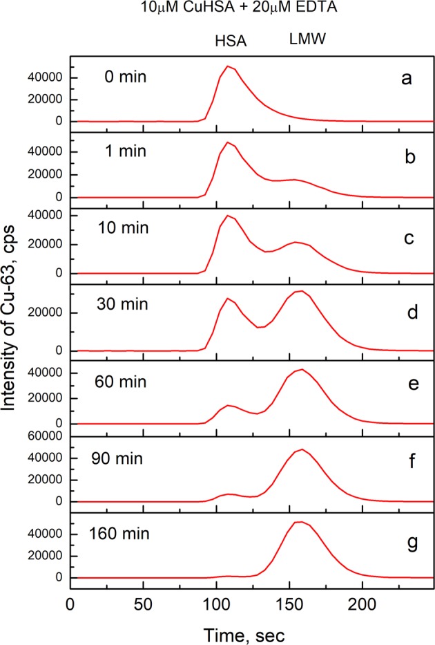
Demetallation of Cu•HSA by EDTA monitored by LC-ICP MS. Conditions: 10 μM Cu•HSA, 20 μM EDTA; incubation buffer 50 mM HEPES, 50 mM NaCl, pH 7.4; incubation times: (a) - 0 min, (b) - 1 min, (c) −10 min, (d) - 30 min, (e) - 60 min, (f) - 90 min, (g) - 160 min; column - 1 ml Sephadex G25 Superfine; elution buffer 200 mM NH4NO3, pH 7.4; flow rate 0.4 ml/min; injection volume 10 μl. Cu-63 was monitored by ICP MS. Figure was created by program Origin 9 Pro (https://www.originlab.com/).
Adding of Cu(II) to HSA in a twofold excess did not increase the amount of copper in the HMW peak, which confirms that only the binding to the single high-affinity site is seen in the experiment. At the same time, the peak of LMW copper was not observed in the chromatogram, demonstrating that the column material has some capacity to bind the “free” Cu(II) ions. We confirmed that the column-bound copper could be quantitatively removed from the column by a subsequent elution with 10 μl of 1 mM EDTA. In the Cu(II)-competition experiments with LMW ligands like EDTA, NTA, DTPA, the copper was quantitatively eluted from the column, suggesting that the binding affinity of column matrix is sufficiently lower as that for listed ligands. In case of HSA - His competition experiments LMW copper peak was smaller than expected at lower His concentrations. To quarantee similar LC conditions for all experiments, column decoppering with10 μl of 1 mM EDTA was performed before each LC ICP MS experiment.
Demetallation of Cu(II)•HSA with copper-binding ligands
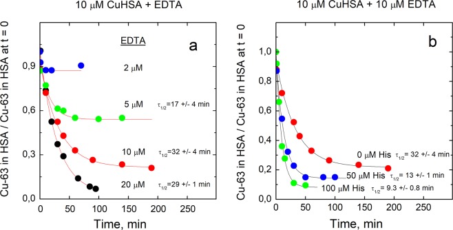
Incubation of Cu•HSA complex with EDTA decreased the HMW copper peak and increased the LMW copper peak (Fig. 1b–g). The half-life of Cu•HSA demetallation in the presence of EDTA was 20–30 min (Fig. 2) and the protein was almost fully demetallated already by an equimolar amount of EDTA. Thus, the dissociation of Cu(II) ions from HSA in the presence of EDTA is relatively slow and the affinity of Cu(II) for HSA is lower than that for EDTA. EDTA binds one Cu(II) ion with KD = 1.26 × 10⁻¹⁶ M at pH 7.430.
Figure 2.
Demetallation kinetics of Cu•HSA by EDTA in the absence and presence of His. Decrease of relative content of Cu-63 in HSA in time (a); effect of His on demetallatiuon of Cu•HSA by EDTA (b). Conditions: 10 μM Cu•HSA, 2–20 μM EDTA (a); 10 μM Cu•HSA, 10 μM EDTA, 0–100 μM His (b); incubation buffer 50 mM HEPES, 50 mM NaCl, pH 7.4; LC-ICP MS: column - 1 ml Sephadex G25 Superfine; elution buffer 200 mM NH4NO3, pH 7.4; flow rate 0.4 ml/min; injection volume 10 μl, Cu-63 was monitored by ICP MS. Figure was created and fittings were performed by program Origin 9 Pro (https://www.originlab.com/).
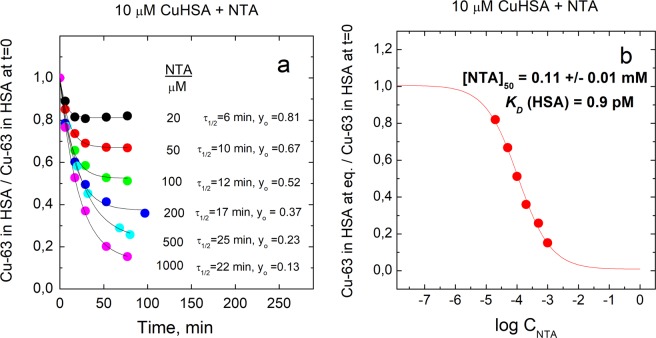
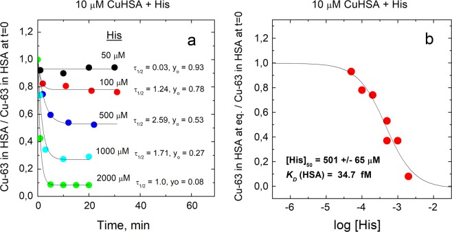
A substantially weaker Cu(II)-binding ligand NTA (KD = 1.99 × 10⁻¹¹ M at pH = 7.431) also demetallated Cu•HSA with the half-life of 20–25 min. As expected, a substantially higher than equimolar concentration of NTA was required for complete demetallation of HSA (Fig. 3a), which allows to determine the KD value for Cu•HSA complex. From the fractional content of Cu•HSA at equilibrium in the presence of different concentrations of NTA (Fig. 3b) the KD value equal to 0.900 ± 0.091 pM was determined by using Eqn. 4 as described in Experimental section. Demetallation of Cu•HSA complex in the presence of His was also studied. His forms 2:1 complex with Cu(II) at pH = 7.4 where KD1 = 3.7 × 10⁻⁹ M and KD2 = 4.7 × 10⁻⁷ M31. In contrast to EDTA and NTA, His demetallated Cu•HSA with fast kinetics (half-life of appr. 1-2 min) and at relatively high concentrations (Fig. 4a), which also enables determination of KD for Cu•HSA complex. By using of fractional content of Cu•HSA at equilibrium conditions at different concentrations of His (Fig. 4b) the KD = 34.7 ± 4.5 fM was determined according to the Eqn. 4 as described in Experimental section.
Figure 3.
Demetallation kinetics of Cu•HSA by NTA and determination of dissociation constant for Cu•HSA. Decrease of relative content of Cu-63 in HSA in time (a); relative content of Cu-63 in HSA at equilibrium at different concentrations of NTA (b). Conditions: 10 μM Cu•HSA, 10–1000 μM NTA; incubation buffer 50 mM HEPES, 50 mM NaCl, pH 7.4; LC-ICP MS: column - 1 ml Sephadex G25 Superfine; elution buffer 200 mM NH4NO3, pH 7.4; flow rate 0.4 ml/min; injection volume 10 μl, Cu-63 was monitored by ICP MS. Figure was created and fittings were performed by program Origin 9 Pro (https://www.originlab.com/).
Figure 4.
Demetallation kinetics of Cu•HSA by His and determination of dissociation constant for Cu•HSA. Decrease of relative content of Cu-63 in HSA in time (a); relative content of Cu-63 in HSA at equilibrium at different concentrations of His (b). Conditions: 10 μM Cu•HSA, 10–2000 μM His; incubation buffer 50 mM HEPES, 50 mM NaCl, pH 7.4; LC-ICP MS: column - 1 ml Sephadex G25 Superfine; elution buffer 200 mM NH4NO3, pH 7.4; flow rate 0.4 ml/min; injection volume 10 μl, Cu-63 was monitored by ICP MS. Figure was created and fittings were performed by program Origin 9 Pro (https://www.originlab.com/).
A separate experiment showed that low concentrations of His that alone did not demetallate Cu•HSA, substantially increased the rate of the HSA demetallation by EDTA: three and sixfold increases were observed in the presence of 50 μM and 100 μM of His, respectively (Fig. 2b). 0.5 mM Glu and Gly were not able to demetallate Cu•HSA complex and did not accelerate its demetallation in the presence of EDTA (Data not shown).
Attempts to demetallate Cu•CP with copper-binding ligands
In Cu•CP samples the copper eluted from the Sephadex G25 column in HMW region confirming that metal is strongly bound to the protein. Adding up to 200 mM of strong Cu(II) chelators (DTPA, EDTA) at pH 7.4 did not decrease the metal content in Cu•CP peak even after 100 min incubation with 200 mM EDTA or 21 h incubation with 100 mM DTPA (Supplementary Information, Figure S1a). At high, non-physiological pH values (pH 11) Cu•CP could be demetallated by EDTA in a dose-dependent manner (Supplementary Information, Figure S1b).
Metallation of α2M with Cu(II) ions
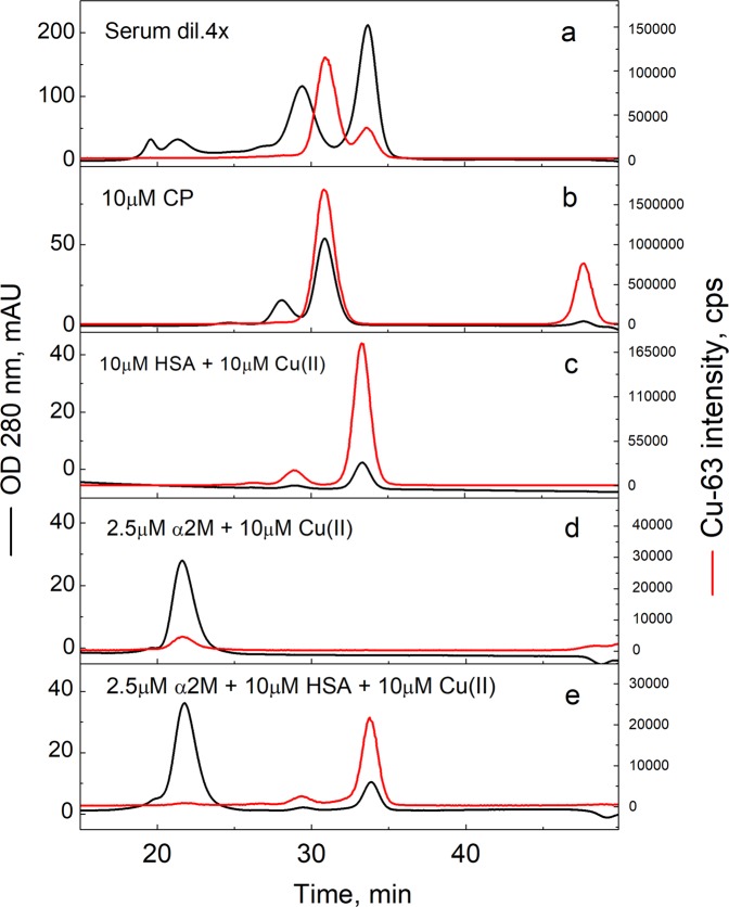
The experiments were carried out with two preparations of normal (“slow form”) human plasma α2M (from Sigma and Athens Research) as well as with the so-called “fast form” of α2M from Athens Research. The “slow form” of α2M, represents more than 99% of total α2M in blood plasma and possesses the ability to bind and inhibit proteases. The “fast form” of α2M arises through a conformational change caused by entrapment of a protease in the α2M bait region, or chemical cleavage of an internal thiol ester bond located near the bait region17. “Fast form” of α2M represents only 0.17–0.7% of the total α2M in blood plasma and is rapidly taken up by the liver. Our LC-ICP MS experiments on Sephadex G25 column showed that both forms of α2M neither contained copper nor were able to bind added Cu(II) ions essentially (Supplementary Information, Figure S2). We tested Cu(II) binding of normal α2M also by ultrafiltration with 10 kD cut-off membranes and confirmed that α2M had only marginal ability (<5%) for retention of Cu(II) ions during ultrafiltration. These results suggest that α2M from human plasma does not bind Cu(II) ions with a biologically significant (e.g. µM or higher) affinity. We also performed a metal competition experiment with α2M and HSA by LC-ICP MS using a Superdex 200 (10 × 300 mm) column, which can separate α2M and HSA peaks. When an equimolar concentration of Cu(II) was added to the equimolar mixture of normal α2M and HSA, almost all copper was eluted in a single peak corresponding to Cu•HSA, demonstrating that α2M cannot compete for Cu(II) ions with HSA even at equimolar concentration (Fig. 5e).
Figure 5.
Separation of human serum proteins by SEC. 4 x diluted human serum (a), 10 μM Cu•CP (b), 10 μM HSA plus 10 μM Cu(II) (c), 2.5 μM α2M tetramer plus 10 μM Cu(II) (d), 10 μM HSA, 2.5 μM α2M tetramer plus 10 μM Cu(II) (e). Conditions: incubation buffer 50 mM HEPES, 50 mM NaCl, pH 7.4; LC-ICP MS: column - 10 × 300 mm Superdex 200; elution buffer 200 mM NH4NO3, pH 7.4; flow rate 0.4 ml/min; injection volume 10 μl; UV at 280 nm (left axis and black lines) and Cu-63 (right axis and red line), monitored by ICP MS. Figure was created by program Origin 9 Pro (https://www.originlab.com/).
Discussion
The affinities of metalloproteins for the metal ions, characterized by KD values, are the key thermodynamic parameters, which direct the metabolism of metal ions and determine their homeostasis. The metal homeostasis can be understood and described quantitatively only if the affinities of the proteins involved are comparable to each other, e.g. they have been determined absolutely correctly or with the same method and using the same reference compounds to ensure the comparability. The full set of comparable metal-binding affinities is available only for the intracellular Cu(I) proteome32. The metal-binding affinities of the individual Cu(II) proteins, determined by different methods vary largely as their accurate estimation is a subject to a number of pitfalls and complicating factors29. Here we have applied an unified approach for determination of Cu(II)-binding affinities for three major blood copper proteins with the aim to get reliable and comparable KD values for individual proteins determining the copper distribution in the blood and also in other extracellular media such as cerebrospinal fluid.
Binding of Cu(II) ions to human serum albumin
The release of Cu(II) ions from Cu•HSA complex in the presence of strong multidentate ligands such as EDTA and NTA is a slow process. The independence of the Cu•HSA demetallation half-life on the multidentate ligands and their concentrations used suggests that the rate-limiting step of the metal release is the dissociation of Cu(II) ions from NTS of HSA. NTS of HSA is highly dynamic33 and no large-scale protein conformational changes or other slow processes are assumed to occur during dissocition. Moreover, the maximal half-life of unassisted dissociation of a complex with pKD 13, is approx. 30 min that is similar to the observed half-life of Cu•HSA decoppering in the presence of EDTA and NTA, which supports our suggestion. The fast release of copper from Cu•HSA in the presence of His points to a different mechanism of demetallation and involvement of His in the rate-limiting step. Based on spectroscopic studies it was suggested that His forms a ternary complex with Cu•HSA34, however, a direct [14C]His binding assay by using ultrafiltration showed that His interaction with Cu•HSA could be only transient8. Nevertheless, even a transient His•Cu•HSA can act as a ligand-exchange complexes enhancing the rate of copper release. This conclusion is also supported by the catalytic effect of physiological concentrations of His (50 - 100 μM) on the demetallation of Cu•HSA by EDTA observed in the current study. The catalytic effect on metal release from Cu•HSA was specific for His as neither 1 mM Glu nor 0.5 mM Gly did not accelerate the reaction. From the X-ray crystal structure of model peptides (DAHK) it is known that Cu(II) ion in NTS (DAH) is equatorially coordinated by N-terminal amine, two deprotonated peptide bond nitrogens and imidazole of His-3, whereas one apical coordination site is occupied with water35. It can be suggested that replacement of this water molecule in Cu•HSA with imidazole from free His facilitates the metal release from the ternary complex formed.
The dissociation constant (KD) value for Cu•HSA complex may substantially depend on many factors including the experimental setup of its determination. First, much attention has to be paid to reaching the equilibrium, which is slow in case of many chelators including NTA. Second, our results demonstrate that the estimated KD value for Cu•HSA complex depends also on the nature and binding mode of the competing ligand used in metal competition experiments. Using of NTA, which forms 1: 1 complex with Cu(II) in our experimental conditions and showed slow demetallation of Cu•HSA, a KD value equal to 0.900 ± 0.091 pM (50 mM Hepes, 50 mM NaCl, pH 7.4) was determined. However, titration with His, which forms 2: 1 complex in our experimental conditions and leads to fast demetallation of Cu•HSA, yielded KD equal to 34.7 ± 4.5 fM (50 mM Hepes, 50 mM NaCl at pH 7.4). The lower KD value determined from competition with His might arise due to the omitting of the formation of a putative ternary complex between HSA, Cu(II) ion and His in the binding scheme, which should result in higher affinity estimates, since the copper in the ternary complex is found in the HMW peak. Thus, the KD value determined with NTA should be considered more reliable. This value is very similar to the KD value 1.0 pM, determined from spectroscopic titration of Cu•HSA with NTA in 100 mM NaCl (pH 7.4)28. At the same time a substantially higher KD value 4.0 pM was obtained in 100 mM HEPES, pH 7.4, which was explained with the formation of ternary HEPES• Cu(II)•NTA complex28. We note that in addition to the effect of ternary complex formation there is also contribution from differences in ionic strength and extinction coefficients in two media, which were not considered. In our LC-ICP MS experiments KD values (determined by NTA and His) in 5 mM HEPES, 50 mM NaCl (data not shown) were similar to those in 50 mM HEPES, 50 mM NaCl, which allows to conclude that HEPES at 50 mM level does not compete with HSA, NTA and His for Cu(II) binding or ternary HEPES complexes are formed with both Cu•HSA and Cu•NTA. At the same time we observed that even a slight decrease in pH has a substantial effect on the HSA demetallation levels, which shows that the usage of buffer is necessary. The KD values for ionic equilibria can be affected also by ionic strength. Indeed, conducting NTA titration in 5 mM HEPES, pH 7.4 we observed that absence of 50 mM NaCl caused fivefold increase of the KD for Cu•HSA, which is, however, apparent and should be corrected by using of KD value for Cu(II)•NTA complex at low ionic strength. To avoid these fluctuations and also for practical reasons it is advisable to keep ionic strength value in metal-binding experiments close to 0.1 M, which is a standard condition, where KD values for reference Cu(II)•Ligand complexes are normally determined36. Thus, there is a 4.4-fold difference between KD values determined from NTA competition by spectroscopic (100 mM HEPES, pH 7.4)28 and LC-ICP MS-based method (50 mM HEPES, 50 mM NaCl, pH 7.4). However, we cannot compare these results directly as kinetic results of copper release and incubation times, necessary for reaching the equilibrium were not presented in the earlier paper and it was only declared that the metal release from Cu•HSA in the presence of NTA is faster than in case of EDTA28. Our KD value of 0.90 pM is comparable also with KD = 6.61 pM, determined in 30 mM Hepes, 250 mM NaCl (pH = 7.0) with equilibrium dialysis37, however, difference could be ascribed to different pH value. Therefore KD value of 0.90 pM for Cu•HSA determined in the present work could serve as a reference KD value for Cu•HSA in up to 50 mM HEPES buffer, 50 mM NaCl, pH 7.4, useful for further research. From a methodological point of view our study highlights the need to use a kinetic approach and direct methods for quantification of metallated protein complexes as well as the neccessity for strict control of buffer, ionic strength and pH value during titration with metal-chelating ligands.
Binding of Cu(II) ions to ceruloplasmin
Our results confirm that at pH 7.4 Cu•CP complex can not be demetallated with high millimolar concentrations of strong Cu(II)-chelating ligands like EDTA or DTPA in a timeframe of up to 20 h, which could be explained with three possibilities: first – the thermodynamic Cu(II)-binding affinity of Cu•CP is much higher as compared to chelators used (for EDTA KD = 1.26 × 10⁻¹⁶ M30; for DTPA KD = 5.0 × 10⁻¹⁷ M38, both at pH 7.4); secondly - dissociation of Cu(II) ions from Cu•CP complex is extremely slow, i.e. complex is kinetically inert at physiological pH values or third - the Cu•CP complex is thermodynamically very stabile and also kinetically inert. Based on available data it is impossible to distinguish between these three possibilities and therefore it is also impossible to determine or estimate KD for Cu•CP complex from our results. It is known that in Cu•CP the copper ions are located in three mononuclear and one trinuclear binding sites, which are not exposed to the environment39. Such encapsulation of metal ions in the protein interior might hinder their dissociation and exclude formation of ternary ligand exchange complexes leading to kinetical inertness of the Cu•CP complex at pH 7.4. Cu•CP could be demetallated by EDTA at non-physiological pH value (pH = 11) in dose-dependent manner, characteristic for fast binding equilibrium. Such behaviour could most probably be explained with partial opening of protein conformation or accelaration of conformational dynamics at alkaline pH values. Thus, our results confirm earlier conclusions that CP-bound copper ions are practically nonexchangeable at physiological pH values and CP does not participate in the regulation of exchangeable copper levels in the blood5.
Binding of Cu(II) ions to alpha-2 macroglobulin
Our results demonstrated that α2M does not bind Cu(II) ions. In earlier experiments α2M has been shown to bind a substoichiometric amount of Cu(II) ions18, which is difficult to interpret in terms of thermodynamics. It has been noticed that purified α2M can be contaminated by HSA40 that binds Cu(II) ions, which can be mistakenly interpreted as binding of Cu(II) to α2M. We performed also LC-ICP MS experiments using Superdex 200 column, where peaks of CP, HSA and α2M elute at different timepoints (Fig. 5b–d). The α2M sample was supplemented with equimolar concentration of Cu(II) ions, however, only substoichiometric traces of copper were detected in the peak corresponding to α2M (Fig. 5d), which confirms that α2M cannot bind Cu(II) ions. In the competition experiment with 2.5 µM α2M, 10 µM HSA and 10 µM Cu(II) only HSA was metallated (Fig. 5e). Moreover, only two protein-bound copper peaks, corresponding to the major CP and minor HSA, were detected in case of a pooled human serum sample by similar SEC experiment, whereas practically no copper was detected in the HMW region corresponding to α2M (Fig. 5a).
Copper equilibrium in blood
The average total concentration of copper in normal human blood serum is 16.7 μM41. Our results suggest that copper is distributed only between two principal serum proteins – CP and HSA, which expose different affinity and mechanisms of metal binding. In Cu•CP complex, which constitutes approximately 75% from the total copper pool (our data and4,5), the copper ions are bound in protein interior, which makes them kinetically inert and practically nonexchangeable at physiological pH values even in the presence of high millimolar concentrations of powerful Cu(II)-chelating ligands like EDTA or DTPA.
Metal-binding mode of HSA differs from that of CP as the NTS of HSA is highly dynamic and exposed to the environment33. Cu(II) binding to this site is reversible, however, characterized by slow unassisted dissociation rate and picomolar affinity (KD = 0.90 ± 0.09 pM), determined with NTA or fast and His-assisted dissociation and apparent subpicomolar affinity (KD = 34.7 ± 4.5 fM). Cu(II) bound to HSA constitutes approximately 25% from total blood copper (on average 4.2 μM) and this copper pool could also be in equilibrium with Cu(II) ions bound to free amino acids, primarily His. The outcome of this competition depends from the Cu(II)-binding affinities and concentrations of HSA, Cu•HSA and His according to the following simplified reaction scheme:
| 1 |
| 2 |
| 3 |
HSA and His are present in the blood serum at average 650 μM and 75 μM concentration respectively22,42–44. Taking into account the KD = 34.7 fM (KD determined from competition with His in the present work), KD1 = 3.7 × 10⁻⁹ M and KD2 = 4.7 × 10⁻⁷ M values for Cu(II)-His complexes at pH 7.4 (taken from31) it can be estimated that approximately 0.7 nM of Cu(II) are bound to His mainly in the form of Cu•His2 complex (Equations are presented in Materials and methods). Thus, His alone can bind in average 0.04% from total copper in the blood serum. However, it should be noted that other free amino acids, which are present in the blood serum at total 3 mM concentration42, can also form tight ternary complexes with Cu(II)-His45. Thus, LMW copper pool in the blood serum might be composed mainly from Cu(II)-His-Xaa complexes and can reach appr. 0.2% level. This estimate agrees relatively well with the range of 1–2.4% for low-molecular weight copper pool in blood serum determined in seven independent studies by ultrafiltration10. Moreover, besides binding of minor fraction of Cu(II) ions, His can also act as a catalyst enhancing the rate of the copper transfer between Cu•HSA and other proteins and cellular destinations, which might be the most important physiological role of His in copper metabolism. The proposed catalytic role of His in Cu(II) release from HSA is in agreement with the finding that HSA inhibits copper transport into liver cells whereas His can specifically mobilize Cu(II) from plasma and facilitate the process46.
Conclusion
Cu(II)-binding affinities for three major blood copper proteins: human serum albumin, ceruloplasmin and alpha-2-macroglobulin, were studied through their competition with a set of low-molecular weight Cu(II)-binding reference ligands (DTPA, EDTA, NTA, His) by using an unified LC-ICP MS-based approach. A reliable KD value for human serum albumin was determined and it was demonstrated that metallated ceruloplasmin is resistant for copper release by chelators used, whereas alpha-2 macroglobulin does not bind Cu(II) ions. Obtained thermodynamic and kinetic data allow determination of the copper distribution in the human blood and also in other extracellular media like for example cerebrospinal fluid. Results allow detection of disturbances in copper metabolism, characteristic for many diseases and provide a rationale for effective metalloregulation.
Methods
Instrumentation
For LC-ICP MS analyses an Agilent Technologies (Santa Clara, USA) Infinity HPLC system, which consisted of 1260 series µ-degasser, 1200 series capillary pump, Micro WPS autosampler and 1200 series MWD VL detector was coupled with Agilent 7800 series ICP-MS instrument (Agilent, USA). For instruments control and data acquisition, ICP-MS MassHunter 4.4 software Version C.01.04 from Agilent was used. ICP MS was operated under following conditions: RF power 1550 W, nebulizer gas flow 1.03 l/min, auxiliary gas flow 0.90 l/min, plasma gas flow 15 l/min, nebulizer type: MicroMist, isotope monitored: Cu-63. A 1 ml gel filtration column, self-filled with HiTrap Desalting resin Sephadex G25 Superfine (Amersham/GE Healthcare, Buckinghamshire, UK), was used for the separation of HMW and LMW pools. Injection volume of 10 ul for HSA and 2 ul for other proteins were used. Separation of pooled plasma and individual copper proteins was conducted using Superdex 200 SEC column (10 × 300 mm) (Amersham Biosciences AB, Uppsala, Sweden) by using injection volume of 40 μl. ICP MS compatible flow rate of 0.4 ml/min was used in all separations. In order to get rid of contaminating metal ions in the buffer, the mobile phase was eluted through the Chelex100 Chelating Ion Exchange resin (Sigma, Merck KGaA, Darmstadt, Germany) prior to liquid chromatographic separation. The demetallation of the SEC columns before each experiment was conducted by injecting of 1 mM EDTA into the columns (injection volumes were the same for all experiments, depending on the column type and size). For ultrafiltration experiments, 0.5 ml Amicon Ultra centrifugal filters with 10 kDa cut-off were used (Merck Millipore Ltd, Ireland).
Materials
Ultrapure milliQ water with a resistivity of 18.2 MΩ/cm, produced by a Merck Millipore Direct-Q & Direct–Q UV water purification system (Merck KGaA, Darmstadt, Germany), was used for all applications.
Mobile phase for gel filtration was 200 mM NH4NO3 at pH 7.5, prepared from TraceMetal Grade nitric acid (Fisher Scientific UK Limited, Leicestershire, UK) and ammonium hydroxide 25% solution (Honeywell Fluka, Seelze, Germany), which is compatible with ICP MS.
Following lyophilized proteins were used: human serum albumin (HSA) from Sigma/Merck (Darmstadt, Germany), alpha-2 macroglobulin (α2M) from human plasma (“slow form” and “fast form”) and ceruloplasmin (CP) from human plasma were from Athens Research and Technology (Athens, USA), α2M was purchased also from Sigma/Merck (Darmstadt, Germany).
All protein stock solutions were prepared in milliQ water and further diluted in reaction buffer solution (containing 50 mM HEPES and 50 mM NaCl, pH 7.4) for all experiments. For metallation of HSA and α2M Cu(II)acetate from Sigma (Sigma/Merck KGaA, Darmstadt, Germany) was used. An equimolar concentration of Cu(II)acetate was added to HSA in 50 mM Hepes, 50 mM NaCl, pH 7.4 and the sample was incubated 10 min at room temperature, which is sufficient for Cu•HSA complex formation (in separate kinetic experiment we established that the half-life of Cu(II) binding to HSA is approx. 1.5 min). An equimolar concentration of Cu(II)acetate was added α2M in 50 mM Hepes, 50 mM NaCl, pH 7.4 and sample was incubated for up to 1 h. Following reagents were used: ethylenediaminetetraacetic acid (EDTA, 99.995% trace metal basis) and diethylenetriaminepentaacetic acid (DTPA) from Sigma/Merck (Merck KGaA, Darmstadt, Germany), nitrilotriacetic acid (NTA) and L-amino acids (His, Glu and Gly) from Fluka (Merck KGaA, Darmstadt, Germany). Stock solutions of amino acids were prepared in pure milliQ water whereas stock solutions of EDTA, DTPA and NTA were neutralized with 0.1 M NaOH to pH 7-8 and further diluted into reaction buffer. Mobile phase and reagent solutions were prepared daily before experiment.
Blood serum of anonymous donors was obtained from West-Tallinn Central Hospital (Estonia) and the study approval was obtained from Tallinn Medical Research Ethics Committee. For current research, blood plasma pool from 15 individuals was prepared, in order to eliminate individual variability and exclude possibility for identification of a participants. We confirm that all experiments were performed in accordance with relevant guidelines and regulations.
SDS PAGE
Commercial protein samples have been analyzed by SDS PAGE performed by the Mini-PROTEAN Tetra System (BioRad) with Laemmli gel (10%T/5%C) consisting of separation and comb gel. Samples were prepared in milliQ water at concentration of 3 mg/ml. Samples (20 µl) were mixed with 5 µl of 5x loading buffer (0.3 M Tris-HCl pH = 6.8, 25% β-mercaptoethanol, 50% glycerol, 10% SDS, 1% bromophenol blue), heated to 95 °C for 5 min, and 15 µl of samples were applied to the gel together with 5 µl of “Thermo Scientific PageRuler Unstained Broad Range Protein Ladder”. Electrophoresis was performed with 25 mM Tris, 192 mM Glycine, 0,1% SDS running buffer for about 1 h (130 V). The gel was stained in Coomassie staining solution for overnight followed by destaining in milliQ water and the results, presented in Figure S3 show, that all commercial proteins used have high purity and are suitable for metal-binding studies.
Calculations for dissociation constants
First, the titration curves (Figs. 3b and 4b) were fitted to a dose-dependent demetallation curve to determine the ligand concentration where the concentrations of copper ions bound to the protein was decreased by 50%. At that point, the equilibrium concentration of the free copper ions [Cu]free, which can be calculated according to Eqn.431 equals to the KD value of the protein.
| 4 |
In the Eqn.4 L is the concentration of the competing ligand, K1 and K2 correspond to the dissociation constants of Cu•L and Cu•L2 complexes, respectively [Cu]total is the concentration of copper and [Cu]bound to protein is the concentration of the copper bound to the protein at IC50.
Calculations for concentrations of free Cu(II) ions and Cu(II)•His2 complex
The concentration of free Cu(II) ions at equilibrium can be calculated as follows:
| 5 |
[Cu]free in blood serum is calculated from the metallation equilibrium of HSA taking into account the total HSA concentration 650 µM and the concentration of non-CP bound copper available for HSA in blood 4.2 µM. [Cu]free is related to the amount of copper bound to LMW ligands like His according to Eqn. 4 and the concentration of LMW copper is given by:
| 6 |
where [L] is the concentration of the competing ligand (His), K1 and K2 correspond to the dissociation constants of CuHis and CuHis2 complexes (K1 = 3.7 × 10⁻⁹ M, K2 = 4.7 × 10⁻⁷ M, pH = 7.431). Considering the average concentration of His in blood 75 µM43, the concentration of Cu•His complexes (mainly Cu•His2) is equal to 0.7 nM. Considering that the total concentration of free amino acids in the blood is approximately 3 mM45, then concentration of Cu•His•Xaa ternary complexes in the blood is appr. 28 nM.
Supplementary information
Acknowledgements
This work was supported during the 2019 by the Estonian Ministry of Education and Research (Grant IUT 19-8 to PP), by a research grant from Wilson Therapeutics AB/Alexion Pharmaceuticals Inc. Support from Estonian Ministry of Education and Research discontinued since 2020. Authors like to thank Andra Noormägi for assistance in LC-ICP MS experiments and anonymous Reviewer 2 for very constructive critics.
Author contributions
P.P. and T.P. conceived the study; T.K., M.F., A.Z. and J.S. carried out the main experiments; T.K., A.Z., J.S., M.F., V.T., T.P. and P.P. analyzed the data; P.P. wrote the main draft of the paper and all authors participated in revising the manuscript.
Competing interests
The authors declare no competing interests.
Footnotes
Publisher’s note Springer Nature remains neutral with regard to jurisdictional claims in published maps and institutional affiliations.
Supplementary information
is available for this paper at 10.1038/s41598-020-62560-4.
References
- 1.de Bie P, Muller P, Wijmenga C, Klomp LW. Molecular pathogenesis of Wilson and Menkes disease: correlation of mutations with molecular defects and disease phenotypes. J. Med. Genet. 2007;44:673–688. doi: 10.1136/jmg.2007.052746. [DOI] [PMC free article] [PubMed] [Google Scholar]
- 2.Linder MC, Hazegh-Azam M. Copper biochemistry and molecular biology. Am. J. Clin. Nutr. 1996;63:797S–811S. doi: 10.1093/ajcn/63.5.797. [DOI] [PubMed] [Google Scholar]
- 3.Donnelly PS, Xiao Z, Wedd AG. Copper and Alzheimer’s disease. Curr. Opin. Chem. Biol. 2007;11:128–133. doi: 10.1016/j.cbpa.2007.01.678. [DOI] [PubMed] [Google Scholar]
- 4.Cabrera A, et al. Copper binding components of blood plasma and organs, and their responses to influx of large doses of (65)Cu, in the mouse. Biometals. 2008;21:525–543. doi: 10.1007/s10534-008-9139-6. [DOI] [PMC free article] [PubMed] [Google Scholar]
- 5.Linder MC. Ceruloplasmin and other copper binding components of blood plasma and their functions: an update. Metallomics. 2016;8:887–905. doi: 10.1039/c6mt00103c. [DOI] [PubMed] [Google Scholar]
- 6.Sarkar, B. K. T. In Biochemistry of copper (ed. J.; Aisen Peisach, P.; Blumberg, W.) 183–196 (Academic Press, 1966).
- 7.Neumann PZ, Sass-Kortsak A. The state of copper in human serum: evidence for an amino acid-bound fraction. J. Clin. Invest. 1967;46:646–658. doi: 10.1172/JCI105566. [DOI] [PMC free article] [PubMed] [Google Scholar]
- 8.Masuoka J, Saltman P. Zinc(II) and copper(II) binding to serum albumin. A comparative study of dog, bovine, and human albumin. J. Biol. Chem. 1994;269:25557–25561. [PubMed] [Google Scholar]
- 9.Valko M, et al. High-affinity binding site for copper(II) in human and dog serum albumins (an EPR study) J. Phys. Chem. B. 1999;103:5591–5597. doi: 10.1021/jp9846532. [DOI] [Google Scholar]
- 10.Catalani S, et al. Free copper in serum: An analytical challenge and its possible applications. J. Trace Elem. Med. Biol. 2018;45:176–180. doi: 10.1016/j.jtemb.2017.11.006. [DOI] [PubMed] [Google Scholar]
- 11.Deschamps P, Kulkarni PP, Gautam-Basak M, Sarkar B. The saga of copper(II)-L-histidine. Coord. Chem. Rev. 2005;249:895–909. doi: 10.1016/j.ccr.2005.09.013. [DOI] [Google Scholar]
- 12.Takahashi N, Ortel TL, Putnam FW. Single-chain structure of human ceruloplasmin: the complete amino acid sequence of the whole molecule. Proc. Natl Acad. Sci. USA. 1984;81:390–394. doi: 10.1073/pnas.81.2.390. [DOI] [PMC free article] [PubMed] [Google Scholar]
- 13.Adamczyk-Sowa M, et al. Changes in Serum Ceruloplasmin Levels Based on Immunomodulatory Treatments and Melatonin Supplementation in Multiple Sclerosis Patients. Med. Sci. Monit. 2016;22:2484–2491. doi: 10.12659/msm.895702. [DOI] [PMC free article] [PubMed] [Google Scholar]
- 14.Hellman NE, Gitlin JD. Ceruloplasmin metabolism and function. Annu. Rev. Nutr. 2002;22:439–458. doi: 10.1146/annurev.nutr.22.012502.114457. [DOI] [PubMed] [Google Scholar]
- 15.Ramos D, et al. Mechanism of Copper Uptake from Blood Plasma Ceruloplasmin by Mammalian Cells. PLoS One. 2016;11:e0149516. doi: 10.1371/journal.pone.0149516. [DOI] [PMC free article] [PubMed] [Google Scholar]
- 16.Marrero A, et al. The crystal structure of human alpha2-macroglobulin reveals a unique molecular cage. Angew. Chem. Int. Ed. Engl. 2012;51:3340–3344. doi: 10.1002/anie.201108015. [DOI] [PubMed] [Google Scholar]
- 17.Sottrup-Jensen L. Alpha-macroglobulins: structure, shape, and mechanism of proteinase complex formation. J. Biol. Chem. 1989;264:11539–11542. [PubMed] [Google Scholar]
- 18.Moriya M, et al. Copper is taken up efficiently from albumin and alpha2-macroglobulin by cultured human cells by more than one mechanism. Am. J. Physiol. Cell Physiol. 2008;295:C708–721. doi: 10.1152/ajpcell.00029.2008. [DOI] [PMC free article] [PubMed] [Google Scholar]
- 19.Xu WH, Dong C, Rundek T, Elkind MS, Sacco RL. Serum albumin levels are associated with cardioembolic and cryptogenic ischemic strokes: Northern Manhattan Study. Stroke. 2014;45:973–978. doi: 10.1161/STROKEAHA.113.003835. [DOI] [PMC free article] [PubMed] [Google Scholar]
- 20.Jun JE, et al. Increase in serum albumin concentration is associated with prediabetes development and progression to overt diabetes independently of metabolic syndrome. PLoS One. 2017;12:e0176209. doi: 10.1371/journal.pone.0176209. [DOI] [PMC free article] [PubMed] [Google Scholar]
- 21.Fasano M, et al. The extraordinary ligand binding properties of human serum albumin. IUBMB Life. 2005;57:787–796. doi: 10.1080/15216540500404093. [DOI] [PubMed] [Google Scholar]
- 22.Fanali G, et al. Human serum albumin: from bench to bedside. Mol. Asp. Med. 2012;33:209–290. doi: 10.1016/j.mam.2011.12.002. [DOI] [PubMed] [Google Scholar]
- 23.Bal W, Sokolowska M, Kurowska E, Faller P. Binding of transition metal ions to albumin: sites, affinities and rates. Biochim. Biophys. Acta. 2013;1830:5444–5455. doi: 10.1016/j.bbagen.2013.06.018. [DOI] [PubMed] [Google Scholar]
- 24.Sokolowska M, Krezel A, Dyba M, Szewczuk Z, Bal W. Short peptides are not reliable models of thermodynamic and kinetic properties of the N-terminal metal binding site in serum albumin. Eur. J. Biochem. 2002;269:1323–1331. doi: 10.1046/j.1432-1033.2002.02772.x. [DOI] [PubMed] [Google Scholar]
- 25.Harford C, Sarkar B. Amino terminal Cu(II)- and Ni(II)-binding (ATCUN) motif of proteins and peptides: Metal binding, DNA cleavage, and other properties. Acc. Chem. Res. 1997;30:123–130. doi: 10.1021/ar9501535. [DOI] [Google Scholar]
- 26.Roche M, Rondeau P, Singh NR, Tarnus E, Bourdon E. The antioxidant properties of serum albumin. FEBS Lett. 2008;582:1783–1787. doi: 10.1016/j.febslet.2008.04.057. [DOI] [PubMed] [Google Scholar]
- 27.Bossak-Ahmad K, Fraczyk T, Bal W, Drew SC. The Sub-picomolar Cu(2+) Dissociation Constant of Human Serum Albumin. Chembiochem. 2019 doi: 10.1002/cbic.201900435. [DOI] [PubMed] [Google Scholar]
- 28.Rozga M, Sokolowska M, Protas AM, Bal W. Human serum albumin coordinates Cu(II) at its N-terminal binding site with 1 pM affinity. J. Biol. Inorg. Chem. 2007;12:913–918. doi: 10.1007/s00775-007-0244-8. [DOI] [PubMed] [Google Scholar]
- 29.Xiao Z, Wedd AG. The challenges of determining metal-protein affinities. Nat. Prod. Rep. 2010;27:768–789. doi: 10.1039/b906690j. [DOI] [PubMed] [Google Scholar]
- 30.Atwood CS, et al. Characterization of copper interactions with alzheimer amyloid beta peptides: identification of an attomolar-affinity copper binding site on amyloid beta1-42. J. Neurochem. 2000;75:1219–1233. doi: 10.1046/j.1471-4159.2000.0751219.x. [DOI] [PubMed] [Google Scholar]
- 31.Sarell CJ, Syme CD, Rigby SE, Viles JH. Copper(II) binding to amyloid-beta fibrils of Alzheimer’s disease reveals a picomolar affinity: stoichiometry and coordination geometry are independent of Abeta oligomeric form. Biochemistry. 2009;48:4388–4402. doi: 10.1021/bi900254n. [DOI] [PubMed] [Google Scholar]
- 32.Banci L, et al. Affinity gradients drive copper to cellular destinations. Nature. 2010;465:645–648. doi: 10.1038/nature09018. [DOI] [PubMed] [Google Scholar]
- 33.Guizado TR. Analysis of the structure and dynamics of human serum albumin. J. Mol. Model. 2014;20:2450. doi: 10.1007/s00894-014-2450-y. [DOI] [PubMed] [Google Scholar]
- 34.Lau SJ, Sarkar B. Ternary coordination complex between human serum albumin, copper (II), and L-histidine. J. Biol. Chem. 1971;246:5938–5943. [PubMed] [Google Scholar]
- 35.Hureau C, et al. X-ray and solution structures of Cu(II) GHK and Cu(II) DAHK complexes: influence on their redox properties. Chemistry. 2011;17:10151–10160. doi: 10.1002/chem.201100751. [DOI] [PubMed] [Google Scholar]
- 36.Andrderegg G. Critical survey of stability constants of NTA complexes. Pure Appl. Chem. 1982;54:2693–2758. doi: 10.1351/pac198254122693. [DOI] [Google Scholar]
- 37.Masuoka J, Hegenauer J, Van Dyke BR, Saltman P. Intrinsic stoichiometric equilibrium constants for the binding of zinc(II) and copper(II) to the high affinity site of serum albumin. J. Biol. Chem. 1993;268:21533–21537. [PubMed] [Google Scholar]
- 38.Thompsett AR, Abdelraheim SR, Daniels M, Brown DR. High affinity binding between copper and full-length prion protein identified by two different techniques. J. Biol. Chem. 2005;280:42750–42758. doi: 10.1074/jbc.M506521200. [DOI] [PubMed] [Google Scholar]
- 39.Samygina VR, et al. Ceruloplasmin: macromolecular assemblies with iron-containing acute phase proteins. PLoS One. 2013;8:e67145. doi: 10.1371/journal.pone.0067145. [DOI] [PMC free article] [PubMed] [Google Scholar]
- 40.Liu N, et al. Transcuprein is a macroglobulin regulated by copper and iron availability. J. Nutr. Biochem. 2007;18:597–608. doi: 10.1016/j.jnutbio.2006.11.005. [DOI] [PMC free article] [PubMed] [Google Scholar]
- 41.Li DD, Zhang W, Wang ZY, Zhao P. Serum Copper, Zinc, and Iron Levels in Patients with Alzheimer’s Disease: A Meta-Analysis of Case-Control Studies. Front. Aging Neurosci. 2017;9:300. doi: 10.3389/fnagi.2017.00300. [DOI] [PMC free article] [PubMed] [Google Scholar]
- 42.Pitkanen HT, Oja SS, Kemppainen K, Seppa JM, Mero AA. Serum amino acid concentrations in aging men and women. Amino Acids. 2003;24:413–421. doi: 10.1007/s00726-002-0338-0. [DOI] [PubMed] [Google Scholar]
- 43.Lepage N, McDonald N, Dallaire L, Lambert M. Age-specific distribution of plasma amino acid concentrations in a healthy pediatric population. Clin. Chem. 1997;43:2397–2402. doi: 10.1093/clinchem/43.12.2397. [DOI] [PubMed] [Google Scholar]
- 44.Schmidt JA, et al. Plasma concentrations and intakes of amino acids in male meat-eaters, fish-eaters, vegetarians and vegans: a cross-sectional analysis in the EPIC-Oxford cohort. Eur. J. Clin. Nutr. 2016;70:306–312. doi: 10.1038/ejcn.2015.144. [DOI] [PMC free article] [PubMed] [Google Scholar]
- 45.Brumas V, Alliey N, Berthon G. A new investigation of copper(II)-serine, copper(II)-histidine-serine, copper(II)-asparagine, and copper(II)-histidine-asparagine equilibria under physiological conditions, and implications for simulation models relative to blood plasma. J. Inorg. Biochem. 1993;52:287–296. doi: 10.1016/0162-0134(93)80032-5. [DOI] [PubMed] [Google Scholar]
- 46.Darwish HM, Cheney JC, Schmitt RC, Ettinger MJ. Mobilization of copper(II) from plasma components and mechanisms of hepatic copper transport. Am. J. Physiol. 1984;246:G72–79. doi: 10.1152/ajpgi.1984.246.1.G72. [DOI] [PubMed] [Google Scholar]
Associated Data
This section collects any data citations, data availability statements, or supplementary materials included in this article.


