Abstract
Introduction
Oxidative stress (OS) seems to be an important mediator of cellular injury, from which sepsis can proceed. Studies have demonstrated the protective effect of controlled hypothermia in sepsis. This study aimed to evaluate its effects on OS parameters in rat hepatic and renal tissue septic after caecal ligation and puncture (CLP).
Material and Methods
Three groups were appointed (10 rats/group): C (control), SN (sepsis normothermic), and SH (sepsis hypothermic). Ten hours from CLP, the liver and kidneys were harvested and total protein concentration, superoxide dismutase (SOD), glutathione peroxidase (GPx) activities, lipid peroxidation level (malondialdehyde (MDA), carbonylated proteins (2,4-dinitrophenylhydrazine (DNPH), and fatty acid profile were analysed.
Results
Sepsis significantly increased SOD and GPx activities in the liver, regardless of the temperature. In renal tissue, GPx activity increased significantly in normothermic conditions and SOD tended to decrease in hypothermic conditions. MDA and DNPH concentrations increase in both tissues after CLP. Hypothermia significantly lowered MDA in the liver but only changed it insignificantly in the kidneys. The DNPH in the liver and kidneys was significantly lower in hypothermic conditions. The unsaturated-to-saturated fatty acids ratio was significantly lower in sepsis, and the fall in temperature raised this ratio.
Conclusion
Experimentally induced sepsis in rats enhances OS in the liver and kidneys. The effect of hypothermia on OS indices is dependent on the type of tissue.
Keywords: rats, hypothermia, sepsis, oxidative stress
Introduction
Sepsis is the combination of pathological infection and physiological changes which when sufficiently advanced is known as the systemic inflammatory response syndrome (SIRS). Clinical complications of sepsis vary from SIRS to septic shock and even to multiple organ dysfunction syndrome (MODS) (21). Sepsis describes a process that causes SIRS, highlighted by hyperthermia, tachypnoea, tachycardia, and leukocytosis, most often caused by affected ischaemic tissue. The systemic inflammatory response can lead to favourable conditions for the onset of sepsis without being associated with any infectious cause (28). Depending on the aggressiveness of the pathogen and the ability of the cellular and humoral immune systems, the pathology may progress systemically and induce SIRS syndrome. This is the stage in which in attempts to remove the pathogen, the body will produce severe lesions and gradually develop MODS (9). Multiple organ failure induced by sepsis is associated with a high mortality rate and is clinically characterised by liver, pulmonary, cardiovascular, renal, and gastrointestinal dysfunctions (20, 29).
In order to study acute inflammation in animals, several models are commonly used, such as i.p. injections of lipopolysaccharide (LPS) endotoxin, faecal peritonitis including caecal ligation and puncture (CLP), and injection of live bacteria (17). In general, sepsis animal models can be divided into three categories: injection of an exogenous toxin; infusion or installation of exogenous bacteria; and modification of the endogenous barrier of animal protection, such as intestinal leakage, e.g. CLP or colon ascendens stent peritonitis. The major advantages of the CLP method are its early silent period, moderate and delayed peak of mediators, and multiple bacterial flora. As disadvantages, the literature mentions the age and strain variability and an early haemodynamic period in some models (10).
Reactive oxygen species (ROS) are believed to be important mediators of cellular injury contributing to the development of sepsis. The proinflammatory properties of ROS include endothelial cell damage, formation of chemotactic factors, recruitment of neutrophils, lipid peroxidation, DNA damage, release of tumour necrosis factor (TNF)-α, and interleukin (IL) 1β, and formation of peroxynitrite (23). In the early stage of sepsis, pro-inflammatory mediators such as TNF-α and IL-6 are released in large quantities by various immune cell types. This phenomenon may be amplified by an increase in both ROS and reactive nitrogen species (RNS) (16).
In sepsis, there are several potential sources of reactive species, including the mitochondrial respiratory electron transport chain, xanthine oxidase activation as a result of ischaemia and reperfusion, the respiratory burst associated with neutrophil activation, and arachidonic acid metabolism. Activated neutrophils produce superoxide as a cytotoxic agent and free radical nitric oxide (NO). These two reactive species can react and produce peroxynitrite, itself a powerful oxidant, which may decompose to form the hydroxyl radical. The recruitment and activation of neutrophils and their adherence to endothelial cells, which stimulates the formation of xanthine oxidase in the endothelium, represent another source of superoxide (6, 19). Production of NO is increased during sepsis by de novo synthesis of the inducible form of type II nitric oxide synthase (NOS). Although superoxide anions are quickly converted to hydrogen peroxide under the action of the endogenous antioxidant enzyme systems, the reaction between superoxide anions and NO to form peroxynitrite is far more rapid (6, 13).
Several interacting endogenous antioxidant systems exist in order to protect against the negative effects of ROS. These antioxidant defence systems are tightly regulated and consist of a combination of enzyme and non-enzyme pathways. ROS species represent a key element in the deleterious cascade process when endogenous antioxidant defences (superoxide dismutase (SOD), glutathione peroxidase (GPx), and catalase (CAT)) are overcome and/or when a redox imbalance occurs. Oxidative stress results when ROS production and the antioxidant protection mechanisms are imbalanced. In general, the oxidative stress markers in sepsis include reduced levels of antioxidant enzyme activity, increased levels of malondialdehyde (MDA) – a marker of lipid peroxidation – and increased levels of carbonylated proteins – markers of oxidative degradation of proteins (13, 16, 21).
Controlled hypothermia (HT) is one of the most widely accepted ways available to protect tissues from ischaemia. Applying controlled hypothermia variously in medicine and surgery involves knowing the effects of low temperatures on cells. Improving anaesthesia and intensive care methods have made it more effective in protecting vital organs (the heart, central nervous system, etc.). Programmed body temperature reduction entails reducing cellular metabolism, and with decreasing oxygen consumption it prevents hypoxia and anoxia (14). Numerous studies have demonstrated the protective effect of controlled hypothermia in inflammation, a protection that is achieved by inhibiting the expression of proinflammatory cytokines and by reducing cellular metabolism, as well as the formation of tissue lesions in the organs (18). The effect of HT in sepsis is still under debate. Some authors reported that therapeutic HT would have protective effects via modulation of the inflammatory response, attenuation of lung injury, and a decrease in mortality. Conversely, there are reports that the induction of mild HT was detrimental during treatment of sepsis (22).
The objectives of our study were to evaluate the effects of hypothermia on oxidative stress state (the activity of SOD and GPx, lipid peroxidation level, and total protein and protein oxidation levels) in hepatic and renal tissue in rats where CLP sepsis was induced. Also, in order to accurately illustrate the oxidative modifications produced at the lipid level, a profile of the different classes of fatty acids in the total lipid extracts obtained from the tissues was analysed.
Material and Methods
Chemicals and reagents. All chemicals and reagents were purchased from Merck (Darmstadt, Germany), were of analytical grade, and were used without further purification.
The experimental model. A total of 30 female Wistar rats were used. The rats were kept under strictly controlled conditions, had unrestricted access to a regular rodent pelleted diet and tap water ad libitum, and were acclimated to laboratory environments in advance of the study. All animals underwent a preoperative clinical examination to determine if they could be included in the study. This was done by inspection, auscultation, and thermometry. The individuals were declared clinically healthy as the values were within the physiological reference limits.
The experiment was conducted on three equal groups constituted as follows: group C (controls), group SN (sepsis normothermic), and group SH (sepsis hypothermic). Control animals did not undergo any intervention, and their samples were only collected at the end of the experiment. In group SN rats, septicaemia was induced by CLP and the group was then maintained under normothermic conditions, in the physiological range 35.9–37.5°C using an IR lamp in order to compensate for the hypothermic effect of the anaesthetic. Group SH also had septicaemia and was maintained in mean hypothermia in the range of 30–32°C, which was achieved by dorsally trimming the subjects, misting them with alcohol, keeping them at an ambient temperature between 17 and 19°C, and placing frozen refrigerant bricks under their cages.
General anaesthesia was induced with an intramuscular injection of xylazine (5 mg/kg), ketamine (75 mg/kg), and tramadol (20 mg/kg). After the caecum had been exteriorised in the surgical wound, it was gently massaged from the base to the tip to produce a slight distension and to bring content to the portion to be ligated. The caecum ligament was made with a resorbable thread (polyglycolic acid 4.0) distal to the ileo-caecal valve so that no intestinal obstruction occurred. The length of the caecal ligature, defined as the distance from the distal end of the caecum to the ligation point, determines the degree of severity. A distance greater than 1 cm induces a high degree of sepsis, and a ligature positioned at less than 1 cm induces the condition moderately (25). In this experiment, we wanted to induce acute sepsis, and therefore we performed the ligature at 1 cm from the caecal tip. The ligated portion was then punctured once with a 21 g needle from one side to the other, thus making two orifices, avoiding the heavily vascularised areas to reduce the incidence of bleeding. After puncture, the ligated and punctured portion was compressed slightly to express a quantity of caecal content. After that, the caecum was repositioned in the abdominal cavity, avoiding contamination of the incision line with faecal matter.
Animals were monitored clinically to detect outward changes and to observe the development of sepsis. Ten hours after CLP, the rats were euthanised by lethal isoflurane doses followed by cervical dislocation. Necropsy examinations were performed and the left kidney and the right lateral hepatic lobe were taken for biochemical examinations (24).
Determination of oxidative stress markers
Protein extraction. The tissue samples were washed twice with saline buffer solution (potassium phosphate 10 mM, pH 7.4). Afterwards, the proteins were extracted with a potassium phosphate buffer (50 mM, pH 7.35) (12). The obtained protein extracts were analysed for total protein content, SOD and GPx activity, lipid peroxidation, and protein oxidation.
Total protein concentration. It was determined by spectrophotometric assay, using the biuret reaction adapted for an extract obtained from animal tissue (12). Absorbance values were measured at 560 nm with a Jenway 6315 UV–vis spectrophotometer (Bibby Scientific Ltd (now Cole Parmer Ltd.), Stone, UK). The results were interpreted according to a bovine serum albumin standard curve expressed in mg/g of tissue.
GPx and SOD activities. They were determined using the Ransel and Ransod commercial kits, respectively (Randox Laboratories Ltd., Crumlin, UK), according to the manufacturer’s instructions. In both cases, a MasterPlus Screen semiautomatic analyser (Hospitex Diagnostics S.r.l., Firenze, Italy) was used.
Lipid peroxidation level. The assay was based on the interaction between the MDA and thiobarbituric acid (TBA) (12). Basically, 1 mL of protein extract was added to 2 mL of TBA reagent (15% trichloroacetic acid and 0.375 % thiobarbituric acid in 0.25 N HCl). Samples were maintained at 100°C for 15 min, cooled, and centrifuged for 10 min at 1,000 g. The absorbance of pink MDA-TBA adduct was measured at 535 nm with the Jenway 6315 UV–vis spectrophotometer. The results were expressed in μmol/mg of proteins and were obtained using an average absorption of 1.56 × 10⁵ M−1 cm–1/24.
Protein oxidation level (carbonylated proteins). Carbonylated protein concentration was determined using a method based on the reaction of carbonyl groups with 2,4-dinitrophenylhydrazine (20). The assays of protein carbonyls involved derivatisation of the carbonyl group with 2,4-dinitrophenylhydrazine, which forms 2,4-dinitrophenylhydrazone product (DNPH), measured at 370 nm with the same spectrophotometer. Results were expressed in nmoles DNPH/mg of proteins and were obtained using an average absorption of 21 mM−1cm−1.
Total lipid extraction and determination of the fatty acid profile. The extraction of total lipids was performed using a 2:1 mixture of chloroform:methanol (11). The profile of the different classes of fatty acids (saturated fatty acids (SFAs), unsaturated fatty acids (UFAs), monounsaturated fatty acid (MUFAs), and polyunsaturated fatty acids (PUFAs) in the total lipid extracts was analysed. Production of the fatty acid methyl esters (FAMEs) of the lipids was by acid-catalysed transesterification using 1% sulphuric acid in methanol (8). The methylated fatty acids were determined with a gas chromatograph (GC) coupled to a mass spectrometer (MS) (Clarus 600 T GC-MS, Perkin Elmer, Waltham, MA, USA) (26). A volume of 0.5 μL of sample was injected into a 60 m × 0.25 mm i.d., 0.25 μm film thickness SUPELCOWAX 10 capillary column (Supelco Inc., part of MilliporeSigma, St. Louis, MO, USA). The operating conditions were as follows: injector temperature 210°C, helium carrier gas flow rate 0.8 ml/min, split ratio 1:24, oven temperature 140°C held for 2 min and thereafter increased to 220°C at 7°C/min held for 23 min, electron impact ionisation voltage 70 eV, trap current 100 μA, ion source temperature 150°C, and mass range 22–395 m/z (0.14 scans/s with an intermediate time of 0.02 s between them). The FAMEs were identified by comparing their retention times with those of known standards in the 37-component FAME Mix (Supelco no. 47885-U) and comparing the resulting mass spectra to those in the NIST MS Search 2.0 database. The amount of each fatty acid was expressed as the percentage of total fatty acid content.
Statistical analysis. The results are presented as means ± standard deviation of measurements of the samples analysed individually in triplicate. Statistical analysis was performed using Statistic 5.0 for Windows software. Groups were compared by Student’s t-test and two way analysis of variance, and differences were considered significant at p < 0.05.
Results
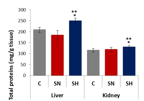
The data obtained in protein concentration analysis (Fig. 1) showed that different variations appeared depending on the type of tissue and temperature conditions. Thus, in the case of hepatic tissue, the sepsis in normothermic conditions induced an insignificant decrease in protein concentration (208.06 ± 11.75 mg/g tissue vs. 183.30 ± 21.35 mg/g tissue, p > 0.05). In rats kept under hypothermic conditions, a normalisation of the concentration was observed, values being produced similar to that in the control group (208.06 ± 11.75 mg/g tissue vs. 249.50 ± 11.80 mg/g tissue, p > 0.05) and significantly higher than that in the normothermic group (249.50 ± 11.80 mg/g tissue vs. 183.30 ± 21.35 mg/g tissue, p < 0.05). Kidney tissue variation was not different to that observed in the liver, with sepsis causing an insignificant increase in protein concentration in the normothermic group (114.57 ± 7.86 mg/g tissue vs. 118.12 ± 9.93 mg/g tissue, p > 0.05). Hypothermia affected the concentration of proteins, the values being significantly increased compared to the normothermic group (129.84 ± 6.56 mg/g tissue vs. 118.12 ± 9.93 mg/g tissue, p < 0.05) and to the control (129.84 ± 6.56 mg/g tissue vs. 114.57 ± 7.86 mg/g tissue, p < 0.05).
Fig. 1.

Total proteins concentration in the liver and kidneys for each experimental condition (C – control; SN – sepsis normothermic; SH– sepsis hypothermic). * – different from control (P < 0.05); ** – different from SN (p < 0.05). Values are expressed as means ± SD
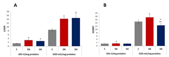
Figs 2A and 2B present changes in the profiles of antioxidant enzymes activity in the liver and kidneys. In the liver, GPx activity increased statistically significantly in both groups with sepsis versus the control group, regardless of the temperature (3.05 ± 0.49 U/mg of proteins vs. 1.42 ± 0.15 U/mg of proteins, p < 0.05 for SN and 2.58 ± 0.16 vs. 1.42 ± 0.15, p < 0.05 for SH). SOD activity was increased significantly in the liver by inducing sepsis, both in normal and hypothermic conditions (14.17 ± 0.73 mU/mg of proteins vs. 8.44 ± 0.65 mU/mg of proteins, p < 0.05 for SN and 14.60 ± 2.49 vs. 8.44 ± 0.65, p < 0.05 for SH). It has also been observed that hypothermia did not induce significant changes in hepatic GPx or SOD activities.
Fig. 2.
GPx and SOD activity in the liver (A) and kidneys (B) for each experimental condition (C – control; SN – sepsis normothermic; SH – sepsis hypothermic). * – different from control (p < 0.05); ** – different from SN (p < 0.05). Values are expressed as means ± SD
In renal tissue, GPx activity increased statistically significantly only in the group with sepsis maintained in normothermic conditions versus the control (1.42 ± 0.009 U/mg of proteins vs. 1.25 ± 0.10 U/mg of proteins, p < 0.05 for SN and 1.28 ± 0.11 U/mg of proteins vs. 1.25 ± 0.10 U/mg of proteins, p > 0.05 for SH). SOD activity increased statistically significantly in sepsis versus the control in normothermic conditions (14.72 ± 1.33 mU/mg of proteins vs. 17.35 ± 1.32 mU/mg of proteins, p < 0.05 for SN) but decreased in hypothermic conditions (14.72 ± 1.33 mU/mg of proteins vs. 12.43 ± 1.30 mU/mg of proteins, p < 0.05 for SH compared with the control and 17.35 ± 1.33 mU/mg of proteins vs. 12.43 ± 1.30 mU/mg of proteins, p < 0.05 for SH compared with SN).
Figs 3A and 3B show changes between the profiles of MDA and DNPH concentrations in the liver and kidneys during the experiment. An increase in MDA concentration was seen in all organs after CLP. For the liver, the determined values were 0.93 ± 0.12 μmol/mg of proteins vs. 3.59 ± 0.13 μmol/mg of proteins, (p < 0.05) in normothermic conditions and 0.928 ± 0.12 μmol/mg of proteins vs. 2.94 ± 0.051 μmol/mg of proteins (p < 0.05) in hypothermic conditions. For renal tissue, the values were 1.39 ± 0.05 μmol/mg of proteins vs. 5.43 ± 0.10 μmol/mg of proteins (p < 0.05) in the SN group and 1.39 ± 0.05 μmol/mg of proteins vs. 5.48 ± 0.30 μmol/mg of proteins (p < 0.05) in the SH group. Under hypothermia the SH group showed significantly lower MDA compared with the SN group in the liver (2.94 ± 0.051 μmol/mg of proteins vs. 3.59 ± 0.13 μmol/mg of proteins, p < 0.05) but no statistically significant differences were observed in the kidneys (5.48 ± 0.30 μmol/mg of proteins vs. 5.43 ± 0.10 μmol/mg of proteins, p > 0.05).
Fig. 3.
MDA and DNPH concentrations in the liver (A) and kidneys (B) for each experimental condition (C – control; SN – sepsis normothermic; SH – sepsis hypothermic). * – different from control; ** – different from SN (p < 0.05). Values are expressed as means ± SD
In the liver, the DNPH concentration increased after CLP, shown in the values 6.93 ± 0.27 nmol/mg of proteins vs. 15.46 ± 0.32 nmol/mg of proteins (p < 0.05) in the SN group and 6.93 ± 0.27 nmol/mg of proteins vs. 12.15 ± 0.15 nmol/mg of proteins (p < 0.05) in the SH group. In the kidneys, the values were 6.22 ± 0.17 nmol/mg of proteins vs. 23.58 ± 0.40 nmol/mg of proteins (p < 0.05) in the SN group and 6.22 ± 0.17 nmol/mg of proteins vs. 12.01 ± 0.10 nmol/mg of proteins (p < 0.05) in the SH group. Under hypothermic conditions, the level of carbonylated proteins was statistically significantly lower compared to this level under normothermic conditions for both tissues (Fig. 3).
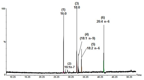
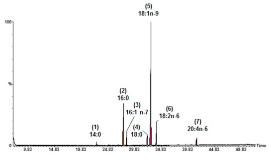
The results of fatty acid analysis in total lipid extracts are detailed in Fig. 4, Fig. 5, Table 1, and Table 2. From Table 1 and the chromatograms it is clear that the main fatty acids determined in lipid extracts obtained from tissues varied with both the type of tissue analysed and the experimental conditions.
Fig. 4.

GC-MS chromatogram of the FAMEs from total lipids of the liver (SH – sepsis hypothermic group) analysed with a BPx-70 column. Peaks of fatty acids: (1) palmitic (16:0); (2) palmitoleic (16:1 n–7); (3) stearic (18:0); (4) oleic (18:1 n–9); (5) linoleic (18:2 n–9); and (6) arachidonic (20:4 n–6)
Fig. 5.

GC-MS chromatogram of the FAMEs from total lipids of kidneys (SN – sepsis normothermic group) analysed with a BPx-70 column. Peaks of fatty acids: (1) myristic (14:0); (2) palmitic (16:0); (3) palmitoleic (16:1 n–7); (4) stearic (18:0); (5) oleic (18:1 n–9); (6) linoleic (18:2 n–6); and (7) arachidonic (20:4 n–6)
Table 1.
Fatty acid composition of total lipids determined by GC-MS in the liver and kidneys for each experimental condition (C – control; SN – sepsis normothermic; SH – sepsis hypothermic)
| Fatty acid % a | ||||||
|---|---|---|---|---|---|---|
| Fatty acid | C | Liver SN | SH | C | Kidneys SN | SH |
| Myristic (14:0) | nd | nd | nd | 0.17 ± 0.09 | 1.01 ± 0.01 | 0.15 ± 0.01 |
| Palmitic (16:0) | 26.61 ± 1.21 | 24.48 ± 1.03 | 23.17 ± 0.98 | 7.17 ± 0.21 | 24.53 ± 1.06 | 9.71 ± 0.42 |
| Palmitoleic (16:1 n–9) | 0.16 ± 0.08 | 0.19 ± 0.06 | 0.17 ± 0.11 | 0.8 ± 0.09 | 0.21 ± 0.05 | 0.78 ± 1.01 |
| Palmitoleic (16:1 n–7) | 0.96 ± 0.12 | 0.92 ± 0.19 | 0.98 ± 0.11 | 0.26 ± 0.07 | 3.92 ± 0.24 | nd |
| Stearic (18:0) | 18.02 ± 1.13 | 29.69 ± 1.65 | 26.65 ± 1.16 | 0.53 ± 0.03 | 6.88 ± 0.29 | 0.62 ± 0.13 |
| Oleic (18:1 n–9) | 8.48 ± 0.32 | 5.15 ± 0.19 | 8.43 ± 0.28 | 69.22 ± 1.69 | 50.16 ± 1.53 | 67.62 ± 0.63 |
| Oleic (18:1 n–7) | 2.71 ± 0.13 | 1.94 ± 0.16 | 1.4 ± 0.09 | 4.01 ± 0.24 | 3.3 ± 0.52 | 3.59 ± 0.12 |
| Linoleic (18:2 n–9) | 14.12 ± 0.5 | 13.49 ± 0.29 | 12.88 ± 0.33 | 13.01 ± 0.31 | 9.85 ± 0.22 | 13.4 ± 0.52 |
| Arachidonic (20:4 n–6) | 28.94 ± 0.89 | 24.15 ± 0.91 | 26.32 ± 1.41 | 4.83 ± 0.12 | 0.14 ± 0.06 | 4.12 ± 0.16 |
– % of total fatty acids; nd – not detected. Values are mean ± standard deviation of samples analysed individually in triplicate
Table 2.
Statistical analysis of fatty acid distribution in total lipids for each experimental condition (C – control; SN – sepsis normothermic; SH – sepsis hypothermic)
| Fatty acid/% a | ||||||
|---|---|---|---|---|---|---|
| Type of tissues/ | group | Σ SFAs | Σ MUFAs | Σ PUFAs | Σ UFAs | Σ UFAs / Σ SFAs |
| C | 44.63 ± 1.79 | 12.31 ± 0.46 | 43.06 ± 1.46 | 55.37 ± 2.13 | 1.24 | |
| Liver | SN | 54.17 ± 1.65* | 8.19 ± 0.29* | 37.63 ± 1.38* | 50.19 ± 1.98* | 0.84* |
| SH | 49.82 ± 1.84*,** | 10.98 ± 0.36*,** | 39.19 ± 1.65 | 45.83 ± 1.73*,** | 1.007** | |
| C | 7.87 ± 0.53 | 74.29 ± 2.93 | 17.84 ± 0.55 | 92.13 ± 3.69 | 11.70 | |
| Kidneys | SN | 32.42 ± 1.27* | 57.59 ± 1.95* | 9.99 ± 0.37* | 67.58 ± 2.24* | 2.08* |
| SH | 10.48 ± 0.67*,** | 72.00 ± 2.63** | 17.52 ± 0.51 | 89.52 ± 3.33** | 8.54** | |
– % of total fatty acids; * – different from control; ** – different from SN (P < 0.05); SFAs – saturated fatty acids; MUFAs – monounsaturated fatty acid; PUFAs – polyunsaturated fatty acids; UFAs – unsaturated fatty acids. Values are mean ± standard deviation of samples analysed individually in triplicate
Discussion
Some of the postulated molecular mechanisms of sepsis progression are linked to the imbalance between ROS production and cellular antioxidant systems. Pro-inflammatory effects of ROS include endothelial damage, neutrophil recruitment, cytokine release, and mitochondrial damage. Accumulation of neutrophils is a phenomenon that occurs relatively early in the liver during the development of sepsis. Liver neutrophil recruitment is the result of a complex interaction between neutrophils, sinusoidal endothelium, hepatocytes, and Kupffer cells. Neutrophils accumulated in the liver cause the secretion of proteolytic enzymes and ROS. They may also induce the production of proinflammatory cytokines and chemokines, all contributing to an oxidant–antioxidant imbalance. Thus, the sepsis state induces changes in the activity of the enzymatic antioxidant systems in the target organs, including the liver (2, 21, 27).
SOD is the enzyme that catalyses the conversion of the superoxide anion into hydrogen peroxide, the substrate specific for catalase and GPx. If a high level of SOD activity occurs at the cellular level without a proportional increase in the activity of catalase and/or GPx, a large amount of hydrogen peroxide accumulates intracellularly and in metallic catalysis can generate the hydroxyl radical, known as the most harmful ROS. Two possible mechanisms can explain this asymmetric growth. The first mechanism assumes that in the CLP sepsis model there is an increase in the level of IL-1, TNF, and LPS in the peritoneal fluid and plasma, pro-inflammatory molecules that increase mRNA-SOD expression. The second mechanism is based on the idea that catalase activity can be inhibited by superoxide, which is produced in large amounts in sepsis. It was demonstrated that after sepsis induction, there was an imbalance between the antioxidant enzymes CAT and SOD, which was followed by oxidative damage in the major organ systems (the lungs, diaphragm, heart, liver, and kidneys). Also, a significant increase in SOD after CLP in the lungs and kidneys was observed. In addition, it was demonstrated that sepsis did not alter GPx activity in the liver (2). Our data are consistent with these in terms of the activity of SOD in the kidneys but are contradictory in terms of GPx renal activity.
MDA is one of the most frequently used indicators of lipid peroxidation mediated by free oxygen radicals, and is thought to damage cell membrane integrity substantially (12, 22, 23). Our data are in accordance with reports that indicate there is a marked increase in the level of lipid and protein degradation in all target organs in rats where sepsis has been induced by the CLP technique (29). This amplification of MDA formation can be explained by the fact that the GPx enzyme has lower activity, lipid peroxides being the specific substrate of this enzyme. Another study determined the level of lipid and protein oxidation as well as the production of superoxide in liver cells after CLP. It was observed that the lipid oxidation level increased from 6 to 48 h. In addition, the level of carbonylated proteins showed the same profile as lipid degradation, increasing significantly at 6 h after the procedure (20).
The effects of hypothermia in sepsis have been studied in various models, and the results obtained in the present study are consistent with those in the literature. Hypothermia reduced the level of tissue lipid peroxidation in the liver and lung tissue, supporting the idea that the beneficial effect of hypothermic therapy is partially mediated by its antioxidant effect (22).
Oxidative degradation of proteins due to ROS accumulation at the cellular level leads to various functional consequences such as the inactivation of some enzymes and increased susceptibility to aggregation (precipitation) and proteolysis. The oxidative modification of proteins is primarily initiated by reaction with hydroxyl radicals, but it is further dependent on the concentration of the radical superoxide anion and the hydrogen peroxide in the medium. In cellular systems, oxidation of proteins can also occur under the action of some carbonyl radicals or products formed by lipid peroxidation processes. An increase in the level of oxidation of unsaturated fatty acids therefore leads to an increase in the degree of oxidation of proteins (4). Protein oxidation can occur along with membrane damage due to lipid peroxidation, leading to altered membrane permeability, protein structure modification, and functional alterations. Also, oxidative lesions of the mitochondrial membrane may occur, leading to membrane depolarisation and decoupling of oxidative phosphorylation with alteration of cellular respiration (19).
Metabolic dysfunctions during sepsis include alterations in lipid metabolism and decreased fatty acid β-oxidation. Consequently, plasma free fatty acids increase, resulting in intensified tissue lipolysis in organs such as the liver, kidneys, heart, and skeletal muscles. This brings in harmful consequences for patients with diverse pathological conditions (15).
A marked increase in the levels of stearic acid (18:0) in the liver of rats with sepsis maintained in normothermic and hypothermic conditions was found. In contrast, there was a significant decrease in oleic acid (18:1), linolenic acid (18:3), and arachidonic acid (20:4). In renal tissues a marked increase in the levels of palmitic acid (16:0), stearic acid (18:0), and oleic acid (18:1) was only observed in rats with sepsis maintained in normothermic conditions. In parallel, there was a significant decrease in oleic acid (18:1), linolenic acid (18:3), and arachidonic acid (20:4). However, following the hypothermia the fatty acid composition was brought back to near normal.
Lipid accumulation in cells that are not equipped with the molecular tools to handle large lipid loads has been associated with cellular injury and dysfunction. This process has been termed lipotoxicity and has been linked to dysfunction in multiple organs, including the liver, heart, skeletal muscles, and pancreas. The principal determinant of lipotoxicity seems to be excessive intracellular free fatty acid content, leading to accumulation of potentially toxic metabolites such as fatty acyl-CoA, diacylglycerol, and ceramides. Lipotoxic cellular dysfunction and injury occur through several mechanisms, including the generation of ROS, multiple organellar damage, disruption of intra-cellular signalling pathways, release of proinflammatory and profibrotic factors, and lipid-induced apoptosis (lipoapoptosis) (7).
From the statistical analysis of data obtained for different types of fatty acids, it is observed that induced sepsis in rats maintained under normal temperature conditions (SN) raised SFAs significantly and concomitantly diminished all types of UFAs, so that the ΣUFA:ΣSFA ratio was significantly lower than that of the control group. In the hypothermic group (SH), there were statistically significant decreases in SFAs and MUFAs. In the case of PUFAs, the reduction was not statistically significant compared with the control group. The decrease in temperature caused changes in the fatty acid profile compared to normothermic rats. Thus, the content of SFAs and MUFAs and the ΣUFA:ΣSFA ratio increased to a statistically significant extent.
For both types of analysed septic tissue, the most severe decrease was observed in the PUFA content. Long chain PUFAs such as arachidonic acid play an important role in various biological functions, being essential components in membranes of eukaryotic cells and maintaining their fluidity, flexibility, and selective permeability. All cell membranes are particularly vulnerable to oxidation due to high concentrations of PUFAs. The main substrate of lipid peroxidation is constituted by PUFA components of subcellular and cellular membranes. Most associated studies employed the traditional TBA assay for estimation of the degree of lipid peroxidation. It is now known that the TBA assay should be conducted in the presence of an antioxidant in order to avoid artificial lipid peroxidation during the assay. In the heated acidic medium of the TBA assay under aerobic conditions, biological samples would be more susceptible to artificial oxidation. The determination of PUFA content and the ratio of UFAs to SFAs are the other methods that can be used in order to determine the extent of lipid peroxidation (1, 3, 5). Our study results obtained by analysing peroxidation level by the MDA method correlate with those obtained by determining the ΣUFA:ΣSFA ratio.
The increase in oxidative stress in septic rat livers and kidneys was confirmed by an increase in SOD and GPx activity and correlated with an increase in the concentration of MDA, a decrease in UFA/SFA ratio, and an increased level of protein oxidation. The effects of hypothermia on the oxidative stress indices in experimentally induced sepsis in rats are dependent on the type of tissue. Hypothermia does not induce significant changes in enzyme antioxidant activity, with differences being insignificant between septic rats under normal temperature conditions and those under low temperature conditions, both in the liver and kidneys. In the liver, a slight decrease in MDA concentration in the hypothermic group was observed compared to the normothermic group. In the kidneys, regardless of the temperature at which the animals were kept, the MDA concentration increased significantly compared to the control. Under hypothermic conditions the decrease in the DNPH concentration was significantly higher in the case of renal tissue than in that of the liver.
Footnotes
Conflict of Interest
Conflict of Interests Statement: The authors declare that there is no conflict of interests regarding the publication of this article.
Financial Disclosure Statement: This project is funded by the Ministry of Research and Innovation of Romania, Projects for Financing the Excellence in CDI, contract no. 37PFE/06.11.2018.
Animal Rights Statement: All experiments were carried out according to the practices mandated by the Romanian Board of Animal Research, in agreement with the European Union regulation on animal testing, and were permitted by the Committee of Animal Ethics at the Faculty of Veterinary Medicine Cluj-Napoca, Romania.
References
- 1.Ando K., Nagata K., Yoshida R., Kikugawa K., Suzuki M.. Effect of n-3 polyunsaturated fatty acid supplementation on lipid peroxidation of rat organs. Lipids. 2000;35:401–407. doi: 10.1007/s11745-000-538-6. [DOI] [PubMed] [Google Scholar]
- 2.Andrades M., Ritter C., de Oliveira M.R., Streck E.L., Fonseca Moreira J.C., Dal-Pizzol R.. Antioxidant treatment reverses organ failure in rat model of sepsis: role of antioxidant enzymes imbalance, neutrophil infiltration, and oxidative stress. J Surg Res. 2011;167:e307–e313. doi: 10.1016/j.jss.2009.08.005. [DOI] [PubMed] [Google Scholar]
- 3.Andrei S., Bunea A., Pintea A. Oxidative stress and natural antioxidants. edited by Academic Press Cluj; 2014. pp. 89–95. [Google Scholar]
- 4.Andrei S., Joanta A., Pintea A., Varga A.. Studies regarding antioxidant administration on fatty acids and cholesterol content in rats i.p. treated with L-thyroxin. Bulletin UASVM Vet Med. 2004;61:16–22. [Google Scholar]
- 5.Andrei S., Pintea A., Rugină D., Ştefănuţ C., Bunea A., Gal A.F.. Protective activity of Polyphenon E against liver protein oxidation in rats after MNU i.p. administration. Bulletin UASVM Vet Med. 2014;71:15–18. [Google Scholar]
- 6.Bar-Or D., Carrick M., Mains C., Rael L.T., Slone D., Brody E.N.. Sepsis, oxidative stress, and hypoxia: are there clues to better treatment? Redox Rep. 2015;20:193–197. doi: 10.1179/1351000215Y.0000000005. [DOI] [PMC free article] [PubMed] [Google Scholar]
- 7.Bobulescu I.A.. Renal lipid metabolism and lipotoxicity. Curr Opin Nephrol Hypertens. 2010;19:393–402. doi: 10.1097/MNH.0b013e32833aa4ac. [DOI] [PMC free article] [PubMed] [Google Scholar]
- 8.Christie W.W. William W.C. Gas Chromatography and Lipids. A Practical Guide. Oily Press; Glasgow: 1989. Preparation of methyl ester and other derivatives; pp. 36–47. edited by. [Google Scholar]
- 9.Dare A.J., Phillips A.R., Hickey A.J., Mittal A., Loveday B., Thompson N., Windsor J.A.. A systematic review of experimental treatments for mitochondrial dysfunction in sepsis and multiple organ dysfunction syndrome. Free Radic Biol Med. 2009;47:1517–1525. doi: 10.1016/j.freeradbiomed.2009.08.019. [DOI] [PubMed] [Google Scholar]
- 10.Doi K., Leelahavanichkul A., Yuen S.T., Star R.A.. Animal models of sepsis and sepsis-induced kidney injury. J Clin Invest. 2009;119:2868–2878. doi: 10.1172/JCI39421. [DOI] [PMC free article] [PubMed] [Google Scholar]
- 11.Folch J., Lees M., Sloane Stanley G.H.. A simple method for the isolation and purification of total lipides from animal tissues. J Biol Chem. 1957;226:497–509. [PubMed] [Google Scholar]
- 12.Gal A.F., Andrei S., Cernea C., Taulescu M., Catoi C.. Effects of astaxanthin supplementation on chemically induced tumorigenesis in Wistar rats. Acta Vet Scand. 2013;54:50. doi: 10.1186/1751-0147-54-50. [DOI] [PMC free article] [PubMed] [Google Scholar]
- 13.Galley H.F.. Oxidative stress and mitochondrial dysfunction in sepsis. Br J Anaesth. 2011;107:57–64. doi: 10.1093/bja/aer093. [DOI] [PubMed] [Google Scholar]
- 14.Ginsburg M.D., Belayev L. Mayer S.A., Sessler D.I. Therapeutic Hypothermia. Marcel Dekker; New York: 2005. Biological and Molecular Mechanisms of Hypothermic Neuroprotection; pp. 85–119. edited by. [Google Scholar]
- 15.Gonçalves-de-Albuquerque C.F., Medeiros-de-Moraes I.M., Oliveira F.M., Burth P., Bozza P.T., Castro Faria M.V., Silva A.R., Castro-Faria-Neto H.C. Omega-9 oleic acid induces fatty acid oxidation and decreases organ dysfunction and mortality in experimental sepsis. PLOS One. 2016. DOI, 10.1371/journal.pone. 0153607. [DOI] [PMC free article] [PubMed]
- 16.Karelle L., Moisan C., Amérand A., Poupon G., L’Her E.. Effect of induced mild hypothermia on two pro-inflammatory cytokines and oxidative parameters during experimental acute sepsis. Redox Rep. 2013;18:120–126. doi: 10.1179/1351000213Y.0000000049. [DOI] [PMC free article] [PubMed] [Google Scholar]
- 17.Lee I., Hüttemann M.. Energy crisis: the role of oxidative phosphorylation in acute inflammation and sepsis. Biochim Biophys Acta. 2014;1842:1579–1586. doi: 10.1016/j.bbadis.2014.05.031. [DOI] [PMC free article] [PubMed] [Google Scholar]
- 18.Lee J.H., Kim K., Jo Y.H., Kim M.A., Rim K.P., Kang K.W., Rhee J.E., Lee M.J., Lee H.S., Kwon W.Y., Suh G.J.. Therapeutic hypothermia attenuates liver injury in polymicrobial sepsis model of rats via akt survival pathway. J Surg Res. 2013;181:114–120. doi: 10.1016/j.jss.2012.05.045. [DOI] [PubMed] [Google Scholar]
- 19.Macdonald J., Galley H.F., Webster N.R.. Oxidative stress and gene expression in sepsis. Br J Anaesth. 2003;90:221–232. doi: 10.1093/bja/aeg034. [DOI] [PubMed] [Google Scholar]
- 20.Nakamura A., Goto S.. Analysis of protein carbonyls with 2,4-dinitrophenyl hydrazine and its antibodies by immunoblot in two-dimensional gel electrophoresis. J. Biochem. 1996;119:768–774. doi: 10.1093/oxfordjournals.jbchem.a021306. [DOI] [PubMed] [Google Scholar]
- 21.Prauchner C.A.. Oxidative stress in sepsis: pathophysiological implications justifying antioxidant co-therapy. Burns. 2017;43:471–485. doi: 10.1016/j.burns.2016.09.023. [DOI] [PubMed] [Google Scholar]
- 22.Rim K.P., Kim K., Jo Y.H., Lee J.H., Rhee J.E., Kang K.W., Suh G.J., Kwon W.Y., Lee M.J., Lee H.S.. Effect of therapeutic hypothermia according to severity of sepsis in a septic rat model. Cytokine. 2012;60:755–761. doi: 10.1016/j.cyto.2012.08.013. [DOI] [PubMed] [Google Scholar]
- 23.Ritter C., Andrades M., Frota M.C., Bonatto F., Pinho R.A., Polydoro M., Klamt F., Pinheiro C.S., Menna-Barreto S.S., Moreira J.F., Dal-Pizzol F.. Oxidative parameters and mortality in sepsis induced by cecal ligation and perforation. Intensive Care Med. 2003;29:1782–1789. doi: 10.1007/s00134-003-1789-9. [DOI] [PubMed] [Google Scholar]
- 24.Ruehl-Fehlert C., Kittel B., Morawietz G.. Revised guides for organ sampling and trimming in rats and mice – Part 1. A joint publication of the RITA and NACAD groups. Exp Toxic Pathol. 2003;55:91–106. doi: 10.1078/0940-2993-00350. [DOI] [PubMed] [Google Scholar]
- 25.Toscano M.G., Ganea D., Gamero A.M.. Cecal ligation puncture procedure. J Vis Exp. 2011;7:2860. doi: 10.3791/2860. [DOI] [PMC free article] [PubMed] [Google Scholar]
- 26.Vodnar D.C., Călinoiu L.F., Dulf F.V., Ştefănescu B.E., Crişan G., Socaciu C.. Identification of the bioactive compounds and antioxidant, antimutagenic and antimicrobial activities of thermally processed agro-industrial waste. Food Chem. 2017;231:131–140. doi: 10.1016/j.foodchem.2017.03.131. [DOI] [PubMed] [Google Scholar]
- 27.Wang D., Yin Y., Yao Y.. Advances in sepsis-associated liver dysfunction. Burns Trauma. 2014;28:97–105. doi: 10.4103/2321-3868.132689. [DOI] [PMC free article] [PubMed] [Google Scholar]
- 28.Wingfield W.E., Raffe M.R. Wingfield W.E., Raffe M.R. The Veterinary ICU Book. Telton NewMedia; 2000. Sepsis; pp. 685–710. edited by. [Google Scholar]
- 29.Zapelini P.H., Rezin G.T., Cardoso M.R., Ritter C., Klamt F., Moreira J.F., Streck E.L., Dal-Pizzol F.. Antioxidant treatment reverses mitochondrial dysfunction in a sepsis animal model. Mitochondrion. 2008;8:211–218. doi: 10.1016/j.mito.2008.03.002. [DOI] [PubMed] [Google Scholar]


