

Abstract
In this study, respiratory viral pathogens were screened using real-time RT-PCR in 86 broiler chicken flocks suffering from respiratory diseases problems in 4 Egyptian governorates between January 2012 and February 2014. The mortality rates in the investigated flocks ranged from 1 to 47%. Results showed that mixed infection represented 66.3% of the examined flocks. Mixed infectious bronchitis (IBV) and avian influenza (AI)-H9N2 viruses were the most common infection (41.7%). Lack of AI-H9N2 vaccination and high rates of mixed infections in which AI-H9N2 is involved indicate an early AI-H9N2 infection with a potential immunosuppressive effect that predisposes for other viral infections. High pathogenic AI-H5N1 and virulent Newcastle disease virus (vNDV) infections were also detected (26.7% and 8.1%, respectively). Interestingly, co-infection of AI-H9N2 with either AIV-H5N1 or vNDV rarely resulted in high mortality. Partial cell-mediated immunity against similar internal AI genes, as well as virus interference between AI and vNDV, could be an explanation for this. Highly prevalent IBV and AI-H9N2 were isolated and were molecularly characterized based on S1 gene hypervariable region 3 (HVR3) and hemagglutinin gene (HA) sequences, respectively. IBV strains were related to the variant group of IBV with multiple mutations in HVR3. Though AI-H9N2 viruses showed low rate of evolution in comparison to recent strains, few amino acid substitutions indicative of antibody selection pressure were observed in the HA gene. In conclusion, mixed viral infections, especially with IBV and AI-H9N2 viruses, are the predominant etiology of respiratory disease problems in broiler chickens in Egypt. Further investigations of the role of AI, IBV, and ND viruses’ co-infections and interference in terms of altering the severity of clinical signs and lesions and/or generating novel reassortants within each virus are needed.
Key words: chicken, Egypt, H9N2, infectious bronchitis, respiratory outbreaks
INTRODUCTION
Respiratory disease outbreaks with variable mortality rates and different clinical manifestations have been increasing in Egyptian commercial chicken flocks during the last few years. Respiratory diseases represent a large problem to the poultry industry because of their multifactorial nature (Roussan et al., 2008). Different pathogens such as avian influenza (AI), virulent Newcastle disease virus (vNDV), and infectious bronchitis (IBV) are associated with high mortality rates in broiler chicken flocks (Haghighat-Jahromi et al., 2008). These pathogens are of major significance and have a large economic impact because they are able to induce disease independently or in association with each other (Roussan et al., 2008). The failure of control of poultry diseases in Egypt is a final result of retarded biosecurity, shortage of vaccine coverage, and/or the mismatch between circulating and vaccine strains.
Extensive surveillance and genetic studies revealed that AI-H5N1 viruses became endemic in poultry in many countries including Egypt. Clinically unapparent, infected free-ranging ducks and geese, as well as mixed species backyard holdings, are suspected to play a pivotal role in avian influenza epidemiology in Egypt (Aly et al., 2008). The detection of the AI-H9N2 subtype during 2010 and 2011 in chickens and commercial quail was an additional challenge facing the poultry industry in Egypt (El-Zoghby et al., 2012). As both H5N1 and H9N2 viruses are endemic in Egypt, the genetic characterization of AIVs is necessary for screening the dynamic evolution of each subtype and potential reassortment events.
One of the major components of mixed infections is IBV that produces air saculitis in chickens, which may result in condemnation of broilers during processing (Hofstad, 1984). In spite of regular vaccinations with Massachusetts (Mass) strains, IBV still has diverse effects on the poultry industry in the country. In Egypt, IBV has been recognized since the 1950s (Sheble et al., 1986) with the isolation of an IB variant shown by neutralization tests to be closely related to the Dutch variant D3128 and subsequently variants related to Mass (Eid, 1998), D3128, D274, D-08880, 4/91 were characterized. Novel genotypes endemic in Egypt represented by the isolate Egypt/Beni-Suef/ 01 and nephropathogenic IBV strains Egypt/Beni-Suef/03, which are closely related to Israeli variant 2 and Mass serotype, respectively, were isolated from poultry (Abdel-Moneim et al., 2006). Recently, other novel genotypes were reported in Egypt and classified as Egyptian variant I (e.g., CK/Eg/BSU-1,4,5/2011) and Egyptian variant II (e.g., CK/Eg/BSU-2,3/2011) (Abdel-Moneim et al., 2012).
In spite of strict vaccination regimes, the NDV has been a very serious problem for poultry production in many countries and enormous efforts have been made for controlling the disease. NDV strains from recent outbreaks affecting poultry farms in Egypt between 2011 and 2012 were confirmed to belong to class II, genotype VII, sub genotype d (lineage 5 sub lineage d). Genotype VII is thought to be spreading in Egypt through the trading of poultry and poultry products with Middle Eastern countries and China (Mohamed et al., 2011; Radwan et al., 2013).
For avian respiratory viruses, virus isolation is the standard test for diagnosis although it tends to be costly, labor intensive, and slow (Suarez et al., 2007). Rapid diagnostic tests have been developed for detection of either the viral nucleic acids or viral antigens including rapid chromatographic tests for antigen detection, reverse transcriptase polymerase chain reaction (RT-PCR) and real-time RT-PCR (rRT-PCR) (Spackman et al., 2002).
In this study the rRT-PCR technique was utilized to investigate the current field situation of avian respiratory pathogens especially AI, vNDV, and IBV viruses in Egypt. Moreover, selected respiratory viruses were molecularly identified to monitor the genetic properties of the circulating viruses.
MATERIAL AND METHODS
Field Samples
Tracheal and/or oropharyngeal swabs and tissues were collected from 86 broiler chicken flocks (10 samples per flock) suffering from respiratory disease problems between January 2012 and February 2014. Samples collected varied between seasons and the largest numbers of samples (39 flocks) were collected during winter of 2012 to 2013. The area of investigation included the Beni-Suef, Fayoum, Menia, and Qalubia governorates in Egypt. Tissue samples were ground in phosphate buffer saline pH 7.0 to 7.4 containing gentamycin (50 μg/mL) and mycostatin (1,000 units/mL) in a 1:5 (w/v) dilution, centrifuged, and tissue supernatant was collected (OIE, 2014).
Viral RNA Extraction and Real-Time RT-PCR (rRT-PCR)
The viral RNA was extracted by Bioflux viral RNA Mini Spin column kit (Bioflux, China) in accordance with manufacturer's instructions. Single step rRT-PCR assays using Verso 1-Step qRT-PCR Kit Plus ROX Vial (Thermo Scientific, US) were conducted using specific oligonucleotide primers and probes for each target virus. The final reaction volume was 25 μL including; 5 μL RNA template, 12.5 μL 2X 1-step PCR ready mix, 1.25 μL RT-enhancer, 0.25 μL Verso enzyme mix, 1 μL of each of the forward and reverse primers, and 0.25 μL probe for AI-H5N1 (Slomka et al., 2007), AI-H9N2 (Ben Shabat et al., 2010), IBV (Callison et al., 2006), and vNDV (Wise et al., 2004), together with 3.75 μL nuclease free water. Thermocycling rRT-PCR conditions were 50°C for 15 min, 95°C for 15 min, followed by 40 cycles at 95°C for 15 sec and 30 sec at 60°C (for AI-H9N2 and IBV) or at 54°C (for AI-H5N1 and NDV) with reading of fluorescence in this step.
Virus Isolation
Five samples were selected for each of the highly prevalent respiratory viruses (i.e., IBV and AI-H9N2) based on rRT-PCR detection, seasonal and geographical distribution, and mortality rates observed (Table 1 ). Tracheal swabs and/or tissue suspensions were collected from diseased birds and prepared for inoculation through allantoic route of 10-day-old specific pathogen free embryonated chicken eggs) (SPF-ECE) (OIE, 2014). Inoculated eggs were incubated at 37°C for 96 hours and candled daily for embryo viability. In IBV positive rRT-PCR samples, 7 serial blind passages in SPF-ECE were done for isolation. All inoculated egg allantoic fluids from dead and surviving embryos were harvested and tested for hemagglutination using 1% washed chicken red blood cells (OIE, 2014).
Table 1.
Isolated viruses geographic distribution and history.
| Virus | Isolate (acc. number) | Governorate | Age (Day) | Vaccination | Mortality | Lesions |
|---|---|---|---|---|---|---|
| IBV | Ck/Eg/BSU-FA-KB23/13 (KR010940) | Fayoum | 30 | IB Hitchner (D1), H5N1(D9) IB Clone (D15) | 10% | Tracheal caseous plug, Enteritis |
| Ck/Eg/BSU-FA-KB27/13 (KR010941) | Fayoum | 38 | 4% | |||
| Ck/Eg/BSU-MN-KB44/13 (KR010942) | Menia | 21 | IB Hitchner (D7), H5N1(D10) | 1% | ||
| Ck/Eg/BSU-BS-KB60/14 (KR010943) | Beni-Suef | 29 | IB (4/91) (D1), H9ND (D5), H5N1(D9), NDV Lasota (D17) | 30% | ||
| Ck/Eg/BSU-BS-KB61/14 (KR010944) | Beni-Suef | 16 | Vaccitec Hatchery, IB (H120) (D1) H9ND (D5), IB (4/91) (D14) | 4% | ||
| AIV-H9 | A/Ck/Eg/BSU-FA-KB1/12 (KR010954) | Fayoum | 27 | IB (Ma 5) (D1), IB Hitchner (D7), H5N1 (D9), IB (4/91) (D14) | 6% | Caseous tracheitis, caseous plug, Enteritis Congested trachea, |
| A/Ck/Eg/BSU-MN-KB15/13 (KR010955) | Menia | 34 | IB Hitchner (D1), H5N1 (D9), IB-Clone (D15) | 6% | ||
| A/Ck/Eg/BSU-FA-KB55/13 (KR010956) | Fayoum | 30 | IB Hitchner (D7), IB (Ma 5) (D14), NDV Avinew (D17) | 2% | ||
| A/Ck/Eg/BSU-BS-KB59/13 (KR010957) | Beni-Suef | 28 | IB (Ma 5) (D1), H5N1 (D10) | 20% | ||
| A/Ck/Eg/BSU-BS-K7T/12 (KF998212) | Beni-Suef | 26 | IB (Ma 5) (D1), IB Hitchner (D7), H5N1(D9), IB-Clone (D15) | 18% | ||
| AIV-H5 | A/Ck/Eg/BSU-BS-KF27/12 (KR010950) | Fayoum | 31 | IB (Ma 5) (D1), IB Hitchner (D7) | 16% | General congestion, trachitis Shank Hemorrhage |
| A/Ck/Eg/BSU-BS-KF28/12 (KR010951) | Fayoum | 42 | IB Hitchner (D7), IB-Clone (D15) | 15% | ||
| A/Ck/Eg/BSU-BS-KB56/13 (KR010952) | Beni-Suef | 34 | IB Primer (D1), H9ND (D5), H5N1(D10), NDV Lasota (D17) | 30% | ||
| A/Ck/Eg/BSU-BS-KB62/14 (KR010953) | Beni-Suef | 32 | IB Hitchner (D7), NDV Avinew (D17) | 23% | ||
| vNDV | VII/Ck/Eg/BSU-BS-KN1/13 (KR010945) | Beni-Suef | 34 | IB (Ma 5) (D1), IB Hitchner (D6), IB-Clone (D14), NDV Avinew (D20) | 13% | Hemorrhage on proventiculur glands |
| VII/Ck/Eg/BSU-BS-KN2/13 (KR010946) | Beni-Suef | 27 | IB Hitchner (D1), H5N1(D9) IB Clone (D17) | 3% | ||
| VII/Ck/Eg/BSU-BS-KB57/13 (KR010947) | Beni-Suef | 27 | IB Hitchner (D7), NDV Lasota (D17) | 13% | ||
| VII/Ck/Eg/BSU-BS-KB58/13 (KR010948) | Beni-Suef | 30 | 8% |
Conventional RT-PCR and Gene Sequencing
The viral RNA was extracted from harvested allantoic fluids by Bioflux viral RNA Mini Spin column kit (Bioflux, China) according to the manufacturer's instructions. RT-PCR used for molecular characterizations of isolates using specific oligonucleotide primers for partial amplification of target genes of AI viruses (hemagglutinin gene (HA) gene) (Shany, 2015), IBV (S1 gene) (Selim et al., 2013), and vNDV genes (Wise et al., 2004). Quant One Step RT-PCR Kit (TIANGEN Biotech, China) was used with 25 μL reaction volume including: 7 μL RNA template, 5 μL 5X RT-PCR enhancer, 2.5 μL 10X RT-PCR buffer, 1.5 μL of each of the forward and reverse primers (Table 1), 1.25 μL Hot master Taq-polymerase, 1 μL Super Pure dNTPS, 0.25 μL RNasin, 0.25 μL Quant RTase, and 4.75 μL RNase free water. Thermocycling RT-PCR conditions were 50°C for 30 min, 95°C for 2 min, followed by 35 cycles at 95°C for 15 sec and 30 sec at 50 to 52°C according to the oligonucleotide primers, final extension was performed at 65°C for 10 min.
Amplified RT-PCR products were purified using PCR purification Kit (Thermo Scientific) according to manufacturer's instructions and were then sequenced directly using the ABI Prism 3100 automated sequencing machine (Applied Biosystems, Foster City, CA). A BLAST search was conducted for each sequence (http://www.ncbi.nlm.nih.gov/BLAST). Sequence comparisons and phylogenetic relationships through a bootstrap of 1,000 trials were determined with the MEGA version 6 program using the Clustal W alignment algorithm (Tamura et al., 2013), also Nucleotide and amino acid identities were determined using Geneious 7.1.3 (Biomatters Ltd, New Zealand).
RESULTS
Clinical Finding and Mortality Rates of Examined Flocks
Sixty-eight broiler chicken flocks suffering from respiratory affections from the Beni-Suef, Fayoum, Menia, and Qalubia governorates were examined (supplementary Table 1). Clinical manifestations and postmortem lesions in investigated respiratory disease outbreaks varied according to the infecting virus strain, the vaccination program of the flock, and whether the disease was due to single or multiple infections. The main clinical manifestations were respiratory distress in the form of gasping, rales, and nasal discharges. In some flocks nervous manifestations were observed in the form of head tilting. Tracheal caseation at tracheal bifurcation, tracheitis, and congested viscera were the main pathological features noticed upon postmortem examination. Typical clinical findings of AI-H5N1 and vNDV infected flocks were observed (supplementary Table 1) The incidence of clinical disease was 59.3% in young birds (20–30 days old), which was higher than that of birds aged 31–40 days or those above 40 days old (34.8% and 5.8%, respectively).
Mortality rates in the flocks under investigation ranged from 1 to 47% with the highest mortality rate found in a flock suffering triple IBV, AI-H5N1, AI-H9N2 infection though previous vaccinations against IBV (at 1 and 14 days-old), AI-H5N1 (at 5 days), and AI-H9N2 (at 9 days). Mortality rates in flocks co-infected with IBV and AI-H9N2 were as high as 30%, even in flocks vaccinated against both pathogens. Also AI-H9N2 single infection in some flocks resulted in respiratory signs, tracheal caseation, and 20% mortality rate. Notably, co-infection with AI-H5N1 and H9N2 did not result in very high mortality rates in 11 flocks that were found to be co-infected with both subtypes beside other viruses. Also natural infection with vNDV has resulted in 8 to 13% mortality rates.
Avian respiratory Diseases Viruses’ Detection by rRT- PCR
The results of rRT-PCR revealed that mixed infection is the most common cause of respiratory affection in Egypt and that mixed infection with IB and AIV-H9 viruses was the most common situation in the examined flocks (Table 2 ). Mixed infection represented 66.3% (57 flocks) while single viral infection of the tested viruses was found in 33.7% (29 flocks). High prevalence of both IBV and AI-H9N2 was observed in investigated flocks (83.7 and 61.6%, respectively). Subsequently, mixed infection with AI-H9N2 with IBV has been found to be the most common mixed infection (36 flocks) representing 41.9% of the total investigated flocks.
Table 2.
Real-time RT-PCR detection of IBV, H9N2, H5N1, and vNDV viruses from broiler flocks.
| Single viral infection |
Mixed viral infection |
|||||||||||
|---|---|---|---|---|---|---|---|---|---|---|---|---|
| IB | H9 | H5 | VNDV | IB-H9 | IB-VND | IB-H5 | H9-H5 | IB-H9-H5 | IB-H5-ND | IB-H5-H9-VND | Total | |
| Total IBV | 16 | – | – | – | 36 | 2 | 7 | – | 9 | 1 | 1 | 72 (83.7) |
| Total H9 | – | 6 | – | – | 36 | – | – | 1 | 9 | – | 1 | 53 (61.6%) |
| Total H5 | – | 4 | – | – | – | 7 | 1 | 9 | 1 | 1 | 23 (26.7%) | |
| Total vNDV | – | – | – | 3 | – | 2 | – | – | – | 1 | 1 | 7 (8.1%) |
| No. (%) | 16 (18.6%) | 6 (7%) | 4 (4.6%) | 3 (3.5%) | 36 (41.9%) | 2 (2.3%) | 7 (8%) | 1 (1.2%) | 9 (10.5%) | 1 (1.2%) | 1 (1.2%) | 86 (100%) |
Molecular Identification of Avian Respiratory Viruses
Five isolates of IBV and AI-H9N2 and 4 isolates of AI-H5N1 and vNDV viruses were molecularly identified. The IBV S1 gene, AI virus hemagglutinin gene, and the vNDV F gene sequences were submitted to GenBank and accession numbers were assigned (Table 1).
Avian IB Viruses.
Sequences encoding S1 spike glycoprotein subunit hyper variable region (HVR 3) of IBV were analyzed. The phylogenetic analysis revealed that four of obtained IBV isolates were closely related to each other and to recent Egyptian isolates belonging to variant 2 Egyptian IBV. Only the isolate Ck/Eg/BSU-FA-KB23/2013 was distant from the other 4 isolates and clustered alone (Figure 1 ). Deduced amino acid alignments showed that 4 of IBV isolates obtained; Ck/Eg/BSU-FA-KB27/2013, Ck/Eg/BSU-MN-KB44/2013, Ck/Eg/BSU-BS-KB60/2014, and Ck/Eg/BSU-BS-KB61/2014 (accessions KR010941 to 44, respectively) shared 99.2 to 100% and 98.2 to 99.4% S1 nucleotide and amino acid identities, respectively. The isolates were also 98.8 to 100% and 96.3 to 100% similar to the recent Egyptian isolates based on S1 gene nucleotides and amino acid identity, respectively.
Figure 1.
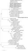
Phylogenetic analysis of partial S1 gene sequence of isolated IBV strains (Bold and ◂). Abbreviations: EG, Egypt; CK, chicken). Phylogenetic relationships through a bootstrap trial of 1,000 were determined with the MEGA version 6 using the Clustal W alignment algorithm and neighbor-joining method for tree construction.
The fifth isolate Ck/Eg/BSU-FA-KB23/2013 was genetically distant from the other isolates of the current study and from currently circulating IBV variants in Egypt (83.6 to 86.4% and 82.4 to 82.9% nucleotide and amino acid identities, respectively). This isolate showed 18 amino acid substitutions at the HVR3, of which 3 successive amino acid substitutions at positions 304 to 306 (asparagine to lysine, phenylalanine to tyrosine, and asparagine to aspartic acid, respectively) (Figure 2 ). It showed relatively higher nucleotides and amino acid identity to the IS/885/00 isolate (88.4 and 85.7% nucleotide and amino acid identities, respectively).
Figure 2.
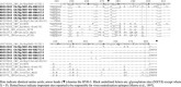
Deduced amino acid sequences of S1 gene of IBV strains in comparison to selected variant and classical strains and vaccines. IBV numbering is according to IBV strain H120 Strain (accession M21970).
Amino acid substitutions observed in IBV isolates include glutamine to histidine at position 296 (QTA/HTA), while isolate Eg/BSU-FAY-B23/2013 showed glutamine to serine (QTA/STA). In another VN epitope, amino acid substitution arginine to leucine was observed at position 378 (PRG/PRL).
AIV-H9N2 Viruses.
The partial HA gene sequences of AI-H9N2 viruses showed that all isolates obtained were related to each other and also related to recent Egyptian and Middle East circulating H9N2 strains, which belong to G1-like lineage (Figure 3 ). Results also showed that all AI-H9N2 isolates share 99.2% nucleotide and amino acid identities; while they share 97.1 to 97.9% nucleotide and 96.2 to 98.5% amino acid identities with recent Egyptian strain A/Ck/Egypt/S4454E/2011 (accession CY110927) and currently used vaccine strain A/Ck/Egypt/114940v/2011(accession JQ440373).
Figure 3.
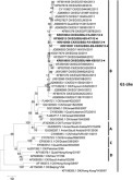
Phylogenetic analysis of partial the HA gene sequence of isolated AIV-H9N2 strains (Bold and ◂). Abbreviations: EG, Egypt; CK, chicken). Phylogenetic relationships through a bootstrap trial of 1,000 were determined with the MEGA version 6 using the Clustal W alignment algorithm and neighbor-joining method for tree construction.
Deduced amino acid sequence analysis of the HA of AI-H9N2 showed only 4 amino acid substitutions in the signal peptide. Other mutations including serine to threonine in the overlapping area of antigenic epitope at position 127 and glycine to alanine at the receptor binding site at position 128 were observed in the HA of the isolate Ck/Eg/BSU-FA-KB1/2012 (Figure 4 ). Another isolate Ck/Eg/BSU-BS-KB59/2013 (accession KR010957) also had an amino acid substitution threonine to arginine at the antigenic epitope 2 at position 182 and alanine to valine in the previously reported receptor binding site at position 180 (H9 numbering).
Figure 4.
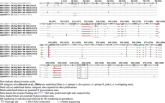
Deduced amino acid sequences of the HA protein of Egyptian AIV-H9N2 strains in comparison to selected Egyptian strains.
AIV-H5N1 and vNDV Viruses.
All AI-H5N1 isolates obtained in the current study (Table 1) were closely related to each other and recent Egyptian strains which belong to clade 2.2.1 (class C). They shared 97.2 to 99% nucleotide identities and 99.2 to 100% amino acid identities with the recent Egyptian strains. The vNDV isolates phylogeny revealed that all isolates were classified as genotype VIId (lineage 5d class II) responsible of vNDV outbreaks in Egypt with no significant genetic changes (data not shown).
DISCUSSION
The rapid development of the poultry industry in Egypt as well as international trade and movement of poultry has been accompanied with the emergence and spread of many viral diseases (Abdelwhab el et al., 2010). Thus ecology and epidemiology of different avian pathogens are now of worldwide concern. In this study, the prevalence of viral pathogens, with special focus on AI, IBV and NDV, was investigated in broiler chickens suffering respiratory disease problems.
There were widespread respiratory disease problems in the winter season where the cold climate is thought to favor virus survival, a high ammonia level, and bad ventilation in different farms, and at least in part, is associated with increased poultry stocking capacity in Egypt (Seififi et al., 2010). The onset of clinical respiratory disease and the highest mortalities in investigated flocks were mainly reported as early as 20 days of age. The lack of vaccination against AI-H9N2 in the majority of the investigated flocks could explain the appearance of disease signs at early ages (supplementary Table 1). Moreover, the high rate of mixed respiratory viral infections observed further indicates a potential immunosuppressive role of AI-H9N2. It was reported that early H9N2 infection causes atrophy and lymphoid depletion of the thymus, and probably also some other lymphoid organs, causing immunosuppression and predispose the chickens to secondary infections (Hadipour et al., 2011).
Data in the current study revealed that IBV alone can cause high mortality (up to 19%) even in vaccinated flocks. Mortalities increased with co-infection of IBV and AIV-H9 (≥30%) even in flocks vaccinated against both pathogens. Also AI-H9N2 infection alone resulted in severe respiratory signs, tracheal caseation, and 20% mortality rate. These data highlight the pathogenicity of AI-H9N2 as previously reported (Nili and Asasi, 2002). Mixed infections of AI-H9N2 with other respiratory pathogens (particularly IBV, Mycoplasma gallisepticum, and Escherichia coli) are thought to be responsible for severe clinical disease with subsequent high mortality (Nili and Asasi, 2003; Haghighat-Jahromi et al., 2008). Though not analyzed in this study, the role of secondary agents as bacteria, and environmental factors in the AI-H9N2 pathogenesis, cannot be neglected.
Interestingly, co-infection with AI-H5N1 and H9N2 infrequently resulted in very high mortality rates. The early exposure to H9N2 viruses possibly provoked cell-mediated immunity against H5N1 due to their similar internal genes. Therefore, partial protection by what is called the “protected window” after H9N2 infection can mask the lethal infection of H5N1 that may go unnoticed while the virus is shed by infected birds. This is a potentially important problem for countries such as Egypt that use H5N1 vaccines and cull flocks only when clinical disease is evident (Khalenkov et al., 2009). Although co-circulation of H5N1 and H9N2 has been suggested to limit the spread and the epizootiologic pattern of the infections for both subtypes (Arafa et al., 2013), their co-circulation in susceptible host populations can increase the likelihood of generating novel reassortant viruses with public health implications (Guan et al., 1999).
Low mortality rates in vNDV infected flocks may be the result of partial protection offered by previous vaccination with live lentogenic NDV vaccines (Hadipour et al., 2011; Munir et al., 2012). Moreover, speculation regarding viral interference exerted by wide and rapid spread of AI-H9N2 LPAI may explain the low mortalities caused by vNDV. Viral interference could be explained by different mechanisms including: competing by attachment interference or blocking of receptor sites for the superinfecting virus; competing intracellularily for replication host machinery; and virus-induced interferon (Ge et al., 2007).
The virus-specific rRT-PCR showed that mixed infections are the predominant etiology of respiratory diseases, especially co-infection with the IBV and AI-H9N2 viruses. The high prevalence of IBV (Abdel-Moneim et al., 2006) and AI-H9N2 (El-Zoghby et al., 2012) in Egyptian poultry has previously been reported. Higher rates of co-infection with both viruses further indicate a potential role for IBV in increasing the severity of AI-H9N2 infection through HA cleavage activation by providing trypsin-like proteases encoded by coronavirus IBV (Klenk and Garten, 1994; Ng and Liu, 2000; Perk et al., 2004; Haghighat-Jahromi et al., 2008).
It is worth mentioning that rRT-PCR assay alone cannot differentiate between field and vaccine strains of IBV and S1 gene sequencing is thought to be the only method used to discriminate between all IBV strains. Therefore, partial IBV S1 gene phylogenetics were analyzed and it was revealed that 4 isolates were closely related to recent Egyptian IBV isolate Eg/BSU-2/2011 (accession JX174185) that was isolated in northern and middle Egypt and showed a unique genetic signature and subsequently was suggested as a new genotype variant 2 (Abdel-Moneim et al., 2012). One isolate, Ck/Eg/BSU-FA-KB23/2013 (accession KR010940) was distant from the other 4 isolates and it was relatively closer to the nephropathogenic Israeli isolate IS/885/00 (Meir et al., 2004). The strain (IS/885/00) also shared 96.6% S1 similarity to the Egyptian isolate Eg/Beni-Suef/01 (accession JX174183).
Avian IBV serotypic determinants have been identified in the first 395 amino acid region of the S1 subunit, which contains three major hyper variable regions (HVRs). The HVRs are associated with virus-neutralizing antigenic sites and are located between the amino acid residues 38 to 67 (HVR-1), 97 to 141 (HVR-2), and 274 to 387 (HVR-3) (Moore et al., 1997). In this study, amino acid substitutions at virus neutralization (VN) critical antigenic epitopes were observed in IBV isolates. At position 296; QTA/HTA or QTA/STA and position 378; PRG/PRL substitutions were previously documented to cause reduced vaccine protection and generation of IBV escape mutant (Yu et al., 2001). Additionally, the widespread use of various vaccines, including illegally imported heterologous IBVs vaccines, may also exert pressure resulting in the increase of new genetic variants. In addition to serotype changes, the genetic variation may result in changes of the tissue tropism and pathogenicity of the virus (Cavanagh et al., 1992).
Both groups A and B of the G1-like lineage of AI-H9N2 viruses have been circulating extensively in Middle Eastern countries since 1999 (Bashashati et al., 2013). All Egyptian isolates, including those of the current study, are grouped within group B of G1-like lineage according to (Monne et al., 2012). Though the isolated H9N2 viruses showed minor evolution based on genetic analysis of their HA gene, few amino acid substitutions, which are indicative of antibody selection pressure on H9N2 viruses in the field, were determined. Among those substitutions which were observed were that in the overlapping area of antigenic epitope at position 127 previously reported by (Kaverin et al., 2004), receptor binding sites 128 and 180 (H9 numbering) (Wan et al., 2014), and antigenic epitope 2 at position 182 (Figure 4). The later substitution (i.e., position 182) is unique and was reported as indicative of antibody selection pressure on H9N2 viruses in the field (Wan et al., 2014).
To conclude, the co-infection of IBV and AI-H9N2 are the main causes of increased severity and high mortality rates of field outbreaks of respiratory infections in broiler chickens in Egypt. Circulation of variant IBV strains with multiple mutations at the virus neutralizing epitopes in the S1 genes with continuous emergence of new variants were observed. Public health implications may arise due to the co-circulation of AI-H9N2 with H5N1 in susceptible host populations. The AI, IBV, and NDV viruses’ interference and co-infections in terms of altering the severity of clinical signs and lesions need further investigation. Identification of factors that influence avian respiratory virus interference will provide new insights in the pathogenesis and subsequently improvement of control programs could be achieved.
Acknowledgments
This study was supported by a small project research grant from the Project Support and Financing Unit, Research Center, Beni-Suef University, Egypt.
Supplementary material
REFERENCES
- Abdel-Moneim A.S., Afifi M.A., El-Kady M.F. Emergence of a novel genotype of avian infectious bronchitis virus in Egypt. Arch. Virol. 2012;157:2453–2457. doi: 10.1007/s00705-012-1445-1. [DOI] [PubMed] [Google Scholar]
- Abdel-Moneim A.S., El-Kady M.F., Ladman B.S., Gelb J., Jr. S1 gene sequence analysis of a nephropathogenic strain of avian infectious bronchitis virus in Egypt. Virol J. 2006;3:78. doi: 10.1186/1743-422X-3-78. doi: 10.1186/1743–422X-3–78. [DOI] [PMC free article] [PubMed] [Google Scholar]
- Abdelwhab el S.M., Arafa A.S., Erfan A.M., Aly M.M., Hafez H.M. Modified H5 real-time reverse transcriptase-PCR oligonucleotides for detection of divergent avian influenza H5N1 viruses in Egypt. Avian Dis. 2010;54:1301–1305. doi: 10.1637/9412-053110-ResNote.1. [DOI] [PubMed] [Google Scholar]
- Aly M.M., Arafa A., Hassan M.K. Epidemiological findings of outbreaks of disease caused by highly pathogenic H5N1 avian influenza virus in poultry in Egypt during 2006. Avian Dis. 2008;52:269–277. doi: 10.1637/8166-103007-Reg.1. [DOI] [PubMed] [Google Scholar]
- Arafa A.S., Hagag N.M., Yehia N., Zanaty A.M., Naguib M.M., Nasef S.A. Effect of cocirculation of highly pathogenic avian influenza H5N1 subtype with low pathogenic H9N2 subtype on the spread of infections. Avian Dis. 2013;56:849–857. doi: 10.1637/10152-040812-Reg.1. [DOI] [PubMed] [Google Scholar]
- Bashashati M., Vasfi Marandi M., Sabouri F. Genetic diversity of early (1998) and recent (2010) avian influenza H9N2 virus strains isolated from poultry in Iran. Arch. Virol. 2013;158:2089–2100. doi: 10.1007/s00705-013-1699-2. [DOI] [PubMed] [Google Scholar]
- Ben Shabat M., Meir R., Haddas R., Lapin E., Shkoda I., Raibstein I., Perk S., Davidson I. Development of a real-time TaqMan RT-PCR assay for the detection of H9N2 avian influenza viruses. J. Virol. Methods. 2010;168:72–77. doi: 10.1016/j.jviromet.2010.04.019. [DOI] [PubMed] [Google Scholar]
- Callison S.A., Hilt D.A., Boynton T.O., Sample B.F., Robison R., Swayne D.E., Jackwood M.W. Development and evaluation of a real-time Taqman RT-PCR assay for the detection of infectious bronchitis virus from infected chickens. J. Virol. Methods. 2006;138:60–65. doi: 10.1016/j.jviromet.2006.07.018. [DOI] [PMC free article] [PubMed] [Google Scholar]
- Cavanagh D., Davis P.J., Cook J.K.A. Infectious bronchitis virus: evidence for recombination within the Massachusetts serotype. Avian Pathol. 1992;21:401–408. doi: 10.1080/03079459208418858. [DOI] [PubMed] [Google Scholar]
- Eid A. M. Infectious bronchitis virus infection in Egypt Proc. the International Symposium on infectious bronchitis and pneumovirus infections in Poultry 1998 Germany Rauischholzhausen 145 156
- El-Zoghby E.F., Arafa A.S., Hassan M.K., Aly M.M., Selim A., Kilany W.H., Selim U., Nasef S., Aggor M.G., Abdelwhab E.M., Hafez H.M. Isolation of H9N2 avian influenza virus from bobwhite quail (Colinus virginianus) in Egypt. Arch. Virol. 2012;157:1167–1172. doi: 10.1007/s00705-012-1269-z. [DOI] [PubMed] [Google Scholar]
- Ge J., Deng G., Wen Z., Tian G., Wang Y., Shi J., Wang X., Li Y., Hu S., Jiang Y., Yang C., Yu K., Bu Z., Chen H. Newcastle disease virus-based live attenuated vaccine completely protects chickens and mice from lethal challenge of homologous and heterologous H5N1 avian influenza viruses. J. Virol. 2007;81:150–158. doi: 10.1128/JVI.01514-06. [DOI] [PMC free article] [PubMed] [Google Scholar]
- Guan Y., Shortridge K.F., Krauss S., Webster R.G. Molecular characterization of H9N2 influenza viruses: were they the donors of the “internal” genes of H5N1 viruses in Hong Kong? Proc. Natl. Acad. Sci. USA. 1999;96:9363–9367. doi: 10.1073/pnas.96.16.9363. [DOI] [PMC free article] [PubMed] [Google Scholar]
- Hadipour M.M., Habibi G.H., Golchin P., Hadipourfard M.R., Shayanpour N. The role of avian influenza, newcastle disease and infectious bronchitis viruses during the respiratory disease outbreak in commercial broiler farms of Iran. Int. J. Anim. Vet. Adv. 2011;3:69–72. [Google Scholar]
- Haghighat-Jahromi M., Asasi K., Nili H., Dadras H., Shooshtari A.H. Coinfection of avian influenza virus (H9N2 subtype) with infectious bronchitis live vaccine. Arch. Virol. 2008;153:651–655. doi: 10.1007/s00705-008-0033-x. [DOI] [PMC free article] [PubMed] [Google Scholar]
- Hofstad M.S. Avian infectious bronchitis. In: Hofstad M.S., Barnes H.J., Calnek B.W., Reid W.M., Yoder H.W., editors. Diseases of Poultry. Iowa State University Press; Ames, IA: 1984. pp. 429–443. [Google Scholar]
- Kaverin N.V., Rudneva I.A., Ilyushina N.A., Lipatov A.S., Krauss S., Webster R.G. Structural differences among hemagglutinins of influenza A virus subtypes are reflected in their antigenic architecture: analysis of H9 escape mutants. Virol. 2004;78:240–249. doi: 10.1128/JVI.78.1.240-249.2004. [DOI] [PMC free article] [PubMed] [Google Scholar]
- Khalenkov A., Perk S., Panshin A., Golender N., Webster R.G. Modulation of the severity of highly pathogenic H5N1 influenza in chickens previously inoculated with Israeli H9N2 influenza viruses. Virology. 2009;383:32–38. doi: 10.1016/j.virol.2008.09.026. [DOI] [PMC free article] [PubMed] [Google Scholar]
- Klenk H.D., Garten W. Host cell proteases controlling virus pathogenicity. Trends Microbiol. 1994;2:39–43. doi: 10.1016/0966-842x(94)90123-6. [DOI] [PubMed] [Google Scholar]
- Meir R., Rosenblut E., Perl S., Kass N., Ayali G., Perk S., Hemsani E. Identification of a novel nephropathogenic infectious bronchitis virus in Israel. Avian Dis. 2004;48:635–641. doi: 10.1637/7107. [DOI] [PubMed] [Google Scholar]
- Mohamed M.H., Kumar S., Paldurai A., Samal S.K. Sequence analysis of fusion protein gene of Newcastle disease virus isolated from outbreaks in Egypt during 2006. Virol. J. 2011;18:237. doi: 10.1186/1743-422X-8-237. doi: 10.1186/1743–422X-8–237. [DOI] [PMC free article] [PubMed] [Google Scholar]
- Monne I., Hussein H.A., Fusaro A., Valastro V., Hamoud M.M., Khalefa R.A., Dardir S.N., Radwan M.I., Capua I., Cattoli G. H9N2 influenza A virus circulates in H5N1 endemically infected poultry population in Egypt. Influenza Other Respir. Viruses. 2012;7:240–243. doi: 10.1111/j.1750-2659.2012.00399.x. [DOI] [PMC free article] [PubMed] [Google Scholar]
- Moore K.M., Jackwood M.W., Hilt D.A. Identification of amino acids involved in a serotype and neutralization specific epitope within the S1 subunit of avian infectious bronchitis virus. Arch. Virol. J. 1997;142:2249–2256. doi: 10.1007/s007050050239. [DOI] [PMC free article] [PubMed] [Google Scholar]
- Munir M., Abbas M., Khan M.T., Zohari S., Berg M. Genomic and biological characterization of a velogenic Newcastle disease virus isolated from a healthy backyard poultry flock in 2010. Virol. J. 2012;9:01–11. doi: 10.1186/1743-422X-9-46. [DOI] [PMC free article] [PubMed] [Google Scholar]
- Ng L.F., Liu D.X. Further characterization of the coronavirus infectious bronchitis virus C-like proteinase and determination of a new cleavage site. Virology. 2000;272:27–39. doi: 10.1006/viro.2000.0330. [DOI] [PMC free article] [PubMed] [Google Scholar]
- Nili H., Asasi K. Natural cases and an experimental study of H9N2 avian influenza in commercial broiler chickens of Iran. Avian Pathol. 2002;31:247–252. doi: 10.1080/03079450220136567. [DOI] [PubMed] [Google Scholar]
- Nili H., Asasi K. Avian influenza (H9N2) outbreak in Iran. Avian Dis. 2003;47:828–831. doi: 10.1637/0005-2086-47.s3.828. [DOI] [PubMed] [Google Scholar]
- OIE Manual of Diagnostic Tests and Vaccines for Terrestrial Animals, chapter 2.3.4. Avian influenza 2014 Accessed Nov. 2015 http://www.oie.int/fileadmin/Home/fr/Health_standards/tahm/2.03.04_AI.pdf
- Perk S. Pokamunski S. Elkin N. Perelman B. Low pathogenicity avian influenza H9N2 in Israel a threat to the poultry industry Proc. 5th International Symposium on Turkey Diseases 2004 72 80
- Radwan M.M., Darwish S.F., El-Sabagh I.M., El-Sanousi A.A., Shalaby M.A. Isolation and molecular characterization of Newcastle disease virus genotypes II and VIId in Egypt between 2011 and 2012. Virus Genes. 2013;47:311–316. doi: 10.1007/s11262-013-0950-y. [DOI] [PubMed] [Google Scholar]
- Roussan D.A., Haddad R., Khawaldeh G. Molecular survey of avian respiratory pathogens in commercial broiler chicken flocks with respiratory diseases in Jordan. Poult. Sci. 2008;87:444–448. doi: 10.3382/ps.2007-00415. [DOI] [PubMed] [Google Scholar]
- Seififi S., Asasi K., Mohammadi A. Natural co-infection caused by avian influenza H9 subtype and infectious bronchitis viruses in broiler chicken farms. Veterinarski Arhiv. 2010;80:269–281. [Google Scholar]
- Selim K., Arafa A., Hussein A., El-Sanousi A.A. Molecular characterization of infectious bronchitis viruses isolated from broiler and layer chicken farms in Egypt during 2012. Int. J. Vet. Sci. Med. 2013;1:102–108. doi: 10.1016/j.ijvsm.2013.10.002. [DOI] [PMC free article] [PubMed] [Google Scholar]
- Shany S. A. Further Studies on the Current Situation of Avian Influenza in Egypt 2015 Egypt PhD Diss. Beni-Suef University
- Sheble A., Sabry M.Z., Davelaar F.G., Burger A.G., Khafagy A.R., Moustafa M.M., Henna M. Present status of infectious bronchitis in Egypt. Journal of the Egyptian Veterinary Medical Association. 1986;46:393–411. [Google Scholar]
- Slomka M.J., Pavlidis T., Banks J., Shell W., McNally A., Essen S., Brown I.H. Validated H5 Eurasian real-time reverse transcriptase-polymerase chain reaction and its application in H5N1 outbreaks in 2005–2006. Avian Dis. 2007;51:373–377. doi: 10.1637/7664-060906R1.1. [DOI] [PubMed] [Google Scholar]
- Spackman E., Senne D.A., Myers T.J., Bulaga L.L., Garber L.P., Perdue M.L., Lohman K., Daum L.T., Suarez D.L. Development of a real-time reverse transcriptase PCR assay for type A influenza virus and the avian H5 and H7 hemagglutinin subtypes. J. Clin. Microbiol. 2002;40:3256–3260. doi: 10.1128/JCM.40.9.3256-3260.2002. [DOI] [PMC free article] [PubMed] [Google Scholar]
- Suarez D.L., Das A., Ellis E. Review of rapid molecular diagnostic tools for avian influenza virus. Avian Dis. 2007;51:201–208. doi: 10.1637/7732-101006-REGR.1. [DOI] [PubMed] [Google Scholar]
- Tamura K., Stecher G., Peterson D., Filipski A., Kumar S. MEGA6: Molecular Evolutionary Genetics Analysis version 6.0. Mol. Biol. Evol. 2013;30:2725–2729. doi: 10.1093/molbev/mst197. [DOI] [PMC free article] [PubMed] [Google Scholar]
- Wan Z., Ye J., Xu L., Shao H., Jin W., Qian K., Wan H., Qin A. Antigenic mapping of the hemagglutinin of an H9N2 avian influenza virus reveals novel critical amino acid positions in antigenic sites. J. Virol. 2014;88:3898–3901. doi: 10.1128/JVI.03440-13. [DOI] [PMC free article] [PubMed] [Google Scholar]
- Wise M.G., Suarez D.L., Seal B.S., Pedersen J.C., Senne D.A., King D.J., Kapczynski D.R., Spackman E. Development of a real-time reverse-transcription PCR for detection of Newcastle disease virus RNA in clinical samples. J. Clin. Microbiol. 2004;42:329–338. doi: 10.1128/JCM.42.1.329-338.2004. [DOI] [PMC free article] [PubMed] [Google Scholar]
- Yu L., Jiang Y., Low S., Wang Z., Nam S.J., Liu W., Kwangac J. Characterization of three infectious bronchitis virus isolates from China associated with proventriculus in vaccinated chickens. Avian Dis. 2001;45:416–424. [PubMed] [Google Scholar]
Associated Data
This section collects any data citations, data availability statements, or supplementary materials included in this article.