Correction to: Magnetic Resonance Materials in Physics, Biology and Medicine 10.1007/s10334-019-00778-8
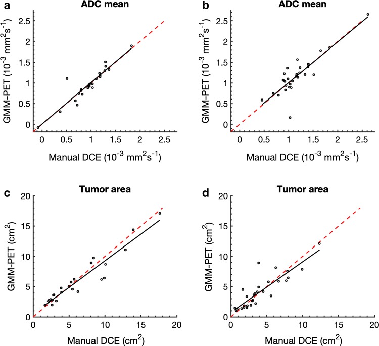
The original version of this article unfortunately contained a mistake in Fig. 6.
The corrected Fig. 6 is placed in the following page.
Fig. 6.
Relationship between the resulting metrics from manual DCE and GMM–PET for a ADC mean for untreated lesions (r = 0.866, p < 0.001) and b treated lesions (r = 0.895, p < 0.001) and m tumor area from c untreated (r = 0.870, p < 0.0001) and d treated (r = 0.928, p < 0.001) lesions. Red identity lines included show that area from GMM–PET is slightly smaller than from manual DCE
Footnotes
Publisher's Note
Springer Nature remains neutral with regard to jurisdictional claims in published maps and institutional affiliations.


