Abstract
Targeting chemoresistant malignant cells is one of the current major challenges in oncology. Therefore, it is mandatory to refine the characteristics of these cells to monitor their survival and develop adapted therapies. This is of particular interest in acute myeloid leukemia (AML), for which the 5-year survival rate only reaches 30%, regardless of the prognosis. The role of the microenvironment is increasingly reported to be a key regulator for blast survival. In this context, we demonstrate that contact with mesenchymal stromal cells promotes a better survival of blasts in culture in the presence of anthracycline through the activation of ABC transporters. Stroma-dependent ABC transporter activation leads to the induction of a Side Population (SP) phenotype in a subpopulation of primary leukemia blasts through alpha (α)4 engagement. The stroma-promoting effect is reversible and is observed with stromal cells isolated from either healthy donors or leukemia patients. Blasts expressing an SP phenotype are mostly quiescent and are chemoresistant in vitro and in vivo in patient-derived xenograft mouse models. At the transcriptomic level, blasts from the SP are specifically enriched in the drug metabolism program. This detoxification signature engaged in contact with mesenchymal stromal cells represents promising ways to target stroma-induced chemoresistance of AML cells.
Introduction
Acute myeloid leukemias (AML) represent a set of hemopathies characterized by a clonal expansion in bone marrow (BM) and blood of immature myeloid cells, called blasts, blocked at different stages of differentiation. AML can be classified according to the degree of immaturity [as according to the French-American-British (FAB) classification] or depending on the cytogenetic or molecular events observed in patients (according to the World Health Organization 2016 criteria).1 AML are also subdivided into three groups that condition therapy: favorable AML, which may be cured without hematopoietic stem cell transplant, and intermediate and adverse AML which may require an allogenic graft. Despite the significant progress made in supportive care, there has been no radical change in the prognosis of AML; the 5-year survival rate is 30% for all groups of AML and 10% for adverse AML. The conventional chemotherapy based on the injection of a nucleoside analog combined with an anthracycline is used to kill AML cells. However, many patients relapse, mainly due to the persistence of rare chemoresistant AML cells able to re-initiate the disease; these are likely to correspond to leukemia stem cells (LSC).
As a mirror of normal hematopoiesis, several studies reported a specific phenotype for LSC or leukemia-initiating cells. Although this generated heterogeneous results,2 it was nevertheless reported that cells able to engraft de novo or after a secondary transplant were present in the CD34+CD38–CD123+ hematopoietic population.3–5 However, other studies showed that cells able to initiate leukemia concerned CD34–, CD33+ or CD13+ cells.6 Recently, Farge et al. demonstrated that chemoresistance was more related to a specific oxidative metabolism than to a level of progenitor/stem cell phenotype.7 Moreover, the high oxidative phosphorylation status is associated with elevated fatty acid oxidation and high expression of CD36, a fatty acid translocase recently identified as a marker of an LSC subpopulation.8
Considering hematopoietic neoplasms, it is now mandatory to also include environmental factors in order to understand their development, resistance and dissemination. In adults, hematopoiesis develops in the BM where a dialogue between hematopoietic stem/progenitor cells (HSPC) and the microenvironment, including mesenchymal stromal cells (MSC), extracellular matrix components and soluble factors,9,10 is critical for maintaining stem cell function and homeostasis. Such a protective microenvironment has been reported to be implicated in the stemness and chemoresistance of leukemia blasts at the origin of the process of Environment Mediated-Drug Resistance (EM-DR), and specifically of Cell Adhesion Mediated-Drug Resistance (CAM-DR).8,11 Quiescence, as well as protection against environmental and drug aggressions, are major characteristics of stem cells also characterized by the Side Population (SP) phenotype.12 We have previously shown that, whereas circulating HSPC from heathy donors (HD) do not exhibit a SP phenotype, this functionality can be induced after co-culture with MSC in a VLA4- and CD44-dependent manner.13 Interestingly, this MSC-induced SP population is enriched in HSC, as shown by its engraftment in immunodeficient mice.13
In the present work, conducted on a cohort of 34 AML patients, we show that MSC activate ABC transporters in a subpopulation of primary blasts, resulting in the induction of an SP phenotype. SP blasts are mostly quiescent and exhibit a low reactive oxygen species (ROS) transcriptional pathway compared to non-SP [Main Population (MP)] blasts. Furthermore, they are capable of effluxing chemotherapy agents in vitro in cultures as well as in vivo in patient-derived xenograft models treated by cytarabine, validating a higher chemoresistance of SP cells compared to their MP counterparts. Altogether, our results demonstrate that the stroma-induced SP functionality is a new mechanism of CAM-DR for AML blasts.
Methods
Preparation of primary acute myeloid leukemia cells
Peripheral blood samples were collected at Percy (Clamart, France) and Saint Louis (Paris, France) hospitals after the informed consent of patients in accordance with the principles of the Declaration of Helsinki (IDRCB 2017-A02149-44, CPP 2017-juill.-14644 ND-1eravis, CNIL MR001). The patient cohort represents 34 patients with primary AML (Online Supplementary Figure S1). Patients were untreated at the time of blood uptake. Blood mononuclear cells were isolated on density gradient (1.077g/mL) before freezing.13
Co-culture of primary acute myeloid leukemia cells on mesenchymal stromal cells
Primary AML mononuclear cells were plated at 2×10⁵/cm2 in SynH (Abcell-Bio) supplemented with L-Glutamine, non-essential amino acids and 10% fetal bovine serum for 3-4 days on confluent MSC isolated from the BM of either AML patients (n=9) or HD (n=5) (Online Supplementary Appendix). AML blasts were cultured without MSC feeders for controls. Transwell and neutralization experiments are described in the Online Supplementary Appendix.
In some experiments, mitoxantrone (50nM; Sigma Aldrich) was added to the co-culture for 24 hours (h) and cells were pretreated or not with verapamil (50 mM) for 2 h before adding mitoxantrone and during mitoxantrone treatment.
At the end of co-cultures, non-adherent cells were flushed and stained with anti-CD45 antibody and annexin V (Invitrogen). Counting beads (CountBright, Life Technologies) were added to cell suspension to quantify cell populations by flow cytometry using Fortessa apparatus with Diva software (Becton Dickinson).
Side Population cell detection and characterization
Hoechst staining was performed as previously described13,14 (Online Supplementary Appendix). After Hoechst incubation, cells were placed on ice and stained with anti-CD45 antibody and a viability dye. Flow cytometry analysis was carried out on BD Fortessa apparatus. CD45 staining was use to gate on AML blasts and to avoid stromal contamination for SP analysis.
Drug efflux - to analyze drug efflux concomitantly with SP cell detection, mitoxantrone (90nM) was added to the cell suspension during the last 30 minutes (min) of Hoechst staining.
ABC transporter functionality
Specific probes for ABCB1 (DioC2(3)), ABCC1 (CMFDA), and ABCG2 (Purpurin 18) were incubated during 30 min at 37°C after co-culture or during Hoechst staining. Cells were then stained with CD45 antibodies and with a viability dye (Online Supplementary Table S2).
Transcriptomic analysis
See the Online Supplementary Appendix.
Patient-derived xenograft model
Patient-derived xenografts (PDX) were achieved as previously described7,15 (Online Supplementary Appendix) under French Institutional Animal Care and Use (Committee of “Midi-Pyrénées” region-France) approval.
Statistical analysis
Raw data of each group were analyzed using R (3.3.3) and Rstudio (0.99.896) software. The packages used were stats, coin and multcomp for tests. Graphic representations of data were made for each group using Prism 6 or R (3.3.3) software (package ggplot2). Statistical comparisons between groups on a single quantitative variable were run as follows: resampling tests were used for group versus group comparisons, pairing on AML donor levels or on AML MSC donor level. When multiple comparisons were used inside a single experiment, P-values were corrected using the Benjamini/Hochberg method. P=0.05 was considered statistically significant; tests were bilateral unless stated. Data are expressed as median with 25-75% interquartile intervals.
Results
Leukemia blasts cultivated on mesenchymal stromal cells actively efflux chemotherapy drugs by activating ABC transporters
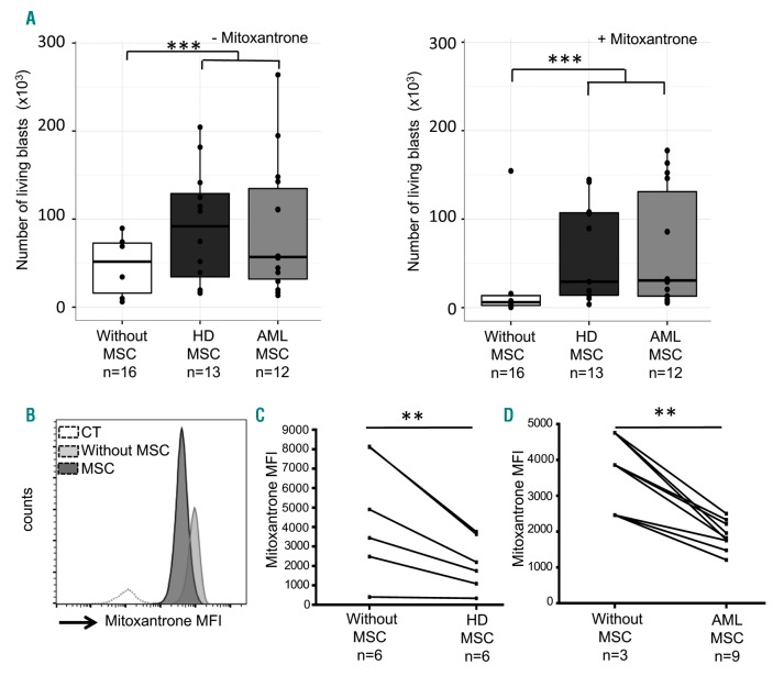
We first characterized MSC isolated from the BM of AML patients (AML MSC) at diagnosis and compared them to MSC isolated from BM of healthy donors (HD MSC). The number of cumulative population doubling was slower for AML MSC than for HD MSC (Online Supplementary Figure S2A). AML MSC clonogenicity was also weaker than that of HD MSC (Online Supplementary Figure S2B). Because AML MSC were morphologically different from HD MSC, we quantified senescent cells using the β-galactosidase activity test. We observed that AML MSC showed 5-10 times more senescent cells than HD MSC as soon as passage 4 (Online Supplementary Figure S2C). This result can explain the limited expansion capacity and clonogenicity of AML MSC. Despite these growth alterations, no differences were observed regarding their phenotype (CD45–CD90+CD73+CD105+) (Online Supplementary Figure S3A) or differentiation capacities, since they were able to differentiate into adipocytes, osteoblasts, and chondrocytes similarly to HD MSC (Online Supplementary Appendix and Online Supplementary Figure S3B and C). We further analyzed their functional roles on AML blast survival by co-culturing blasts on either AML or HD MSC. After a 3-day co-culture, cells were removed and blast viability was evaluated using Annexin V and 7AAD co-staining. Co-culture with HD or AML MSC significantly increases AML blast survival (n>12; P=0.0004) (Figure 1A, left). This difference was also observed when mitoxantrone, a chemotherapy drug, was added to co-cultures (n>12; P=0.0003) (Figure 1A, right).
Figure 1.
Mesenchymal stromal cells (MSC) from heathy donors (HD) or from acute myeloid leukemia (AML) patients confer a better survival to leukemia blasts, even in the presence of chemotherapy agents. (A, left) Histogram representing the number of living AML blasts after a 3-day culture without MSC [median 51,655 (56,830)] or with MSC from HD [median 91,815 (94,556)] or AML patients [median 56,896 (102,863)]. (Right) The same conditions but in the presence of 50nM of mitoxantrone. Culture conditions in the presence of the different types of MSC confer a significantly improved survival to AML blasts (left: ***P=0.0004; right: ***P=0.0003, n>10, represents the number of AML blasts/MSC donor combinations). (B) Histogram showing fluorescence intensity of AML blasts stained with mitoxantrone after a 3-day culture with or without MSC from HD (one representative experiment of the 6 performed). Control (CT) represents non-stained blasts. (C) Mitoxantrone mean fluorescence intensity (MFI) in AML blasts cultivated [median 2,191 (2,032)] or not [median 3,676 (3,546)] on HD MSC (**P=0.01, n=6, Wilcoxon test). (D) Mitoxantrone MFI in AML blasts cultivated [median 3,861 (1,147)] or not [median 1,821 (460)] on AML MSC (**P=0.01, n=3-9, Wilcoxon test). n: number. **P<0.01; ***P<0.001.
To try to explain this increased survival, we evaluated the intracellular amount of mitoxantrone in the blast population co-cultivated with or without MSC isolated from HD or AML patients (Figure 1B). All blasts were positive for mitoxantrone after a 3-day culture without MSC. However, when blasts were co-cultivated in the presence of MSC either from HD (Figure 1C) or AML patients (Figure 1D), the mitoxantrone mean fluorescence intensity (MFI) was lower than that of blasts cultured without MSC. This reduction was observed in most of the patients studied, regardless of the MSC origin (P=0.01, n=6 for HD MSC and n=9 for AML MSC).
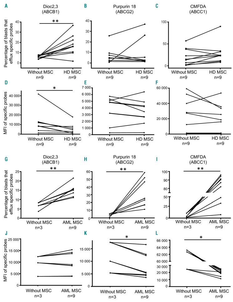
Because blasts co-cultured on MSC exhibit a lower intracellular amount of mitoxantrone compared to cells cultivated without MSC, we studied by which mechanism the amount of intracellular mitoxantrone was reduced. It is now well known that ATP-binding cassette (ABC) transporters can efflux several molecules including chemotherapy drugs.16 Therefore, we used specific probes such as Dioc2(3), CMFDA and purpurin18 to evaluate the activity of three main pump families such as ABCB1 (MDR1), ABCC1 and ABCG2, respectively. There was a significant increase in the percentage of blasts that efflux the Dioc2(3) probe after a 3-day co-culture on HD MSC [median 16.1% (12.8%) with MSC vs. 6.18 (2%) without MSC; P=0.005, n=9] demonstrating the implication of the ABCB1 pump as part of this process (Figure 2A). The Dioc2,3 MFI was also significantly decreased in AML blasts (P=0.02, n=9) (Figure 2D). We also noticed an increase in ABCC1 and ABCG2 pump activities in a small number of AML patients (Figure 2B and C), but with no significant modulation of CMFDA and purpurin18 MFI (Figure 2E and F). However, when activities of these three ABC transporters were analyzed individually, patient per patient, we observed that their activities were systematically modulated by MSC contact (except for one patient, AML 44) (Online Supplementary Figure S4) and that ABC transporters were differentially active from one patient to another. When blasts were co-cultured on MSC isolated from AML patients, we observed an activation of the three ABC transporters (P=0.01, 0.02 and 0.008, respectively; n=3-9) (Online Supplementary Figure S2G-I) associated with a decrease in specific probe MFI (P=0.01, n=3-9) (Figure 2J and L), except for Dioc2,3 MFI which remained high (Figure 2J).
Figure 2.
Co-culture between acute myeloid leukemia (AML) blasts and mesenchymal stromal cells (MSC) modulates the ABC transporter functionality on leukemia blasts in a patient-dependent manner. (A-C) Percentage of AML blasts that efflux specific probes (ABC transporter activity) after a 3-day co-culture on healthy donor (HD) MSC. ABC transporter activities were quantified using specific probes (Dioc2,3, Purpurin 18, CMFDA for ABCB1, ABCG2, ABCC1, respectively) as shown on the histograms. ABCB1 activity is significantly increased (P=0.0059, n=9, Wilcoxon test) by AML blasts after a 3-day co-culture on HD MSC. (D-F) Corresponding probe mean fluorescence intensity (MFI) in AML blasts; Dioc2,3 MFI is significantly decreased (P=0.02, n=9, Wilcoxon test) in blasts after co-culture with HD MSC. (G-I) Percentage of AML blasts that efflux specific probes after a 3-day co-culture on AML MSC. Activity of the ABCB1, ABCG2 and CMFDA transporters was significantly increased (P=0.01, P=0.02 and P=0.008, respectively; n=3 without MSC and n=9 with AML MSC, Wilcoxon test). (J-L) Corresponding MFI in AML blasts. Purpurin 18 and CMFDA MFI are significantly decreased (P=0.01 and P=0.01, respectively; n=3 without MSC and n=9 with AML MSC, Wilcoxon test) in co-culture conditions. *P<0.05; **P<0.01.
Therefore, our results show that leukemia blasts demonstrate a specific pattern of ABC transporter activity that is modulated by contact with stromal cells in a patient-dependent manner. Moreover, the modulation of ABC transporter activities appears higher when blasts are co-cultivated with MSC isolated from AML patients.
Leukemia blasts adopt a Side Population phenotype after close contact with mesenchymal stromal cells through integrin interactions
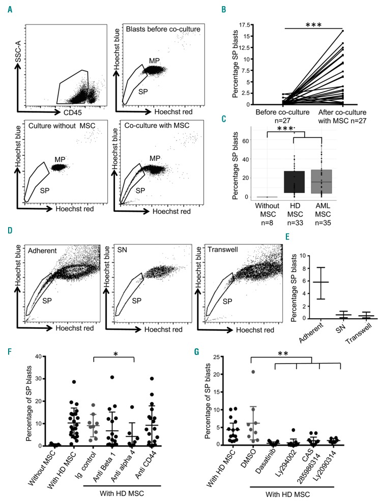
ABC transporter activation is known to be involved in SP phenotype acquisition. We therefore analyzed the SP phenotype of blasts before and after co-culture with MSC. Circulating blasts were gated on their SSC CD45low profile in flow cytometry (Figure 3A). In contrast to circulating Lin– HSPC freshly isolated from HD,13 a small proportion of blasts (0.2%; interquartile 0.44%) isolated from approximately 50% of AML patients expressed a SP phenotype before any stromal co-culture (Figure 3B). However, after a 3-day co-culture on HD MSC, we observed for all AML patients that a SP population had emerged or was increased within blast cells with a median of around 4%, with an interquartile of 8.65% (P<0.001, n=27). We then analyzed whether co-culture with AML MSC also induced or increased the percentage of circulating leukemia blasts with an SP phenotype. Similarly to MSC isolated from HD, co-cultures of blasts with AML MSC systematically increased the percentage of SP blasts (P=10−4, n= 8-35) (Figure 3C).
Figure 3.
Circulating leukemia blasts acquire a Side Population (SP) phenotype in contact with mesenchymal stromal cells (MSC) from healthy donors (HD) or acute myeloid leukemia (AML) patients through α4 integrin interaction. (A) Cytograms illustrating the gating strategy of AML blasts for cytometry analysis (top left) and SP visualization before co-culture (top right) and after a 3-day culture without (bottom left) or with (bottom right) HD MSC. (B) Percentage of SP AML blasts before and after a 3-day co-culture on MSC (either from HD or AML patients) (P<0.001, n=27, Wilcoxon test). (C) Percentage of SP AML blasts after co-culture with HD MSC (P<10−4, n=8-33) or AML patients (P<10−4, n=8-35). (D) Cytograms showing SP phenotype observed on AML blasts from MSC adherent (left) or supernatant fractions (SN, middle) and in transwell experiments (right) after a 3-day co-culture with HD MSC. (E) Percentage of SP blasts in the three experimental conditions. (F and G) Percentage of SP AML blasts after co-culture or not with HD MSC and after inhibition of β1 (n=18) and α4 (n=7) integrins (P=0.04) or CD44 (n=18) (F) and their downstream signaling pathways (G) using dasatinib, LY294002, CAS2859863 and LY209031, inhibiting Src (P=0.002, n=9), AKT (P=0.003, n=9), STAT5 (P=0.009, n=9), and GSK3 pathways (P=0.01, n=6), respectively. *P<0.05; **P<0.01; ***P<0.001.
We further studied whether a close contact between blasts and MSC was required to promote the SP phenotype of blasts. We first addressed this question by analyzing the proportion of SP blasts in the adherent fraction of the co-cultures and compared it with that detected in the supernatants (SN). SP-expressing cells were mostly found in the adherent fraction (Figure 3D). We then confirmed the requirement of cell-cell interactions using transwell experiments in which the percentage of SP blasts was strongly reduced when blasts and stromal cells were separated by the transwell insert (n=3) (Figure 3D and E). Therefore, close contacts between leukemia and stromal cells appear to be essential to the promotion of the SP phenotype in blasts.
We had previously demonstrated that the SP phenotype induced on circulating Lin– HSPC from HD by MSC co-culture was dependent on α4 and β1 integrins as well as CD44 engagement.13 Using blocking antibodies, we tested whether the promotion of the SP phenotype on AML blasts was dependent on MSC interactions through these molecules. Blocking α4 integrin significantly reduced the percentage of SP blasts after MSC co-culture (P=0.04, n=7) while blocking β1 integrin and CD44 interactions had a slight but not significant effect on this proportion (Figure 3F). We then tested the effect of pharmacological inhibitors on the main signaling pathways directly or indirectly activated by integrins. To do this, we first assessed the ‘efficient dose’, corresponding to the highest dose that did not induce toxicity for dasatinib (pSrc inhibitor), LY294002 (pAKT inhibitor), CAS2859863 (pSTAT5 inhibitor) and LY209031 (pGSK3 inhibitor), CPD22 (pILK inhibitor), and simvastatine (HMG coA reductase inhibitor) (data not shown). We then added these inhibitors to co-cultures and quantified the proportion of blasts that have adopted an SP phenotype after a 3-day co-culture on HD MSC. Whereas addition of CPD22 or simvastatine did not affect the percentage of SP blasts (data not shown), a significant decrease in SP blasts was observed when Src (P=0.002, n=9), AKT (P=0.003, n=9), STAT5 (P=0.009, n=9), and GSK3 (P=0.01, n=6) pathways were inhibited (Figure 3G). These data confirm that the stroma-induced SP functionality on AML blasts was partly dependent on integrin interactions, and especially of α4 integrin activation.
Side Population acute myeloid leukemia blasts are quiescent and able to actively efflux chemotherapy through ABCB1 transporters
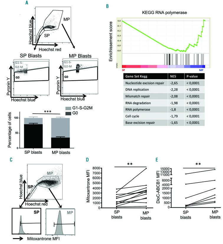
Side Population phenotype is reported to be linked to quiescence.12–14 Therefore, we used flow cytometry to analyze the cell cycle phases of AML blasts expressing or not expressing the SP phenotype (SP vs. non-SP population/MP for Main Population) after a 3-day co-culture on HD MSC. As expected, SP blasts were in the majority in the G0 phase [median 76% (16.5%)] in contrast to MP blasts [median 33.45% (18.6%)], which were mainly in the G1-S-G2-M phases (Figure 4A). In agreement with this result, gene set enrichment analysis (GSEA) performed with the Kyoto Encyclopedia of Genes and Genomes (KEGG) database on data obtained from transcriptomic experiments comparing sorted SP vs. MP blasts showed a repression of cell cycle and DNA replication gene expression in SP than MP cells (Figure 4B). When cultivated on MSC, circulating AML blasts acquire the capability to efflux mitoxantrone through ABC transporter activation (Figure 1B and C). We therefore analyzed whether the drug efflux was restricted to SP blasts. To do this, blasts which were co-cultivated on MSC during three days were incubated with mitoxantrone for the last 30 min of Hoechst incubation. We showed that SP blasts had a lower amount of mitoxantrone compared to MP blasts (Figure 4C). Quantification of mitoxantrone MFI (Figure 4D) in the SP population [median 1,584 (1,545)] showed a 2-fold decrease compared to the MP one [median 3,219 (1,706)] (P=0.0052; n=12) for most of the patients analyzed. We thus evaluated which ABC transporters were activated in SP versus MP cells using specific probes. The ABCB1 transporter was significantly more active in SP [MFI Dioc2(3) 239 (2,422)] than in MP blasts [MFI Dioc2(3) 1,298 (3,182)] (P=0.023, n=7) (Figure 4E). In contrast, ABCG2 and ABCC1 did not appear to be involved in this process (data not shown).
Figure 4.
The mesenchymal stromal cells (MSC)-induced Side Population (SP) functionality in leukemia blasts is associated with quiescence and chemotherapy efflux through ABC transporter activation. (A) Cytograms and histograms showing gating strategies and percentage of SP and MP blasts in G0 and in G1-S-G2-M after a 3-day co-culture on HD MSC. Cell cycle status of SP versus Main Population (MP) blasts was analyzed adding pyronin Y during Hoechst staining. SP blasts are mostly in G0 [median 76% (16.5%)] compared to MP [median 33.45% (18.6%)] blasts which are in G1-S-G2-M (P=0.0009, n=12, Wilcoxon test). (B) Transcriptomic analysis of SP blasts compared to MP blasts. (C) Cytograms showing gating strategy to evaluate mitoxantrone efflux in SP or MP blasts. Quantification of mitoxantrone MFI (D) in SP [median 1,584 (1,845)] and in MP [median 3219 (1,706)] blasts (P=0.0052, n=12, Wilcoxon test). (E) DioC2,3 MFI evaluating ABCB1 activity in SP [median 239 (4,056)] and MP [median 1,298 (5418)] cells (P=0.023, n=7, Wilcoxon test). **P<0.01; ***P<0.001.
Altogether, our results demonstrate that AML blasts adopting SP phenotype after contact with MSC are quiescent and able to actively efflux chemotherapy agents through ABC transporters and, in particular, through ABCB1.
Side Population blasts are more in vitro and in vivo chemoresistant than non-SP cells and this chemoresistance can be partially reversed by ABC transporter inhibition
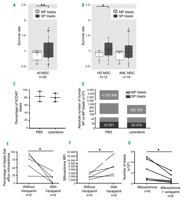
We first tested whether the SP phenotype induced by contact with MSC conferred a better in vitro survival to leukemia blasts in the presence of chemotherapy drugs. We thus co-cultured blasts and MSC isolated from HD or AML patients in the presence or not of mitoxantrone. After a 3-day co-culture, we quantified the absolute number of SP and MP blasts in both conditions and calculated the survival rate of SP and MP cells by dividing the absolute number of SP or MP cells in the treated cultures by their absolute numbers in the non-treated cultures. The survival rate of SP cells was significantly higher than that of MP cells (P=0.041; n=30) when leukemia blasts were co-cultivated on MSC independently of their origin (Figure 5A) or based on their origin (Figure 5B).
Figure 5.
Side Population (SP) functionality of acute myeloid leukemia (AML) blasts induced by mesenchymal stromal cell (MSC) interactions is associated with chemoresistance. (A) Survival rate of SP [median 0.98 (0.46)] or Main Population (MP) [median 0.95 (0.24)] blasts after a 3-day co-culture with MSC in presence of mitoxantrone (P=0.041, n=30, Wilcoxon test). (B) Survival rate of SP or MP blasts after a 3-day co-culture with MSC from healthy donors (HD) or AML patients. (C) Percentage of hCD45+ blasts within the total CD45+ population. (D) Absolute number of human SP and MP blasts in the femur of patient-derived xenograft (PDX) mice after a 5-day treatment with either phosphate buffered saline or cytarabine (30 mg/kg) (n=3 mice per patient). (E) Percentage of blasts that efflux mitoxantrone with or without verapamil after a 3-day co-culture on AML MSC (P=0.05, n=5, Wilcoxon test) and (F) the corresponding mitoxantrone mean fluorescence intensity (MFI) in AML blasts (P=0.05, n=5, Wilcoxon test). (G) Number of living blasts co-cultivated on AML or HD MSC after 24 hours of mitoxantrone and treatment with or without verapamil (P=0.01, n=6, Wilcoxon test) *P<0.05; **P<0.01.
We further compared the in vivo chemoresistance of SP versus MP cells in NSG mice grafted with primary AML blasts (PDX model). Twelve weeks after engraftment, the presence of human CD45+ leukemic cells in mice was controlled by blood analysis. Mice were then treated daily with cytarabine during five days to reduce the human blast population.7 Three days after treatment arrest, mice were euthanized and their BM was harvested. We estimated the cell chimerism and showed that human CD45+ cells represented 70-96% of the whole CD45+ population (Figure 5C). We then quantified the absolute number of total human blasts and of SP versus MP human blasts present in the mouse BM. As expected, there was a large reduction in the global number of human blasts after the cytarabine treatment compared to phosphate buffer solution injection. However, and more interestingly, the absolute number of blasts expressing the SP functionality remained unchanged after cytarabine treatment in contrast to that of MP blasts that was reduced 5-fold (Figure 5D). These results show that cytarabine was active in vivo on MP blasts but less or not active on SP blasts.
Altogether, our results suggest that SP blasts are more resistant than MP cells to chemotherapy agents both in vitro and in vivo.
We then analyzed whether the chemosensitivity of AML blasts could be restored after blocking ABC transporters. We previously showed that ABC transporter activities (modulated on blasts after contact with MSC) were differentially active and were patient dependent (Online Supplementary Figure S4). Thus, we used verapamil, a broad spectrum ABC transporter inhibitor, to inhibit the SP phenotype.12,13 Addition of verapamil into AML blast and MSC co-cultures significantly reduced the percentage of blasts that efflux mitoxantrone (P=0.05, n=5) (Figure 5E) as well as mitoxantrone MFI (P=0.05, n=5) (Figure 5F). Furthermore, when co-cultures were treated with a nontoxic dose of verapamil, we also observed a significant reduction in the number of living blasts compared to that observed with mitoxantrone alone (P=0.01, n=6) (Figure 5G), suggesting that blocking ABC transporters restores blast mitoxantrone chemosensitivity.
Acute myeloid leukemia Side Population blasts exhibit a detoxification transcriptional signature
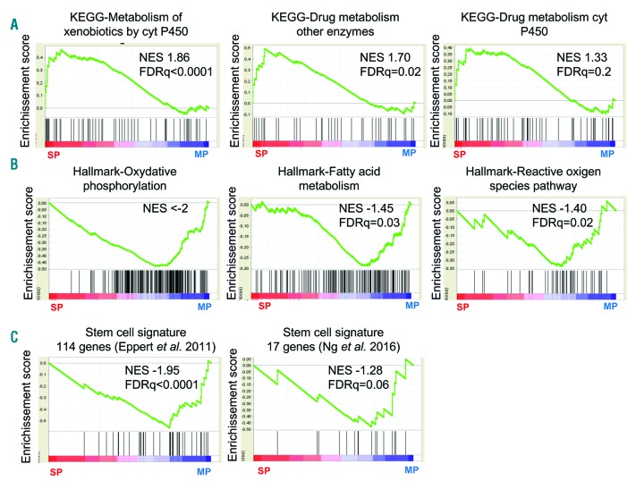
As SP blasts were more chemoresistant in vitro and in vivo than their MP counterparts, we analyzed their transcriptomic profiles to highlight a specific signature of these cells. To do this, we sorted SP versus MP blasts from two AML patients after a 3-day co-culture on HD MSC. A global transcriptomic analysis of sorted SP versus MP AML blasts demonstrated an enrichment of genes implicated in a detoxification program in SP cells. Indeed, using the KEEG database, GSEA analysis showed that the expression of genes involved in xenobiotic and drug metabolism via cytochrome P450 or by other enzymes was up-regulated in SP blasts (Figure 6A). Surprisingly, the expression of ABC transporter transcripts was not homogeneously differentially modulated in SP versus MP cells (Online Supplementary Figure S5), suggesting that, in our experimental conditions, SP phenotype induction is not associated with an increase in ABC transporter mRNA level at steady state, as already suggested in other studies,17 but mainly after drug exposure.18,19 We also observed that, oxidative phosphorylation, fatty acid metabolism and ROS pathway signatures of the Hallmark database were enriched in MP blasts (Figure 6B). Surprisingly, while SP phenotype is a common feature of stem cells,12 and the CD34+ CD38– SP blast population is enriched in cells with a CD123+ stem phenotype (Online Supplementary Figure S6) as compared to CD34+ CD38– MP cells, the stem cell transcriptional signatures proposed by Eppert et al.20 and Ng et al.21 were preferentially observed in the AML MP population (Figure 6C).
Figure 6.
Side Population (SP) blasts exhibit a detoxification program signature. Gene set enrichment analysis with transcriptomics data of sorted SP and Main Population (MP) blasts after a 3-day co-culture on healthy donor (HD) mesenchymal stromal cells (MSC) shows an upregulation in SP cells of genes of xenobiotic metabolism and drug metabolism through the cytochrome P450 or other enzymes (A), an upregulation in MP blasts of genes of oxidative phosphorylation, fatty acid metabolism and reactive oxygen pathway (B), as well as of genes from the stem cell signatures reported by Eppert et al.20 and Ng et al.21 (C).
Discussion
Chemoresistance of leukemia cells is one of the major challenges which hematologists have to face in order to cure AML. Therefore, a better understanding of this process constitutes the keystone of next generation therapies. It is now well known that the BM microenvironment is a key actor in leukemia development and resistance to treatment, but the underlying mechanisms are still unclear.
Our present study shows that MSC that play a central role in BM stromal niches sustain the survival and chemoresistance of AML blasts through the induction of an SP phenotype. This chemoresistance mechanism involves activation of ABC transporters, responsible for drug efflux, in a small proportion of leukemia cells after co-culture with MSC. Whereas these transporters were not active or were poorly active in circulating blasts, interactions with stromal cells constantly increased the proportion of SP cells in the blast population. Interestingly, the ABC transporter activation pattern appears to be patient dependent. While MSC-induced ABC transporter activation is common to all types of AML, we did not found any correlation between specific probe efflux and patient characteristics. However, because AML is an heterogeneous group of diseases, a larger patient study cohort is required in order to identify a potential correlation between ABC transporter activation patterns and AML types. Besides a limited expansion capacity and clonogenicity, the capacity of AML MSC to promote SP phenotype on blasts was similar and even better than that of HD MSC. This observation suggests that the BM stroma of patients may be adapted to promote blast protection/survival and supports the hypothesis of a specific crosstalk between pathological blasts and MSC.
Previous data from our group have demonstrated that BM MSC modulate the SP phenotype of HSC from HD.13 In the present study, we show that they also control the induction of the SP functionality on AML blasts. Interestingly, SP leukemic blasts are mostly found in the MSC-adherent fraction, suggesting that close contacts between blasts and stromal cells are required for SP phenotype induction. As for HD samples, this process is partially dependent on α4 integrin. However, in contrast to HD, for half of the patients studied, this induction is not dependent, or is so at a lower level, on β1 integrin or CD44, two cell-surface glycoproteins known to be involved in BM niche HSC nesting. BM niches are reported to be crucial for HSC long-term maintenance22–24 and SP cell quiescence.25 In patients, we show that the proportion of quiescent blasts is higher in the stroma induced-SP cell population than in their MP counterparts. Quiescence is often related to chemoresistance, as only proliferating cells are sensitive to anthracyclines or nucleoside analogs. In the current study, we show that SP blasts are more chemoresistant than MP cells, either in vitro or in vivo in PDX models. This result is confirmed by our data showing that sorted SP blasts express a specific transcriptomic profile focused on genes involved in drug and environment stress detoxification,16 known to participate in the chemoresistance process.26 Therefore, SP cell detection could allow chemoresistant blasts to be monitored during the course of the disease and, targeting stroma-blast interactions would reinitiate cell cycling and, consequently, blast sensitization to chemotherapy. As VLA4 and CD44 play a role in the stroma-induced SP phenotype in HD HSC, but only partially in AML blasts, it would be interesting to identify specific interactions between MSC and blasts to specifically target leukemia cells. Deep analyses using transcriptomic and interactomic approaches27 focused on MSC, SP and MP cells from AML patients and HD should highlight new pairs of exclusive interactors involved in stroma-blast interactions that would be promising drugable targets.
An important clinical concern is the quest for leukemia stem/initiating cells.28 SP cells exhibit some features of stem cells since they are mostly in G012,14,25 and since their ROS level is lower than that of their MP counterparts.29 In our study, the transcriptomic analysis of SP AML blasts does not correlate with the 114 and 17 gene stemness signatures reported by Eppert et al. and Ng et al., respectively,20,21 suggesting that SP AML cells are not enriched in such stem cells. Interestingly, Farge et al.7 and Boyd et al.30 demonstrated that chemotherapy-resistant human AML cells are not necessarily enriched in LSC, but exhibit increased fatty-acid oxidation, high oxidative phosphorylation (OXPHOS) gene signature, and up-regulated CD36 expression. By associating a reduction in transcripts for oxidative phosphorylation, fatty acid metabolism and ROS in SP as compared to MP blasts, our results are in line with those from Ye et al., suggesting that chemoresistance is not necessarily restricted to the LSC compartment but rather could be associated to metabolic adaptations induced by their microenvironment.8
By promoting a better survival of blasts, the stroma-induced SP functionality we identified in blasts could represent a new mechanism of CAM-DR. Whereas BM MSC from HD or AML patients exhibit similar survival and SP- promoting effects on blasts, preliminary results on four patients suggest that cultivating MSC and blasts in autologous conditions would bring a supplemental advantage on blast survival and chemoresistance (Boutin L et al., 2018, unpublished data). This suggests that an intimate crosstalk has taken place in vivo between MSC and blasts and has been conserved in vitro. Recently, Moschoi et al.31 proposed that mitochondrial transfers from BM stromal cells to AML blasts provided them with a protective effect following chemotherapy. Other communication modes such as exosome exchange, metabolite secretion, and nanotube formation could also be suggested to participate in this process and must be identified to disrupt the stroma-induced chemoresistance of AML blasts.
Induction of SP/chemoresistance phenotype on AML blasts is of major interest to clinicians as ABC transporters implicated in drug/chemotherapy agent efflux could be targeted.32,33 In that context, we have shown that mitoxantrone efflux and blast survival are reversible in vitro by addition of verapamil. However, results from clinical trials using P-glycoprotein inhibitors in AML patients are discouraging.34,35 Our data showing that the functionality of the main ABC transporters implicated in MDR36 is promoted by blast-MSC interactions, and that this process is patient-dependent, suggest that it is time to revisit the role of ABC transporters in therapeutic failure in the context of personalized medicine.37,38
In conclusion, our results show the critical role of stroma interactions in blast chemoresistance through SP phenotype promotion and ABC transporter activation. Therefore, targeting these interactions in combination with the development of new improved inhibitors of ABC transporter function39 could be an attractive therapeutic alternative.
Acknowledgments
We thank the molecular core facility of Cochin Institute for transcriptomic analysis. We are very grateful to the polyclinic of Blois and particularly to Dr Denis Burgot as well as all the orthopedic surgery staff for providing human bone marrow samples from hip replacement. We thank Dr Cedric Thépenier for the statistical analysis and sharpened advices. We also thank Pr Christophe Martinaud for blast analysis on blood smears.
Footnotes
Check the online version for the most updated information on this article, online supplements, and information on authorship & disclosures: www.haematologica.org/content/105/4/987
Funding
This work was supported by grants from Association “Laurette Fugain” (ALF 2013/08) and Association “Vaincre le Cancer-Nouvelles Recherches Biomédicales”.
References
- 1.Arber DA, Orazi A, Hasserjian R, et al. The 2016 revision to the World Health Organization classification of myeloid neoplasms and acute leukemia. Blood. 2016;127(20):2391–2405. [DOI] [PubMed] [Google Scholar]
- 2.Taussig DC, Miraki-Moud F, Anjos-Afonso F, et al. Anti-CD38 antibody-mediated clearance of human repopulating cells masks the heterogeneity of leukemia-initiating cells. Blood. 2008;112(3):568–575. [DOI] [PubMed] [Google Scholar]
- 3.van Rhenen A, Feller N, Kelder A, et al. High stem cell frequency in acute myeloid leukemia at diagnosis predicts high minimal residual disease and poor survival. Clin Cancer Res. 2005;11(18):6520–6527. [DOI] [PubMed] [Google Scholar]
- 4.Vergez F, Green AS, Tamburini J, et al. High levels of CD34+CD38low/−CD123+ blasts are predictive of an adverse outcome in acute myeloid leukemia: a Groupe Ouest-Est des Leucémies Aiguës et Maladies du Sang (GOELAMS) study. Haematologica. 2011;96(12):1792–1798. [DOI] [PMC free article] [PubMed] [Google Scholar]
- 5.Jordan CT, Upchurch D, Szilvassy SJ, et al. The interleukin-3 receptor alpha chain is a unique marker for human acute myelogenous leukemia stem cells. Leukemia. 2000;14(10):1777–1784. [DOI] [PubMed] [Google Scholar]
- 6.Sarry J-E, Murphy K, Perry R, et al. Human acute myelogenous leukemia stem cells are rare and heterogeneous when assayed in NOD/SCID/IL2Rγc-deficient mice. J Clin Invest. 2011;121(1):384–395. [DOI] [PMC free article] [PubMed] [Google Scholar]
- 7.Farge T, Saland E, de Toni F, et al. Chemotherapy-Resistant Human Acute Myeloid Leukemia Cells Are Not Enriched for Leukemic Stem Cells but Require Oxidative Metabolism. Cancer Discov. 2017;7(7):716–735. [DOI] [PMC free article] [PubMed] [Google Scholar]
- 8.Ye H, Adane B, Khan N, et al. Leukemic stem cells evade chemotherapy by metabolic adaptation to an adipose tissue niche. Cell Stem Cell. 2016;19(1):23–37. [DOI] [PMC free article] [PubMed] [Google Scholar]
- 9.Shafat MS, Gnaneswaran B, Bowles KM, Rushworth SA. The bone marrow microenvironment - Home of the leukemic blasts. Blood Rev. 2017;31(5):277–286. [DOI] [PubMed] [Google Scholar]
- 10.Tabe Y, Konopleva M. Leukemia Stem Cells Microenvironment. Adv Exp Med Biol. 2017;1041:19–32. [DOI] [PubMed] [Google Scholar]
- 11.Cukierman E, Bassi DE. The mesenchymal tumor microenvironment. Cell Adhes Migr. 2012;6(3):285–296. [DOI] [PMC free article] [PubMed] [Google Scholar]
- 12.Goodell MA, Brose K, Paradis G, Conner AS, Mulligan RC. Isolation and functional properties of murine hematopoietic stem cells that are replicating in vivo. J Exp Med. 1996;183(4):1797–1806. [DOI] [PMC free article] [PubMed] [Google Scholar]
- 13.Malfuson J-V, Boutin L, Clay D, et al. SP/drug efflux functionality of hematopoietic progenitors is controlled by mesenchymal niche through VLA-4/CD44 axis. Leukemia. 2014;28(4):853–864. [DOI] [PubMed] [Google Scholar]
- 14.Pierre-Louis O, Clay D, Brunet de la Grange P, et al. Dual SP/ALDH functionalities refine the human hematopoietic Lin-CD34+CD38- stem/progenitor cell compartment. Stem Cells. 2009;27(10):2552–2562. [DOI] [PubMed] [Google Scholar]
- 15.Saland E, Boutzen H, Castellano R, et al. A robust and rapid xenograft model to assess efficacy of chemotherapeutic agents for human acute myeloid leukemia. Blood Cancer J. 2015;5(3):e297. [DOI] [PMC free article] [PubMed] [Google Scholar]
- 16.Challen GA, Little MH. A side order of stem cells: the SP phenotype. Stem Cells. 2006;24(1):3–12. [DOI] [PubMed] [Google Scholar]
- 17.Svirnovski AI, Shman TV, Serhiyenka TF, Savitski VP, Smolnikova VV, Fedasenka UU. ABCB1 and ABCG2 proteins, their functional activity and gene expression in concert with drug sensitivity of leukemia cells. Hematol Amst Neth. 2009;14(4):204–212. [DOI] [PubMed] [Google Scholar]
- 18.Imrichova D, Messingerova L, Seres M, et al. Selection of resistant acute myeloid leukemia SKM-1 and MOLM-13 cells by vincristine-, mitoxantrone- and lenalidomide-induced upregulation of P-glycoprotein activity and downregulation of CD33 cell surface exposure. Eur J Pharm Sci. 2015;77:29–39. [DOI] [PubMed] [Google Scholar]
- 19.Nieth C, Lage H. Induction of the ABC-transporters Mdr1/P-gp (Abcb1), mrpl (Abcc1), and bcrp (Abcg2) during establishment of multidrug resistance following exposure to mitoxantrone. J Chemother. 2005;17(2):215–223. [DOI] [PubMed] [Google Scholar]
- 20.Eppert K, Takenaka K, Lechman ER, et al. Stem cell gene expression programs influence clinical outcome in human leukemia. Nat Med. 2011;17(9):1086–1093. [DOI] [PubMed] [Google Scholar]
- 21.Ng SWK, Mitchell A, Kennedy JA, et al. A 17-gene stemness score for rapid determination of risk in acute leukaemia. Nature. 2016;540(7633):433–437. [DOI] [PubMed] [Google Scholar]
- 22.Birbrair A, Frenette PS. Niche heterogeneity in the bone marrow. Ann N Y Acad Sci. 2016;1370(1):82–96. [DOI] [PMC free article] [PubMed] [Google Scholar]
- 23.Crane GM, Jeffery E, Morrison SJ. Adult haematopoietic stem cell niches. Nat Rev Immunol. 2017;17(9):573–590. [DOI] [PubMed] [Google Scholar]
- 24.Wei Q, Frenette PS. Niches for Hematopoietic Stem Cells and Their Progeny. Immunity. 2018;48(4):632–648. [DOI] [PMC free article] [PubMed] [Google Scholar]
- 25.Arai F, Hirao A, Suda T. Regulation of hematopoietic stem cells by the niche. Trends Cardiovasc Med. 2005;15(2):75–79. [DOI] [PubMed] [Google Scholar]
- 26.Gillet J-P, Gottesman MM. Overcoming multidrug resistance in cancer: 35 years after the discovery of ABCB1. Drug Resist Updat. 2012;15(1–2):2–4. [DOI] [PubMed] [Google Scholar]
- 27.Balzano M, De Grandis M, Vu Manh T-P, et al. Nidogen-1 Contributes to the Interaction Network Involved in Pro-B Cell Retention in the Peri-sinusoidal Hematopoietic Stem Cell Niche. Cell Rep. 2019;26(12):3257–3271.e8. [DOI] [PubMed] [Google Scholar]
- 28.De Grandis M, Mancini SJ, Aurrand-Lions M. In quest for leukemia initiating cells in AML. Oncoscience. 2018;5(1–2):9–10. [DOI] [PMC free article] [PubMed] [Google Scholar]
- 29.Hosokawa K, Arai F, Yoshihara H, et al. Function of oxidative stress in the regulation of hematopoietic stem cell-niche interaction. Biochem Biophys Res Commun. 2007;363(3):578–583. [DOI] [PubMed] [Google Scholar]
- 30.Boyd AL, Aslostovar L, Reid J, et al. Identification of Chemotherapy-Induced Leukemic-Regenerating Cells Reveals a Transient Vulnerability of Human AML Recurrence. Cancer Cell. 2018;34(3):483–498.e5. [DOI] [PubMed] [Google Scholar]
- 31.Moschoi R, Imbert V, Nebout M, et al. Protective mitochondrial transfer from bone marrow stromal cells to acute myeloid leukemic cells during chemotherapy. Blood. 2016;128(2):253–264. [DOI] [PubMed] [Google Scholar]
- 32.Saygin C, Matei D, Majeti R, Reizes O, Lathia JD. Targeting Cancer Stemness in the Clinic: From Hype to Hope. Cell Stem Cell. 2019;24(1):25–40. [DOI] [PubMed] [Google Scholar]
- 33.Varatharajan S, Abraham A, Karathedath S, et al. ATP-binding casette transporter expression in acute myeloid leukemia: association with in vitro cytotoxicity and prognostic markers. Pharmacogenomics. 2017;18(3):235–244. [DOI] [PubMed] [Google Scholar]
- 34.Kolitz JE, George SL, Marcucci G, et al. P-glycoprotein inhibition using valspodar (PSC-833) does not improve outcomes for patients younger than age 60 years with newly diagnosed acute myeloid leukemia: Cancer and Leukemia Group B study 19808. Blood. 2010;116(9):1413–1421. [DOI] [PMC free article] [PubMed] [Google Scholar]
- 35.Cripe LD, Uno H, Paietta EM, et al. Zosuquidar, a novel modulator of P-glycoprotein, does not improve the outcome of older patients with newly diagnosed acute myeloid leukemia: a randomized, placebo-controlled trial of the Eastern Cooperative Oncology Group 3999. Blood. 2010;116(20):4077–4085. [DOI] [PMC free article] [PubMed] [Google Scholar]
- 36.Dean M, Hamon Y, Chimini G. The human ATP-binding cassette (ABC) transporter superfamily. J Lipid Res. 2001;42(7):1007–1017. [PubMed] [Google Scholar]
- 37.Sawicki E, Schellens JHM, Beijnen JH, Nuijen B. Pharmaceutical development of an amorphous solid dispersion formulation of elacridar hydrochloride for proof-of-concept clinical studies. Drug Dev Ind Pharm. 2017;43(4):584–594. [DOI] [PubMed] [Google Scholar]
- 38.Robey RW, Pluchino KM, Hall MD, Fojo AT, Bates SE, Gottesman MM. Revisiting the role of ABC transporters in multidrug-resistant cancer. Nat Rev Cancer. 2018;18(7):452–464. [DOI] [PMC free article] [PubMed] [Google Scholar]
- 39.Wijaya J, Fukuda Y, Schuetz JD. Obstacles to Brain Tumor Therapy: Key ABC Transporters. Int J Mol Sci. 2017;18(12). [DOI] [PMC free article] [PubMed] [Google Scholar]


