In 2019, William Kaelin Jr., Peter J. Ratcliffe and Gregg L. Semenza were jointly awarded the Nobel Prize in Physiology or Medicine for their work in elucidating how the transcription factor Hypoxia-inducible factor 1 (HIF-1) senses oxygen availability and adapts cellular response accordingly.1 HIF-1 is a heterodimeric protein complex that consists of two proteins: a constitutively expressed HIF-1β subunit and an inducible HIF-1α subunit. During hypoxia, HIF-1α hydroxylation is reduced, preventing its proteasomal degradation, and the stabilized HIF-1 complex comprised of HIF1α and HIF-1β is transported to the nucleus where it regulates the expression of several hundred genes to counter the lack of oxygen.
In solid cancer, hypoxia is widely involved in tumor biology. Overexpression of HIF-1α is associated with aggressive cancer cell behavior and is correlated with poor overall patient survival. Tumor cells react to low oxygen levels by inducing HIF-1α expression, which results in an activation of many crucial cancer hallmarks, such as angiogenesis, glucose metabolism, cell proliferation/viability, invasion and metastasis.2 Even though hypoxia was initially identified as a driver of HIF-1α expression, it has become clear in recent years that its overexpression in cancer can be also driven by genetic alterations, such as gain-of-function mutations in oncogenes or loss-of-function mutations in tumor-suppressor genes.
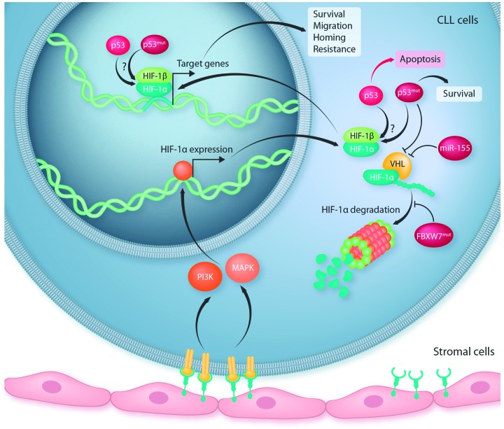
In this issue of Haematologica, Griggio et al. report that disrupting mutations in the tumor-suppressor gene TP53 are associated with higher HIF-1α levels and activity in patients with chronic lymphocytic leukemia (CLL).3 They further show that elevated HIF-1α levels are due to an increased transcription as well as a decreased protein degradation of HIF-1α. Evidence for both regulatory mechanisms have already been described in CLL and are summarized in Figure 1.
Figure 1.
Schematic summary of mechanisms regulating HIF-1α in chronic lymphocytic leukemia (CLL). Contact of CLL cells with stromal cells induces transcriptional upregulation of HIF-1α via PI3K- or MAPK-signaling. Stability of HIF-1α is tightly connected to von Hippel-Lindau (VHL) tumor suppressor protein which triggers proteasomal degration of HIF-1α. In CLL, high levels of miR-155, disrupted p53, and mutant FBXW7 were connected to reduced HIF-1α degradation. Whether direct binding of p53 to HIF-1α is involved in the stability or transcriptional activity of HIF-1α in CLL remains unclear.
A constitutively higher expression and activity of HIF-1α in CLL cells compared to normal B cells was suggested to be due to reduced levels of the von Hippel-Lindau gene product (pVHL), which is responsible for HIF-1α degradation. Downregulation of pVHL was attributed to the inhibitory activity of miR-155, a highly abundant microRNA in CLL which is suggested to contribute to increased HIF-1α stability in CLL cells.4 As pVHL expression levels were lower in CLL patients with disrupted TP53 compared to wild-type TP53 cases, Griggio et al. suggest that the observed accumulation of HIF-1α protein in TP53-disrupted CLL might be partially explained by an increased stability of HIF-1α.
It has recently been suggested that the mutant forms of FBXW7 contribute to HIF-1α stability in CLL cells.5 FBXW7 is known to target proto-oncoproteins including HIF-1α for proteasomal degradation. In silico modeling predicted that mutations in FBXW7 that recurrently occur in CLL change the binding of protein substrates including HIF-1α and are therefore involved in the increased stability of HIF-1α in patients with these mutations.
HIF-1α has been suggested to be transcriptionally regulated in CLL cells via signals delivered by the microenvironment. In oxygenated blood, circulating CLL cells were shown to be primed for hypoxia and to undergo a rapid induction of HIF-1α activity when entering lymphoid tissues.6 In these tissues, CLL cells are in constant interaction with accessory cells which deliver essential signals for survival and proliferation of CLL cells and contribute to resistance to drug-induced apoptosis.7 The contact of CLL cells with stromal cells induces HIF-1α expression via induction of phosphatidylinositol 3-kinase (PI3K) and ERK mitogen-activated protein kinase (MAPK) signaling. In addition, hypoxia in lymphoid tissues was shown to modify central metabolic pathways in CLL cells, including increased adenosine generation and signaling via the A2A adenosine receptor.8 Inhibiting the A2A receptor has been shown to counteract the effects of hypoxia in CLL and accessory cells, and to increase their susceptibility to pharmacological agents.
In line with this, Griggio et al. observed increased expression of HIF-1α in CLL cells upon co-culture with stromal cells to a level that is comparable to hypoxic conditions.3 They further showed that HIF-1α transcript levels of primary CLL cells positively correlate with CLL cell survival in culture. This is likely related to the observation that HIF-1α critically regulates the interaction of CLL cells with their microenvironment. It has previously been shown that HIF-1α induces an upregulation of chemokine receptors, such as CXCR4, and cell adhesion molecules that control the interaction of leukemic cells with bone marrow (BM) and lymphoid microenvironments.9 In line with this, inactivation of HIF-1α was shown to impair chemotaxis and cell adhesion to stroma, reduce BM and spleen colonization in xenograft and allograft CLL mouse models, and prolong survival of these mice. In support of a modulation of CLL cell motility, HIF-1α transcript levels were shown to correlate with the expression of CXCR4 and other target genes in CLL patient samples.
Further evidence for stroma-induced activation of HIF-1α in CLL comes from a comparative transcriptome analysis of primary CLL cells co-cultured or not with protective BM stromal cells which revealed that oxidative phosphorylation, mitochondrial function and hypoxic signaling are the most significantly deregulated functions in non-protected CLL cells.10 These relevant transcriptomic changes were compared to the Connectivity Map database,11 which contains transcriptomic responses of cell lines to treatment with small molecules. This comparison identified drugs that act by repressing HIF-1α and disturbing intracellular redox homeostasis as potential compounds blocking stroma-mediated support of CLL cells.
Under hypoxic conditions, the tumor suppressor protein p53 plays an important role in sensing stress and inducing apoptosis in cells. It was suggested that hypoxia in tumors represents a selection pressure for loss of functional p53 to avoid cell death.12 p53 directly interacts with HIF-1α as an unfolded protein, but the biological implications of this interaction have not been completely clarified.13 Recent work showed that hypoxia-induced HIF-1α leads to TP53 expression, where the resulting p53 protein has a reduced capacity to modulate transcription of target genes but is abundantly available for protein-protein interactions.14 Interestingly, both wild-type and mutant p53 proteins were shown to bind and chaperone HIF-1α to stabilize its binding to DNA response elements and therefore impact on HIF-1α transcriptional activity. Whether direct interactions of p53 with HIF-1α play a role in CLL, or whether CLL cells with disrupted TP53 merely have a selective advantage in the hypoxic lymphoid microenvironment is still not clear.
Based on its role and function in cancer, HIF-1 is a promising target for the treatment of many tumor entities, and a multitude of small molecules of diverse chemical composition and promising biological potency have been identified as inhibiting HIF-1 activity.15 Most of these substances are, however, only indirect inhibitors of HIF-1 and exhibit off-target effects by affecting other pathways as well, such as cell signaling, cell division, and DNA replication. To date, no HIF-1 inhibitor has been clinically approved.
The potential of targeting HIF-1α in CLL has previously been tested in immunocompromised mice that were transplanted with the CLL cell line MEC-1, as well as in the syngeneic Em-TCL1 adoptive transfer mouse model of CLL.9 Camptothecin-11 (EZN-2208), a cytotoxic topoisomerase I inhibitor that also inhibits HIF-1α, was used for treatment of these mice which affected homing and retention of CLL cells in protective microenvironments, as shown by reduced spleen weight and decreased colonization of spleen and/or BM with CLL cells, and prolonged survival of mice. As knockdown of HIF-1α did not cause apoptosis of CLL cells in vitro or in vivo, the authors suggested that the pro-apoptotic effect of EZN-2208 towards CLL cells in vivo may be caused by mechanisms that do not depend on HIF-1α inhibition.
To further explore the potential of HIF-1α targeting in CLL, Griggio et al. used the previously described HIF-1α inhibitor BAY 87-2243 which acts by inhibiting mitochondrial complex I activity and hypoxia-induced mitochondrial reactive oxygen species production, thus achieving reduction of HIF-1α level with relatively higher specificity than other drugs.16 BAY 87-2243 was shown to suppress hypoxia-induced HIF-1 target gene expression in vitro at low nanomolar concentrations and to harbor anti-tumor efficacy in xenograft mouse models of lung carcinoma and melanoma.16,17 Pharmacological inhibition of HIF-1α with BAY87-2243 in primary CLL cells induced cell death in vitro, regardless of the TP53 mutational status, with a generally weaker effect under hypoxia or in co-culture with stromal cells.3 Pre-clinical testing of this drug in the Em-TCL1 adoptive transfer model of CLL resulted in lower numbers of malignant B cells in the BM and among them a higher frequency of apoptotic cells. In all other organs that are affected by CLL, including spleen and blood where the majority of leukemia cells accumulate, HIF-1α inhibition had no effect. Combination treatment of BAY87-2243 with fludarabine or ibrutinib showed higher rates of CLL cell death in vitro compared to monotherapy, which was independent of functional TP53. Therefore, the authors suggest that HIF-1α inhibition renders TP53-disrupted CLL cells sensitive to fludarabine treatment.
Even though treatment options for CLL have improved tremendously within the last decade, dysfunctional TP53 is still associated with inferior patient outcome.18 The development of novel treatment strategies is, therefore, extremely important for this patient group, as well as in the light of overcoming or avoiding therapy resistance to novel targeted therapies due to clonal evolution and/or selection of clones with aggressive phenotype, including disrupted TP53 status.
Acknowledgment
I would like to thank Daniel Mertens, Deyan Yosifov, and Michaela Reichenzeller for their critical review and helpful comments on the manuscript.
References
- 1.The Nobel Prize in Physiology or Medicine 2019. NobelPrize.org. Nobel Media AB 2020. Wed. 12 Feb 2020. <https://www.nobel-prize.org/prizes/medicine/2019/press-release/>
- 2.Semenza GL. Targeting HIF-1 for cancer therapy. Nat Rev Cancer. 2003;3(10):721–732. [DOI] [PubMed] [Google Scholar]
- 3.Griggio V, Vitale C, Todaro M, et al. HIF-1α is overexpressed in leukemic cells from TP53-disrupted patients and is a promising therapeutic target in chronic lymphocytic leukemia. Haematologica. 2019;105(4):1042–1054. [DOI] [PMC free article] [PubMed] [Google Scholar]
- 4.Ghosh AK, Shanafelt TD, Cimmino A, et al. Aberrant regulation of pVHL levels by microRNA promotes the HIF/VEGF axis in CLL B cells. Blood. 2009;113(22):5568–5574. [DOI] [PMC free article] [PubMed] [Google Scholar]
- 5.Close V, Close W, Kugler SJ, et al. FBXW7 mutations reduce binding of NOTCH1, leading to cleaved NOTCH1 accumulation and target gene activation in CLL. Blood. 2019;133(8):830–839. [DOI] [PubMed] [Google Scholar]
- 6.Koczula KM, Ludwig C, Hayden R, et al. Metabolic plasticity in CLL: adaptation to the hypoxic niche. Leukemia. 2016;30(1):65–73. [DOI] [PMC free article] [PubMed] [Google Scholar]
- 7.Hanna BS, Öztürk S, Seiffert M. Beyond bystanders: Myeloid cells in chronic lymphocytic leukemia. Mol Immunol. 2019;110:77–87. [DOI] [PubMed] [Google Scholar]
- 8.Serra S, Vaisitti T, Audrito V, et al. Adenosine signaling mediates hypoxic responses in the chronic lymphocytic leukemia microenvironment. Blood Adv. 2016;1(1):47–61. [DOI] [PMC free article] [PubMed] [Google Scholar]
- 9.Valsecchi R, Coltella N, Belloni D, et al. HIF-1α regulates the interaction of chronic lymphocytic leukemia cells with the tumor microenvironment. Blood. 2016;127(16):1987–1997. [DOI] [PMC free article] [PubMed] [Google Scholar]
- 10.Yosifov DY, Idler I, Bhattacharya N, et al. Oxidative stress as candidate therapeutic target to overcome microenvironmental protection of CLL. Leukemia. 2020;34(1):115–127. [DOI] [PubMed] [Google Scholar]
- 11.Lamb J, Crawford ED, Peck D, et al. The Connectivity Map: Using Gene-Expression Signatures to Connect Small Molecules, Genes, and Disease. Science. 2006;313(5795):1929–1935. [DOI] [PubMed] [Google Scholar]
- 12.Hammond EM, Giaccia AJ. The role of p53 in hypoxia-induced apoptosis. Biochem Biophys Res Commun. 2005;331(3):718–725. [DOI] [PubMed] [Google Scholar]
- 13.Sánchez-Puig N, Veprintsev DB, Fersht AR. Binding of Natively Unfolded HIF-1α ODD Domain to p53. Mol Cell. 2005;17(1):11–21. [DOI] [PubMed] [Google Scholar]
- 14.Madan E, Parker TM, Pelham CJ, et al. HIF-transcribed p53 chaperones HIF-1α. Nucleic Acids Res. 2019;47(19):10212–10234. [DOI] [PMC free article] [PubMed] [Google Scholar]
- 15.Bhattarai D, Xu X, Lee K. Hypoxia-inducible factor-1 (HIF-1) inhibitors from the last decade (2007 to 2016): A “structure–activity relationship” perspective. Med Res Rev. 2018;38(4):1404–1442. [DOI] [PubMed] [Google Scholar]
- 16.Ellinghaus P, Heisler I, Unterschemmann K, et al. BAY 87-2243, a highly potent and selective inhibitor of hypoxia-induced gene activation has antitumor activities by inhibition of mitochondrial complex I. Cancer Med. 2013;2(5):611–624. [DOI] [PMC free article] [PubMed] [Google Scholar]
- 17.Schöckel L, Glasauer A, Basit F, et al. Targeting mitochondrial complex I using BAY 87-2243 reduces melanoma tumor growth. Cancer Metab. 2015;3:11. [DOI] [PMC free article] [PubMed] [Google Scholar]
- 18.Tausch E, Beck P, Schlenk RF, et al. Prognostic and predictive role of gene mutations in chronic lymphocytic leukemia: results from the pivotal phase III study COMPLEMENT1. Haematologica. 2020. January 9 [Epub ahead of print] [DOI] [PMC free article] [PubMed] [Google Scholar]


