Abstract
The aerosol form of the bacterium Yersinia pestis causes the pneumonic plague, a rapidly fatal disease. At present, no plague vaccines are available for use in the United States. One candidate for the development of a subunit vaccine is the Y. pestis virulence (V) antigen, a protein that mediates the function of the Yersinia outer protein virulence factors and suppresses inflammatory responses in the host. On the basis of the knowledge that adenovirus (Ad) gene-transfer vectors act as adjuvants in eliciting host immunity against the transgene they carry, we tested the hypothesis that a single administration of a replication-defective Ad gene-transfer vector encoding the Y. pestis V antigen (AdsecV) could stimulate strong protective immune responses without a requirement for repeat administration. AdsecV elicited specific T cell responses and high IgG titers in serum within 2 weeks after a single intramuscular immunization. Importantly, the mice were protected from a lethal intranasal challenge of Y. pestis CO92 from 4 weeks up to 6 months after immunization with a single intramuscular dose of AdsecV. These observations suggest that an Ad gene-transfer vector expressing V antigen is a candidate for development of an effective anti-plague vaccine
The gram-negative bacterium Yersinia pestis is the etiological agent of plague and is classified as a category A pathogen that is a potential agent of bioterrorism [1, 2]. There are 3 forms of the human disease: bubonic, septicemic, and pneumonic [2, 3]. Of these, the pneumonic plague is of most concern as a biological threat, because of the rapid onset, high mortality, and rapid spread. Although antibiotics can successfully treat plague, the fatality rate is high when treatment is delayed >24 h after the onset of symptoms [2–4]
At present, no plague vaccines are available in the United States. Several vaccines have been developed, including killed whole-cell formulations and the live attenuated EV76 vaccine [5–8]. Although these vaccines have been used in humans, they offer low levels of protection, have numerous adverse side effects, and require frequent immunizations with consequent prolonged time to develop immunity [5–8]. A promising subunit vaccine is based on the virulence (V) antigen (also referred to as “LcrV”) [9–11]. V antigen is a 37-kDa multifunctional protein of Yersinia species encoded by the 70-kb low calcium response (lcr) plasmid. V antigen participates in the type III secretion system in Y. pestis regulating the production and facilitating the translocation of Yersinia outer proteins (Yops) with anti-host activity into the host cell [12, 13]. Active immunization with purified V antigen or passive immunization with antiserum against V antigen provides protection against plague in mice [9, 10, 14, 15]. V antigen–based DNA vaccines are also being developed [16, 17]. These vaccines elicit low antibody titers, and protection against a Y. pestis challenge is reached only after several immunizations [18]
The present study is focused on using replication-deficient adenovirus (Ad) gene-transfer vectors encoding V antigen to elicit protective immune responses against Y. pestis. Ad vectors are excellent candidates for vaccine platforms as they transfer genes effectively to antigen-presenting cells (APCs) in vivo, with consequent activation of APCs, thus conveying immune adjuvant properties and inducing strong, rapid humoral and cellular immune responses against the transgene product [19–26]. On the basis of these considerations, we constructed an E1−E3− Ad-based vaccine vector that encodes a secreted human codon–optimized V antigen (AdsecV). The data demonstrate that AdsecV induces high IgG titers within 2 weeks after a single intramuscular immunization in mice. Importantly, mice immunized with a single intramuscular dose of AdsecV are protected from a lethal intranasal challenge of Y. pestis
Methods
Ad vectorsThe Y. pestis V antigen gene (NCBI accession no. B33601) with mammalian-preferred codons was synthesized by overlap polymerase chain reaction and fused to the human Igκ signal sequence for extracellular secretion. The V antigen gene was cloned into a recombinant Ad5–based vector (E1a, partial E1b, and partial E3 deletion), to generate AdsecV. AdNull was used as a control vector with identical backbone but no transgene [27]. The vectors were produced in 293 cells and were purified by double CsCl gradient centrifugation [28]. Dosing was based on particle units (pu), the physical number of Ad particles as measured by spectrophotometry [29]
Purification of recombinant V antigenRecombinant V antigen was produced as a reagent for assessment of antibodies against V antigen. The V antigen gene was cloned into the pRSET expression plasmid (Invitrogen), and V antigen was purified as a histidine-tag fusion by use of a Ni-NTA Superflow Column (Qiagen) under native conditions
Expression of V antigen by AdsecV in vitroThe A549 lung epithelial cell line (CCL185; American Type Culture Collection) was maintained in complete Dulbecco’s modified essential medium. Cells were infected with AdsecV or AdNull (500 pu/cell) in low-serum medium. Twenty-four hours after infection, cells and medium were collected, and proteins were separated by SDS-PAGE (Invitrogen) and transferred to a polyvinylidene fluoride membrane (BioRad Laboratories). For Western blot analysis, the membrane was probed with a 1:1000 dilution of a rabbit anti–V antigen antibody (provided by S. Bavari, US Army Medical Research Institute of Infectious Diseases, Fort Detrick, MD). A peroxidase-conjugated anti-rabbit antibody (Santa Cruz Biotechnology) and a chemiluminescent peroxidase substrate (ECL+ reagent; Amersham Biosciences) were used for detection
To assess V antigen expression by immunofluorescence, 24 h after infection, cells were fixed with 4% paraformaldehyde and blocked with 5% goat serum (Jackson ImmunoLabs), 1% bovine serum albumin in PBS with 0.05% saponin (Sigma-Aldrich), and 0.1% Triton X-100 (Sigma-Aldrich), followed by incubation with the rabbit anti–V antigen antibody diluted 1:1000. A goat anti-rabbit secondary antibody conjugated to Alexa 488 fluorophore (Jackson ImmunoLabs) was used at a final concentration of 10 μg/mL. Nuclei were counterstained with 4′,6-diamidino-2-phenylindole (Molecular Probes). The samples were observed by fluorescence microscopy with an Olympus IX70 inverted microscope (New York/New Jersey Scientific) equipped with a ×60 PlanApo NA 1.4 objective, and digital image analysis was performed with Metamorph imaging software (version 4.6r9; Universal Imaging)
MiceFemale BALB/c mice were obtained from Taconic. Mice were housed under specific-pathogen-free conditions and were used at 7 weeks of age. Mice were immunized in a single vaccination by 2 intramuscular injections, with 50 μL of the vaccine preparation divided evenly between the quadriceps on each side. Ad vectors were diluted with saline to the specified dose
Transgene-specific humoral responsesSerum samples from immunized mice were obtained from the tail vein, and anti–V antigen serum antibody titers were determined by ELISA. For vector dose response, serum samples were obtained from mice 4 weeks after immunization with AdsecV at doses ranging from 108 to 1011 pu. For time-dependent anti–V antigen antibody titers, mice were immunized with a single administration of 109 pu of AdsecV, and serum samples were obtained before and at 1, 2, 4, 6, and 8 weeks after vaccination. For IgG subtypes, mice were vaccinated with 109 pu of AdsecV, and serum samples were obtained 5 weeks after immunization
For ELISAs, microtiter plates (Corning) were coated with 0.5 μg of recombinant V antigen per well. After blocking, serum samples were added in sequential 2-fold dilutions starting at 1:20 and were incubated for 1 h at 23°C. An anti–mouse IgG–horseradish peroxidase conjugate (Sigma-Aldrich) was used at 1:10,000 dilution. Detection was accomplished using a peroxidase substrate (BioRad Laboratories). Absorbance at 415 nm was read using a microplate reader (BioRad Laboratories). Class-specific anti-IgG antibodies (IgG1, IgG2a, IgG2b, and IgG3) were determined using the Mouse Typer isotyping panel (Bio-Rad Laboratories). Antibody titers were calculated on the basis of a log(optical density) − log(dilution) interpolation model and a cutoff value equal to 2-fold the absorbance of the background [30, 31]
Transgene-specific cellular responsesMice were immunized intramuscularly (n=5) with either saline, AdNull (1011 pu), or AdsecV (1011 pu). The frequency of antigen-specific T lymphocytes was determined using an interleukin (IL)–2–, interferon (IFN)–γ–, and IL-4–specific enzyme-linked immunospot (ELISPOT) assay (R&D Systems). Six days after administration of Ad, CD4+ or CD8+ T cells were purified by negative depletion using SpinSep T cell subset purification kits (StemCell Technologies). Splenic dendritic cells (DCs) were purified from naive mice by positive selection using CD11c MACS beads (Milentyi Biotec) and double purification over 2 MACS-LS columns (Milentyi Biotec). The purity of CD4+ T cells, CD8+ T cells, and DCs was assessed by staining with anti–CD4–phycoerythrin (PE), anti–CD8-PE, and anti–CD11c-PE antibodies (BD Biosciences), respectively. Cell purity evaluation and cell counts were performed using a FACScalibur flow cytometer running at a constant flow rate. For ELISPOT assays, 105 CD4+ or CD8+ T cells were incubated for 36 h with splenic DCs at a ratio of 4:1, with or without purified V antigen. Spots were counted by computer-assisted ELISPOT image analysis (Zellnet Consulting)
Y. pestis CO92 challengeThe Y. pestis challenge studies were conducted at the Public Health Research Institute at the International Center for Public Health under biosafety level 3 conditions. Four weeks or 6 months after immunization, mice (10/group) were challenged intranasally with Y. pestis CO92. Y. pestis CO92 was grown aerobically in heart infusion broth (Difco) at 30°C and was diluted in saline solution at doses ranging from 103 to 106 cfu. Fifty microliters of bacterial suspension was used for intranasal infection of mice. Bacterial dose was controlled by plating on Yersinia selective agar (YSA; Oxoid). Survival was monitored daily for 15 days. From a subset of the mice that died after challenge, liver, spleen, and lungs were removed, homogenized in saline solution, and plated on YSA, to confirm that plague was the cause of death. A subset of the vaccinated mice that survived the challenge were killed 15 days after infection; liver, spleen, and lungs were removed, homogenized in saline solution, and plated on YSA to confirm that bacteria were not present in internal organs
Statistical analysesData are presented as mean±SE values. For ELISPOT assays, statistical analyses were performed using 1-way analysis of variance followed by Fisher’s protected least significant difference test. For survival comparison, Kaplan-Meier analysis was performed; reported P values are from Mantal-Cox analysis. Statistical significance was determined at P<.05
Results
Expression of V antigen by AdsecVV antigen expression by AdsecV was analyzed in vitro by Western blot analysis. Twenty-four hours after infection of A549 cells with AdsecV, a protein with the expected size for V antigen (37 kDa) was identified in medium and cell lysates by use of an anti–V antigen antibody (figure 1A). The protein was not detected in cells infected with AdNull (the control vector) or in uninfected cells. The localization of V antigen in the AdsecV-infected cells was evaluated by indirect immunofluorescence. At 24 h, V antigen (figure 1C) exhibits a broad, diffuse staining pattern as well as a bright, punctate, perinuclear staining pattern consistent with localization of V antigen in the endoplasmic reticulum and Golgi apparatus of the secretory pathway. The protein was not present in AdNull-infected cells (figure 1B)
Figure 1.
Expression of virulence (V) antigen in cells infected with AdsecV. A549 cells were assessed 24 h after infection with AdsecV or the control vector AdNull at 500 particle units/cell. Data are representative of results from 2 independent experiments. A Western blot analysis of medium and cell lysate. V antigen was detected by use of anti–V antigen antibody. Lane 1 medium, naive cells; lane 2 medium, AdNull-infected cells; lane 3 medium, AdsecV-infected cells; lane 4 cell lysate, naive cells; lane 5 lysate, AdNull-infected cells; lane 6 lysate, AdsecV-infected cells. The extra band visible in the supernatant lanes is the result of the cross-reactivity of other antibodies in the polyclonal preparation with other proteins in the medium. B and C Indirect immunofluorescence detection of V antigen. After 24 h, cells were fixed with 4% paraformaldehyde and were stained with anti–V antigen antibody. Nuclei were counterstained with 4′,6-diamidino-2-phenylindole. Panel B shows AdNull-infected cells, and panel C shows AdsecV-infected cells. The bar is 10 μm
Humoral immune responses to AdsecVTo evaluate humoral immune responses in AdsecV-immunized mice, anti–V antigen IgG titers were assessed in serum. Four weeks after a single intramuscular administration of 108–1011 pu of AdsecV, a dose response in the total anti–V antigen IgG titers was observed, with the total anti–V antigen IgG titers for immunized mice reaching a mean±SE titer of 76,000±16,000 for the mice vaccinated with 1011 pu of AdsecV (figure 2A). No anti–V antigen IgG titers were detected in the naive mice (which received saline) or in the AdNull-immunized mice
Figure 2.
Anti–virulence (V) antigen antibodies in serum evoked by AdsecV after intramuscular immunization of mice. Antibody levels were quantified by ELISA. A Dose-dependent induction of anti–V antigen IgG 4 weeks after immunization with AdsecV (n=10 mice/group). B Time course of induction of anti–V antigen IgG after immunization with AdsecV (109 particle units [pu]) or the control vector AdNull (109 pu). Serum samples were collected before and 1, 2, 4, 6, and 8 weeks after immunization (n=5 mice/group). C Anti–V antigen IgG subtypes at 5 weeks in pooled serum samples (n=10 mice/group). Data are mean±SE values except for those in panel C, in which data are from pooled serum samples. For all panels, black triangles indicate AdsecV-immunized mice, white circles indicate AdNull-immunized mice, and black diamonds indicate naive mice; dashed lines indicate the limit of detection of the assay. Data are representative of results from 2 independent experiments
To evaluate the kinetics of the anti–V antibody response, total IgG titers were measured at different time points after a single vaccination. After administration of a 109-pu dose of AdsecV, anti–V antigen titers were detected in serum of immunized mice as early as 1 week (figure 2B). The antibody titer reached a maximum level at 2 weeks and remained high through 8 weeks. Analysis of IgG subclasses at the 109-pu AdsecV dose showed a strong response for both IgG1 and IgG2a and a lesser response for IgG2b and IgG3 (figure 2C). Similar antibody titers and subtypes were observed with an Ad vector expressing a nonsecreted form of V antigen (data not shown). Anti–V antigen antibody after immunization with 109 pu of the nonsecreted form could be detected in serum at 2 weeks, with a mean±SE titer of 13,700±2,900. Antibody levels remained high through week 8, as reported for AdsecV (figure 2B). Because anti–V antigen antibodies in serum could already be detected 1 week after immunization with AdsecV and because titers were similar for both forms of the vaccine, we focused our study on the responses evoked by AdsecV
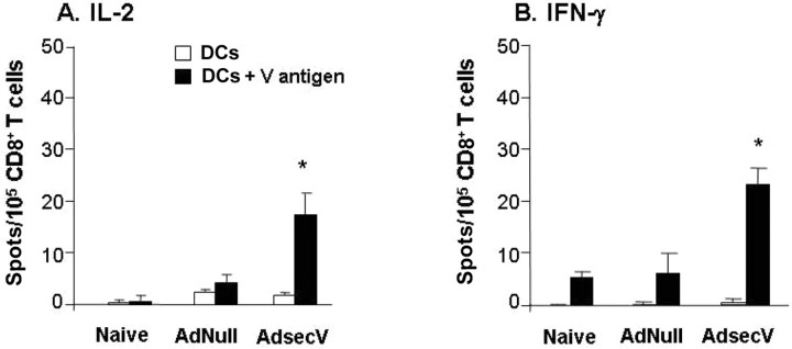
Cellular immune responses to AdsecVThe frequency of T cell responses to V antigen in vaccinated mice was analyzed by ELISPOT assay. Six days after mice were immunized with 1011 pu of AdsecV, purified CD4+ and CD8+ T cells from the spleens of the vaccinated mice were stimulated with syngeneic DCs pulsed with V antigen, and cytokine production was assessed. V antigen–specific IL-2 secretion (mean±SE, 52±6 spots/105 CD4+ T cells) (figure 3A) as well as IFN-γ secretion (mean ± SE, 91±3 spots/105 CD4+ T cells) (figure 3C) was significantly higher (P<.005 and P<.0001, respectively) in CD4+ T cells from the AdsecV-immunized mice than in the 2 control groups. In contrast, no significant differences were observed for V antigen–specific IL-4 production (figure 3B). CD8+ T cell activation was evaluated by V antigen–specific IL-2 and IFN-γ secretion. Both IL-2 (mean±SE, 19±4 spots/105 CD8+ T cells) (figure 4A) and IFN-γ (mean±SE, 24±3 spots/105 CD8+ T cells) (figure 4B) responses were higher in the AdsecV-vaccinated mice than in the 2 control groups (P<.005). The naive mice and the mice immunized with AdNull showed no significant signal of V antigen–induced cytokine production above background
Figure 3.
Virulence (V) antigen–stimulated cytokine production by CD4+ T cells from mice immunized with AdsecV. Mice were immunized intramuscularly with either saline (naive mice), AdNull (1011 particle units [pu]), or AdsecV (1011 pu). Six days after immunization, CD4+ T cells were isolated from spleens and were stimulated for 36 h with syngeneic dendritic cells (DCs) pulsed with 100 μg/mL purified V antigen. Cytokine expression was assessed by enzyme-linked immunospot assay. Panel A shows interleukin (IL)–2 expression, panel B shows IL-4 expression, and panel C shows interferon (IFN)–γ expression, both for stimulation with DCs alone and for stimulation with DCs plus V antigen. Data are mean±SE values (n=5 mice/group) and are representative of results from 3 independent experiments. *P<.005 and **P<.0001, compared with the 2 control groups (analysis of variance followed by Fisher’s protected least significant difference test)
Figure 4.
Virulence (V) antigen-stimulated cytokine production by CD8+ T cells from mice immunized with AdsecV. Mice were immunized intramuscularly with either saline (naive mice), the control vector AdNull (1011 particle units [pu]), or AdsecV (1011 pu). Six days after immunization, CD8+ T cells were isolated from spleens and stimulated for 36 h with syngeneic dendritic cells (DCs) pulsed with 100 μg/mL purified V antigen. Cytokine expression was assessed by enzyme-linked immunospot assay. Panel A shows interleukin (IL)–2 expression and panel B shows interferon (IFN)–γ expression, both for stimulation with DCs alone and for stimulation with DCs plus V antigen. Data are mean±SE values (n=5 mice/group) and are representative of results from 3 independent experiments. *P<.005, compared with the 2 control groups (analysis of variance followed by Fisher’s protected least significant difference test)
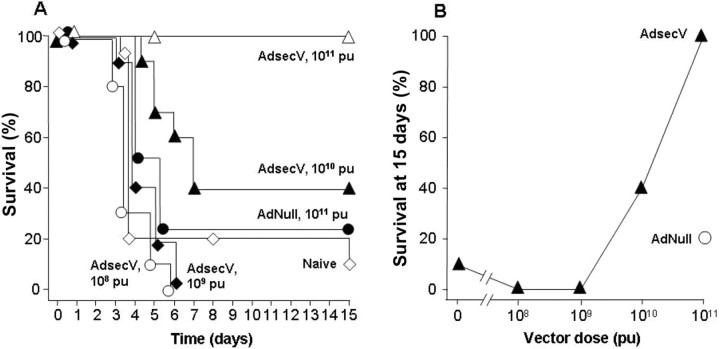
Protection against intranasal challenge with Y. pestis CO92The ability of AdsecV to confer protective immunity was evaluated by challenging immunized mice with the fully virulent Y. pestis strain CO92. The mice received a single intramuscular administration of AdsecV at doses ranging from 108 to 1011 pu. Four weeks after vaccination, the mice were infected intranasally with 3×10³ cfu of Y. pestis strain CO92. All (10/10) of the mice in the group vaccinated with 1011 pu of AdsecV survived the Y. pestis challenge (P<.005) (figure 5A). The survival of mice that were immunized with 1010 pu of AdsecV ranged from 40% (4/10; P<.05) (figure 5A) to 60% (6/10) in a different experiment (data not shown). The 109- and 108-pu doses were not protective; the mice in those groups died according to the same time frame as did the mice in the control groups that received either saline or 1011 pu of AdNull. Assessment of the data at 15 days after challenge showed that the mortality of mice was dependent on the vaccine dose (figure 5B)
Figure 5.
Survival of mice after intranasal challenge with Yersinia pestis CO92 after a single intramuscular administration of AdsecV. Mice were immunized intramuscularly with either saline (naive mice), the control vector AdNull (1011 particle units [pu]), or AdsecV (108, 109, 1010, or 1011 pu) (n=10 mice/group) and were challenged 4 weeks later by intranasal administration of 3×10³ cfu of Y. pestis CO92. Data are representative of results from 2 independent experiments. A Time course of survival after challenge. For the mice that received 1010 or 1011 pu of AdsecV (n=10 mice), P<.05 and P<.005, respectively, compared with the 2 control groups. B Dose-dependent survival of mice immunized with AdsecV at day 15 after challenge
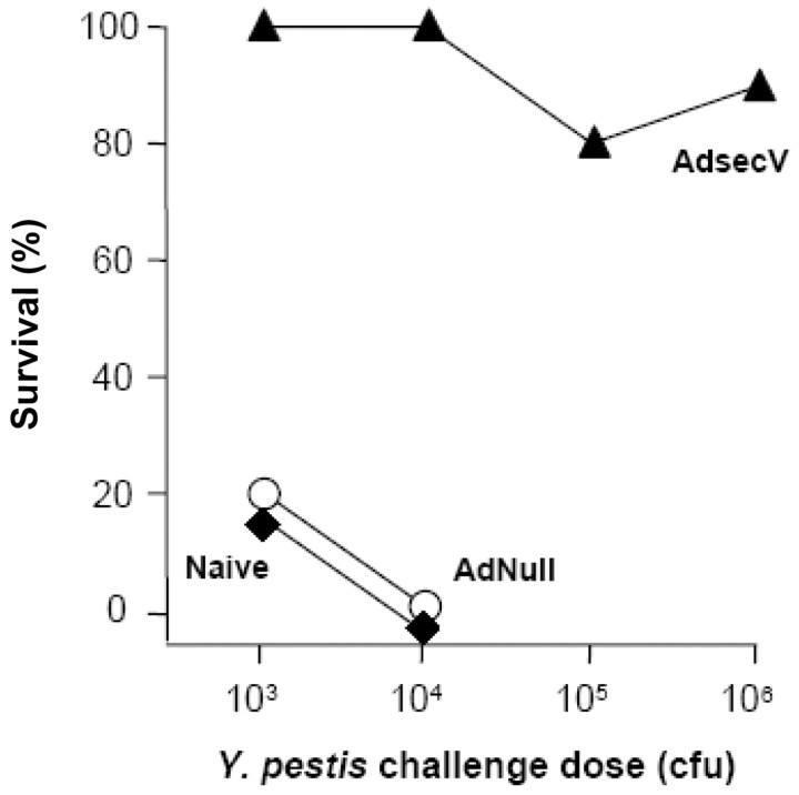
To evaluate the protective capacity of the vaccine at different challenge doses, mice were infected intranasally with 103–106 cfu of Y. pestis CO92 4 weeks after a single administration of 1011 pu of AdsecV. Mice were protected at all doses (figure 6). All AdsecV-immunized mice (10/10) survived the challenge with 103 (P<.005) and 104 (P<.0001) cfu, whereas the naive mice and the mice immunized with 1011 pu of AdNull died within 3–5 days. At higher challenge doses, 80% (8/10) and 90% (9/10) of the mice survived after intranasal infection with 105 and 106 cfu Y. pestis CO92, respectively (P<.0001)
Figure 6.
Yersinia pestis CO92 challenge dose response of AdsecV-immunized mice. Mice were immunized intramuscularly with either saline (naive mice), the control vector AdNull (1011 particle units [pu]), or AdsecV (1011 pu) and were challenged 4 weeks later by intranasal administration of 103–106 cfu of Y. pestis CO92. Data are from 1 experiment (n=10 mice/group). Survival at 15 days is plotted against dose. Because all naive and AdNull-immunized mice died at a dose of 104 cfu, higher doses of Y. pestis were not assessed in the 2 control groups. For the mice that received 103 cfu and for the mice that received the higher challenge doses, P<.005 and P<.0001, respectively, compared with the 2 control groups
The capacity of the vaccine to confer long-term protection was evaluated by challenge at 6 months after a single immunization with 1011 pu of AdsecV. Anti–V antigen total IgG titers in immunized mouse serum before challenge were a mean±SE of 106,000±16,500 (n=20). Mice were infected intranasally with 104 or 106 cfu of Y. pestis CO92. All AdsecV-immunized mice (10/10) survived the challenge with 104 cfu and 90% (9/10) survived the challenge with 106 cfu (P<.0001, for both doses) (figure 7), whereas none of the 10 naive mice survived the challenge with 104 cfu Y. pestis CO92
Figure 7.
Long-term protection of AdsecV-immunized mice. Mice were immunized intramuscularly with either saline (naive mice) or AdsecV (1011 particle units [pu]) and were challenged 6 months later by intranasal administration of 104 or 106 cfu of Yersinia pestis CO92 (P<.0001, compared with the naive mice). Survival is plotted as a function of time. Data are from 1 experiment (n=10 mice/group)
Discussion
In the present study, AdsecV, a replication-defective Ad vector expressing a secreted form of the Y. pestis V antigen, was evaluated as a vaccine against plague. The V antigen sequence in AdsecV was targeted for extracellular expression by an Igκ secretion signal, and V antigen expression in infected cells was confirmed by Western blot analysis and indirect immunofluorescence. After a single intramuscular immunization with AdsecV, mice developed strong humoral responses within 2 weeks, with anti–V antigen IgG titers predominantly of the IgG2a and IgG1 subtypes, suggesting a strong Th1 and Th2 response. The cellular immune responses observed in splenic T cells from vaccinated mice were V antigen–specific Th1 helper (CD4+) and CD8+ responses. Most importantly, immunized mice were protected from an intranasal challenge with a lethal dose of 106 cfu of Y. pestis CO92, from 4 weeks through 6 months after a single administration of the vaccine
Y. pestis vaccinesPlague is one of the most devastating acute infectious diseases experienced by humankind [1–5]. Antibiotics are only marginally effective once symptoms of pneumonic plague develop; moreover, some antibiotic-resistant isolates have been identified. Given these characteristics, there is concern that an aerosolized form of Y. pestis may be exploited as a bioweapon
There is no licensed Y. pestis vaccine for use in the United States. Killed whole-cell vaccines have been used since the late 1890s [5–8]. Although these vaccines have been shown to protect against the bubonic form of the disease, they do not protect against pneumonic plague. These vaccines also have disadvantages, such as a significant incidence of transient local and systemic adverse side effects and the need for frequent boosting to maintain adequate immunity [5, 7, 8]. A live attenuated vaccine based on the pigmentation-negative Y. pestis strain EV76 has been available since 1908 [6]. This vaccine had questionable efficacy in evoking effective immune responses in humans and presents the risk for reversion to virulence in vivo
In recent years, the development of a safe and effective plague vaccine has been focused on using recombinant protein subunits of Y. pestis [5, 9–11]. Several virulence factors have been identified as possible vaccine candidates, but the most promising are the V antigen and the F1 protein [7–11, 32–34]. Antibodies against V and F1 confer protection against both bubonic and pneumonic plague in mice, guinea pigs, and nonhuman primates [9–11, 14, 15, 33, 35]. Recent studies have shown that a single intramuscular immunization with both recombinant antigens delivered with adjuvants and combined in a molar ratio of 2:1 (F1:V antigen) protected mice against an aerosol challenge with Y. pestis [10, 11, 36–39]. Although this protection was correlated with high IgG levels [40], it has been suggested that nonhumoral immune responses also participate, because immunized IL-4–deficient mice, which do not mount effective humoral immune responses, have been shown to be protected against plague [41]. Also, immunization with the combined subunits failed to protect signal transducer and activator of transcription (Stat) 4−/− mice against plague; Stat 4−/− mice are diminished in their capacity to mount type 1 cytokine responses [42]. It was shown recently that cellular immunity in the absence of antibody can protect against pulmonary Y. pestis infection; transfer of Y. pestis–primed T cells to naive B cell–deficient μMT mice protected the mice against a Y. pestis challenge [43]
V antigen–based vaccinesV antigen is a good candidate for Y. pestis vaccine development because it can protect from infection with either F1+ or F1− strains [44]. V antigen plays important roles in the virulence of plague. It participates in the regulation and translocation of the effector proteins Yops into host cells through a type III secretion system. Yops virulence factors produce cytoskeletal rearrangements and apoptosis in macrophage-like cells, allowing bacteria to escape phagocytosis and to proliferate extracellularly [12, 13, 45, 46]. In addition, purified V antigen has been shown to suppress the normal inflammatory response in the host by down-regulating the expression of tumor necrosis factor (TNF)–α and IFN-γ, which promotes the production of IL-10 by macrophages, and by inhibiting the chemotaxis of neutrophils [47–50]
The basis for the protection conferred by the recombinant V antigen vaccine is not well known. The correlation with high IgG titers and protection of immunodeficient SCID/Beige mice against pneumonic plague by passive transfer of anti–V antigen–specific immune serum [14, 15, 51] suggests that anti–V antigen antibodies play a significant role. It has been shown that anti–V antigen antibody administered during infections of mice with Y. pestis restore the production of TNF-α and IFN-γ [49], and in vitro assays have shown that anti-V antigen antibodies can partially block the delivery of Yops (and the consequent downstream effects of Yops) in infected macrophage-like cells [52]. Thus, the protection conferred by a V antigen vaccine might be achieved by opsonization through antibody association with surface V antigen on Y. pestis by blocking the delivery of Yops to host cells, by preventing early bacterial growth in macrophages, and/or by neutralizing the immunomodulatory activity of V antigen, allowing the host to mount an inflammatory response
Ad vectors as vaccine platformsRecombinant Ad vectors are attractive for vaccine strategies against pathogens for many reasons. They are stable, easy to manipulate, can be produced inexpensively at high titer, and can be purified by commonly available methods [53]. Ad vectors are capable of delivering genes to a broad variety of cell types, and relevant to their use as a vaccine is their ability to infect DCs and other APCs in vivo. The Ad vector itself may act as adjuvant by inducing a strong inflammatory response at the injection site and by promoting the differentiation of immature DCs into professional APCs [54–56]. Recombinant Ad vectors expressing a wide variety of pathogen-specific genes have been used in vaccination studies in rodents, canines, and nonhuman primates [57–72]. Ad vectors induce protective adaptive immune responses against the transgene product very rapidly after a single application [19–26]. This feature is particularly useful for postexposure vaccination or to combat infectious agents that cause infrequent but rapidly spreading outbreaks associated with high mortality
The present evaluation of the efficacy of an Ad vaccine, AdsecV, expressing the Y. pestis V antigen demonstrated the induction of rapid protective humoral and cellular immune responses. AdsecV antibody responses were elicited rapidly (within 2 weeks after administration) and showed the characteristic Th1/Th2 responses elicited by Ad vectors. Most importantly, the immune responses evoked by AdsecV were sufficient to protect immunized mice against an intranasal challenge with a fully virulent strain of Y. pestis. Together, these data suggest that AdsecV is a promising vaccine candidate for protection against plague
Acknowledgments
We thank T. Niven, for performing the antibody assays; Dr. D. Perlin and the staff of the Public Health Research Institute at the International Center for Public Health, for help with the biosafety level 3 studies; and N. Mohamed, for help in preparing the manuscript
Footnotes
Potential conflicts of interest: none reported
Financial support: National Institutes of Health (grants U54 AI057158 and R01 AI 55844); Will Rogers Memorial Fund, Los Angeles, California; Robert A. Belfer (gift to support the development of an antibioterrorism vaccine)
M.J.C. and J.L.B. contributed equally to this study
References
- 1.Centers for Disease Control and Prevention
- 2.Perry RD, Fetherston JD. Yersinia pestis—etiologic agent of plague. Clin Microbiol Rev. 10:35–66. doi: 10.1128/cmr.10.1.35. [DOI] [PMC free article] [PubMed] [Google Scholar]
- 3.Inglesby TV, Dennis DT, Henderson DA, et al. Plague as a biological weapon: medical and public health management. Working Group on Civilian Biodefense. JAMA. 283:2281–90. doi: 10.1001/jama.283.17.2281. [DOI] [PubMed] [Google Scholar]
- 4.Smego RA, Frean J, Koornhof HJ. Yersiniosis I: microbiological and clinicoepidemiological aspects of plague and non-plague Yersinia infections. Eur J Clin Microbiol Infect Dis. 18:1–15. doi: 10.1007/s100960050219. [DOI] [PubMed] [Google Scholar]
- 5.Titball RW, Williamson ED, Dennis DT. Plague. In: Plotkin SA, Orenstein WA, editors. Vaccines. p. 999. [Google Scholar]
- 6.Russell P, Eley SM, Hibbs SE, Manchee RJ, Stagg AJ. A comparison of plague vaccine, USP and EV76 vaccine induced protection against Yersinia pestis in a murine model. Vaccine. 13:1551–6. doi: 10.1016/0264-410x(95)00090-n. [DOI] [PubMed] [Google Scholar]
- 7.Titball RW, Williamson ED. Vaccination against bubonic and pneumonic plague. Vaccine. 19:4175–84. doi: 10.1016/s0264-410x(01)00163-3. [DOI] [PubMed] [Google Scholar]
- 8.Titball RW, Williamson ED. Yersinia pestis (plague) vaccines. Expert Opin Biol Ther. 4:965–73. doi: 10.1517/14712598.4.6.965. [DOI] [PubMed] [Google Scholar]
- 9.Anderson GW, Leary SE, Williamson ED, et al. Recombinant V antigen protects mice against pneumonic and bubonic plague caused by F1-capsule-positive and -negative strains of Yersinia pestis. Infect Immun. 64:4580–5. doi: 10.1128/iai.64.11.4580-4585.1996. [DOI] [PMC free article] [PubMed] [Google Scholar]
- 10.Leary SE, Williamson ED, Griffin KF, Russell P, Eley SM. Active immunization with recombinant V antigen from Yersinia pestis protects mice against plague. Infect Immun. 63:2854–8. doi: 10.1128/iai.63.8.2854-2858.1995. [DOI] [PMC free article] [PubMed] [Google Scholar]
- 11.Williamson ED, Eley SM, Griffin KF, et al. A new improved sub-unit vaccine for plague: the basis of protection. FEMS Immunol Med Microbiol. 12:223–30. doi: 10.1111/j.1574-695X.1995.tb00196.x. [DOI] [PubMed] [Google Scholar]
- 12.Pettersson J, Holmstrom A, Hill J, et al. The V-antigen of Yersinia is surface exposed before target cell contact and involved in virulence protein translocation. Mol Microbiol. 32:961–76. doi: 10.1046/j.1365-2958.1999.01408.x. [DOI] [PubMed] [Google Scholar]
- 13.Sarker MR, Neyt C, Stainier I, Cornelis GR. The Yersinia Yop virulon: LcrV is required for extrusion of the translocators YopB and YopD. J Bacteriol. 180:1207–14. doi: 10.1128/jb.180.5.1207-1214.1998. [DOI] [PMC free article] [PubMed] [Google Scholar]
- 14.Green M, Rogers D, Russell P, et al. The SCID/Beige mouse as a model to investigate protection against Yersinia pestis. FEMS Immunol Med Microbiol. 23:107–13. doi: 10.1111/j.1574-695X.1999.tb01229.x. [DOI] [PubMed] [Google Scholar]
- 15.Motin VL, Nakajima R, Smirnov GB, Brubaker RR. Passive immunity to yersiniae mediated by anti-recombinant V antigen and protein A-V antigen fusion peptide. Infect Immun. 62:4192–201. doi: 10.1128/iai.62.10.4192-4201.1994. [DOI] [PMC free article] [PubMed] [Google Scholar]
- 16.Bennett AM, Phillpotts RJ, Perkins SD, Jacobs SC, Williamson ED. Gene gun mediated vaccination is superior to manual delivery for immunisation with DNA vaccines expressing protective antigens from Yersinia pestis or Venezuelan Equine Encephalitis virus. Vaccine. 18:588–96. doi: 10.1016/s0264-410x(99)00317-5. [DOI] [PubMed] [Google Scholar]
- 17.Williamson ED, Bennett AM, Perkins SD, Beedham RJ, Miller J. Co-immunisation with a plasmid DNA cocktail primes mice against anthrax and plague. Vaccine. 20:2933–41. doi: 10.1016/s0264-410x(02)00232-3. [DOI] [PubMed] [Google Scholar]
- 18.Wang S, Heilman D, Liu F, et al. A DNA vaccine producing LcrV antigen in oligomers is effective in protecting mice from lethal mucosal challenge of plague. Vaccine. 22:3348–57. doi: 10.1016/j.vaccine.2004.02.036. [DOI] [PMC free article] [PubMed] [Google Scholar]
- 19.Tripathy SK, Black HB, Goldwasser E, Leiden JM. Immune responses to transgene-encoded proteins limit the stability of gene expression after injection of replication-defective adenovirus vectors. Nat Med. 2:545–50. doi: 10.1038/nm0596-545. [DOI] [PubMed] [Google Scholar]
- 20.Wilson JM. Adenoviruses as gene-delivery vehicles. N Engl J Med. 334:1185–7. doi: 10.1056/NEJM199605023341809. [DOI] [PubMed] [Google Scholar]
- 21.Song W, Kong HL, Traktman P, Crystal RG. Cytotoxic T lymphocyte responses to proteins encoded by heterologous transgenes transferred in vivo by adenoviral vectors. Hum Gene Ther. 8:1207–17. doi: 10.1089/hum.1997.8.10-1207. [DOI] [PubMed] [Google Scholar]
- 22.Hackett NR, Kaminsky SM, Sondhi D, Crystal RG. Antivector and antitransgene host responses in gene therapy. Curr Opin Mol Ther. 2:376–82. [PubMed] [Google Scholar]
- 23.Molinier-Frenkel V, Lengagne R, Gaden F, et al. Adenovirus hexon protein is a potent adjuvant for activation of a cellular immune response. J Virol. 76:127–35. doi: 10.1128/JVI.76.1.127-135.2002. [DOI] [PMC free article] [PubMed] [Google Scholar]
- 24.Bessis N, Garcia Cozar FJ, Boissier MC. Immune responses to gene therapy vectors: influence on vector function and effector mechanisms. Gene Ther. 11(Suppl doi: 10.1038/sj.gt.3302364. 1):S10–7. [DOI] [PubMed] [Google Scholar]
- 25.Barouch DH, Nabel GJ. Adenovirus vector-based vaccines for human immunodeficiency virus type 1. Hum Gene Ther. 16:149–56. doi: 10.1089/hum.2005.16.149. [DOI] [PubMed] [Google Scholar]
- 26.Boyer JL, Kobinger G, Wilson JM, Crystal RG. Adenovirus-based genetic vaccines for biodefense. Hum Gene Ther. 16:157–68. doi: 10.1089/hum.2005.16.157. [DOI] [PubMed] [Google Scholar]
- 27.Hersh J, Crystal RG, Bewig B. Modulation of gene expression after replication-deficient, recombinant adenovirus-mediated gene transfer by the product of a second adenovirus vector. Gene Ther. 2:124–31. [PubMed] [Google Scholar]
- 28.Rosenfeld MA, Yoshimura K, Trapnell BC, et al. In vivo transfer of the human cystic fibrosis transmembrane conductance regulator gene to the airway epithelium. Cell. 68:143–55. doi: 10.1016/0092-8674(92)90213-v. [DOI] [PubMed] [Google Scholar]
- 29.Mittereder N, March KL, Trapnell BC. Evaluation of the concentration and bioactivity of adenovirus vectors for gene therapy. J Virol. 70:7498–509. doi: 10.1128/jvi.70.11.7498-7509.1996. [DOI] [PMC free article] [PubMed] [Google Scholar]
- 30.Plikaytis BD, Turner SH, Gheesling LL, Carlone GM. Comparisons of standard curve-fitting methods to quantitate Neisseria meningitidis group A polysaccharide antibody levels by enzyme-linked immunosorbent assay. J Clin Microbiol. 29:1439–46. doi: 10.1128/jcm.29.7.1439-1446.1991. [DOI] [PMC free article] [PubMed] [Google Scholar]
- 31.Price BM, Liner AL, Park S, Leppla SH, Mateczun A. Protection against anthrax lethal toxin challenge by genetic immunization with a plasmid encoding the lethal factor protein. Infect Immun. 69:4509–15. doi: 10.1128/IAI.69.7.4509-4515.2001. [DOI] [PMC free article] [PubMed] [Google Scholar]
- 32.Andrews GP, Strachan ST, Benner GE, et al. Protective efficacy of recombinant Yersinia outer proteins against bubonic plague caused by encapsulated and nonencapsulated Yersinia pestis. Infect Immun. 67:1533–7. doi: 10.1128/iai.67.3.1533-1537.1999. [DOI] [PMC free article] [PubMed] [Google Scholar]
- 33.Jones SM, Griffin KF, Hodgson I, Williamson ED. Protective efficacy of a fully recombinant plague vaccine in the guinea pig. Vaccine. 21:3912–8. doi: 10.1016/s0264-410x(03)00379-7. [DOI] [PubMed] [Google Scholar]
- 34.Leary SE, Griffin KF, Galyov EE, et al. Yersinia outer proteins (YOPS) E, K and N are antigenic but non-protective compared to V antigen, in a murine model of bubonic plague. Microb Pathog. 26:159–69. doi: 10.1006/mpat.1998.0261. [DOI] [PubMed] [Google Scholar]
- 35.US Food and Drug Administration
- 36.Anderson GW, Heath DG, Bolt CR, Welkos SL, Friedlander AM. Short- and long-term efficacy of single-dose subunit vaccines against Yersinia pestis in mice. Am J Trop Med Hyg. 58:793–9. doi: 10.4269/ajtmh.1998.58.793. [DOI] [PubMed] [Google Scholar]
- 37.Andrews GP, Heath DG, Anderson GW, Welkos SL, Friedlander AM. Fraction 1 capsular antigen (F1) purification from Yersinia pestis CO92 and from an Escherichia coli recombinant strain and efficacy against lethal plague challenge. Infect Immun. 64:2180–7. doi: 10.1128/iai.64.6.2180-2187.1996. [DOI] [PMC free article] [PubMed] [Google Scholar]
- 38.Jones SM, Day F, Stagg AJ, Williamson ED. Protection conferred by a fully recombinant sub-unit vaccine against Yersinia pestis in male and female mice of four inbred strains. Vaccine. 19:358–66. doi: 10.1016/s0264-410x(00)00108-0. [DOI] [PubMed] [Google Scholar]
- 39.Williamson ED, Eley SM, Stagg AJ, Green M, Russell P. A single dose sub-unit vaccine protects against pneumonic plague. Vaccine. 19:566–71. doi: 10.1016/s0264-410x(00)00159-6. [DOI] [PubMed] [Google Scholar]
- 40.Williamson ED, Vesey PM, Gillhespy KJ, Eley SM, Green M. An IgG1 titre to the F1 and V antigens correlates with protection against plague in the mouse model. Clin Exp Immunol. 116:107–14. doi: 10.1046/j.1365-2249.1999.00859.x. [DOI] [PMC free article] [PubMed] [Google Scholar]
- 41.Elvin SJ, Williamson ED. The F1 and V subunit vaccine protects against plague in the absence of IL-4 driven immune responses. Microb Pathog. 29:223–30. doi: 10.1006/mpat.2000.0385. [DOI] [PubMed] [Google Scholar]
- 42.Elvin SJ, Williamson ED. Stat 4 but not Stat 6 mediated immune mechanisms are essential in protection against plague. Microb Pathog. 37:177–84. doi: 10.1016/j.micpath.2004.06.009. [DOI] [PubMed] [Google Scholar]
- 43.Parent MA, Berggren KN, Kummer LW, et al. Cell-mediated protection against pulmonary Yersinia pestis infection. Infect Immun. 73:7304–10. doi: 10.1128/IAI.73.11.7304-7310.2005. [DOI] [PMC free article] [PubMed] [Google Scholar]
- 44.Friedlander AM, Welkos SL, Worsham PL, et al. Relationship between virulence and immunity as revealed in recent studies of the F1 capsule of Yersinia pestis. Clin Infect Dis. 21(Suppl doi: 10.1093/clinids/21.supplement_2.s178. 2):S178–81. [DOI] [PubMed] [Google Scholar]
- 45.Cornelis GR, Boland A, Boyd AP, Geuijen C, et al. The virulence plasmid of Yersinia an antihost genome. Microbiol Mol Biol Rev. 62:1315–52. doi: 10.1128/mmbr.62.4.1315-1352.1998. [DOI] [PMC free article] [PubMed] [Google Scholar]
- 46.Nilles ML, Fields KA, Straley SC. The V antigen of Yersinia pestis regulates Yop vectorial targeting as well as Yop secretion through effects on YopB and LcrG. J Bacteriol. 180:3410–20. doi: 10.1128/jb.180.13.3410-3420.1998. [DOI] [PMC free article] [PubMed] [Google Scholar]
- 47.Nakajima R, Brubaker RR. Association between virulence of Yersinia pestis and suppression of gamma interferon and tumor necrosis factor alpha. Infect Immun. 61:23–31. doi: 10.1128/iai.61.1.23-31.1993. [DOI] [PMC free article] [PubMed] [Google Scholar]
- 48.Welkos S, Friedlander A, McDowell D, Weeks J, Tobery S. V antigen of Yersinia pestis inhibits neutrophil chemotaxis. Microb Pathog. 24:185–96. doi: 10.1006/mpat.1997.0188. [DOI] [PubMed] [Google Scholar]
- 49.Nakajima R, Motin VL, Brubaker RR. Suppression of cytokines in mice by protein A-V antigen fusion peptide and restoration of synthesis by active immunization. Infect Immun. 63:3021–9. doi: 10.1128/iai.63.8.3021-3029.1995. [DOI] [PMC free article] [PubMed] [Google Scholar]
- 50.Sing A, Roggenkamp A, Geiger AM, Heesemann J. Yersinia enterocolitica evasion of the host innate immune response by V antigen-induced IL-10 production of macrophages is abrogated in IL-10-deficient mice. J Immunol. 168:1315–21. doi: 10.4049/jimmunol.168.3.1315. [DOI] [PubMed] [Google Scholar]
- 51.Hill J, Copse C, Leary S, Stagg AJ, Williamson ED. Synergistic protection of mice against plague with monoclonal antibodies specific for the F1 and V antigens of Yersinia pestis. Infect Immun. 71:2234–8. doi: 10.1128/IAI.71.4.2234-2238.2003. [DOI] [PMC free article] [PubMed] [Google Scholar]
- 52.Philipovskiy AV, Cowan C, Wulff-Strobel CR, et al. Antibody against V antigen prevents Yop-dependent growth of Yersinia pestis. Infect Immun. 73:1532–42. doi: 10.1128/IAI.73.3.1532-1542.2005. [DOI] [PMC free article] [PubMed] [Google Scholar]
- 53.Hackett NR, Crystal RG. Adenovirus vectors for gene therapy. In: Lasic D, Templeton NS, editors. Gene therapy: therapeutic mechanisms and strategies. pp. 17–40. [Google Scholar]
- 54.Morelli AE, Larregina AT, Ganster RW, et al. Recombinant adenovirus induces maturation of dendritic cells via an NF-kappaB-dependent pathway. J Virol. 74:9617–28. doi: 10.1128/jvi.74.20.9617-9628.2000. [DOI] [PMC free article] [PubMed] [Google Scholar]
- 55.Worgall S, Busch A, Rivara M, et al. Modification to the capsid of the adenovirus vector that enhances dendritic cell infection and transgene-specific cellular immune responses. J Virol. 78:2572–80. doi: 10.1128/JVI.78.5.2572-2580.2004. [DOI] [PMC free article] [PubMed] [Google Scholar]
- 56.Zhang Y, Chirmule N, Gao GP, et al. Acute cytokine response to systemic adenoviral vectors in mice is mediated by dendritic cells and macrophages. Mol Ther. 3:697–707. doi: 10.1006/mthe.2001.0329. [DOI] [PubMed] [Google Scholar]
- 57.Tatsis N, Ertl HC. Adenoviruses as vaccine vectors. Mol Ther. 10:616–29. doi: 10.1016/j.ymthe.2004.07.013. [DOI] [PMC free article] [PubMed] [Google Scholar]
- 58.Sambor A, Beaudry K, et al. Neutralizing antibodies elicited by immunization of monkeys with DNA plasmids and recombinant adenoviral vectors expressing human immunodeficiency virus type 1 proteins. J Virol. 79:771–9. doi: 10.1128/JVI.79.2.771-779.2005. [DOI] [PMC free article] [PubMed] [Google Scholar]
- 59.Fitzgerald JC, Gao GP, Reyes-Sandoval A, et al. A simian replication-defective adenoviral recombinant vaccine to HIV-1 gag. J Immunol. 170:1416–22. doi: 10.4049/jimmunol.170.3.1416. [DOI] [PubMed] [Google Scholar]
- 60.Natuk RJ, Lubeck MD, Chanda PK, et al. Immunogenicity of recombinant human adenovirus-human immunodeficiency virus vaccines in chimpanzees. AIDS Res Hum Retroviruses. 9:395–404. doi: 10.1089/aid.1993.9.395. [DOI] [PubMed] [Google Scholar]
- 61.Lubeck MD, Natuk RJ, Chengalvala M, et al. Immunogenicity of recombinant adenovirus-human immunodeficiency virus vaccines in chimpanzees following intranasal administration. AIDS Res Hum Retroviruses. 10:1443–9. doi: 10.1089/aid.1994.10.1443. [DOI] [PubMed] [Google Scholar]
- 62.Shiver JW, Fu TM, Chen L, et al. Replication-incompetent adenoviral vaccine vector elicits effective anti-immunodeficiency-virus immunity. Nature. 415:331–5. doi: 10.1038/415331a. [DOI] [PubMed] [Google Scholar]
- 63.Lubeck MD, Davis AR, Chengalvala M, et al. Immunogenicity and efficacy testing in chimpanzees of an oral hepatitis B vaccine based on live recombinant adenovirus. Proc Natl Acad Sci USA. 86:6763–7. doi: 10.1073/pnas.86.17.6763. [DOI] [PMC free article] [PubMed] [Google Scholar]
- 64.Matsui M, Moriya O, Akatsuka T. Enhanced induction of hepatitis C virus-specific cytotoxic T lymphocytes and protective efficacy in mice by DNA vaccination followed by adenovirus boosting in combination with the interleukin-12 expression plasmid. Vaccine. 21:1629–39. doi: 10.1016/s0264-410x(02)00704-1. [DOI] [PubMed] [Google Scholar]
- 65.Vos A, Neubert A, Pommerening E, et al. Immunogenicity of an E1-deleted recombinant human adenovirus against rabies by different routes of administration. J Gen Virol. 82:2191–7. doi: 10.1099/0022-1317-82-9-2191. [DOI] [PubMed] [Google Scholar]
- 66.Xiang ZQ, Yang Y, Wilson JM, Ertl HC. A replication-defective human adenovirus recombinant serves as a highly efficacious vaccine carrier. Virology. 219:220–7. doi: 10.1006/viro.1996.0239. [DOI] [PubMed] [Google Scholar]
- 67.Sullivan NJ, Sanchez A, Rollin PE, Yang ZY, Nabel GJ. Development of a preventive vaccine for Ebola virus infection in primates. Nature. 408:605–9. doi: 10.1038/35046108. [DOI] [PubMed] [Google Scholar]
- 68.Sullivan NJ, Geisbert TW, Geisbert JB, et al. Accelerated vaccination for Ebola virus haemorrhagic fever in non-human primates. Nature. 424:681–4. doi: 10.1038/nature01876. [DOI] [PMC free article] [PubMed] [Google Scholar]
- 69.Gao W, Tamin A, Soloff A, et al. Effects of a SARS-associated coronavirus vaccine in monkeys. Lancet. 362:1895–6. doi: 10.1016/S0140-6736(03)14962-8. [DOI] [PMC free article] [PubMed] [Google Scholar]
- 70.Jaiswal S, Khanna N, Swaminathan S. Replication-defective adenoviral vaccine vector for the induction of immune responses to dengue virus type 2. J Virol. 77:12907–13. doi: 10.1128/JVI.77.23.12907-12913.2003. [DOI] [PMC free article] [PubMed] [Google Scholar]
- 71.Bruna-Romero O, Rocha CD, Tsuji M, Gazzinelli RT. Enhanced protective immunity against malaria by vaccination with a recombinant adenovirus encoding the circumsporozoite protein of Plasmodium lacking the GPI-anchoring motif. Vaccine. 22:3575–84. doi: 10.1016/j.vaccine.2004.03.050. [DOI] [PubMed] [Google Scholar]
- 72.Tan Y, Hackett NR, Boyer JL, Crystal RG. Protective immunity evoked against anthrax lethal toxin after a single intramuscular administration of an adenovirus-based vaccine encoding humanized protective antigen. Hum Gene Ther. 14:1673–82. doi: 10.1089/104303403322542310. [DOI] [PubMed] [Google Scholar]


