

Abstract
Skin disease is an extremely common presenting complaint to the exotic animal practitioner. A systematic diagnostic approach is necessary in these cases to achieve a diagnosis and formulate an effective treatment plan. In all exotic species, husbandry plays a central role in the pathogenesis of cutaneous disease, so a thorough evaluation of the husbandry is critical for successful management. The clinical approach to skin disease in exotic animal patients is reviewed with specific focus on structure and function of the skin, diagnostic testing, and differential diagnoses for commonly encountered cutaneous diseases.
Keywords: Exotic animal dermatology, Reptile dermatology, Avian dermatology, Fish dermatology, Small mammal dermatology
Key points
-
•
Skin disease is an extremely common presenting complaint to the exotic animal practitioner.
-
•
Skin disease cases may be challenging because dermatologic diseases are often multifactorial and many have underlying husbandry or environmental deficiencies that must be identified.
-
•
A thorough diagnostic evaluation is critical for successful management of exotic animal cutaneous disease.
Introduction
Skin disease is an extremely common presenting complaint to the exotic animal practitioner. A systematic diagnostic approach is necessary in these cases to achieve the appropriate diagnosis and formulate an effective treatment plan. In all exotic species, husbandry plays a central role in the pathogenesis of cutaneous disease, so a thorough evaluation of the husbandry is critical for successful management. There are vast differences in the structure and function of the skin in exotic species; an understanding of these unique properties is important when treating skin disease in exotic pets. This article focuses on the clinical approach to skin disease in exotic pets including structure and function of the skin, appropriate diagnostic testing, and differential diagnoses for commonly encountered cutaneous diseases.
Reptiles
Cutaneous disease is common in reptiles, is often multifactorial, and is most often secondary to husbandry and environmental deficiencies. A recent retrospective study of dermatologic lesions in reptiles found that from 29% to 64% (dependent on institution and reptile group) of the cases had underlying husbandry-related deficiencies.1
Skin Structure and Function
Reptile skin is modified into scales and composed of a three-layered epidermis and a dermis that typically is aglandular.2, 3, 4 The three layers of the epidermis are (1) stratum corneum (six to eight cell layers, heavily keratinized); (2) stratum intermedium; and (3) stratum germinativum (deepest).2, 3, 4 Two types of keratins compose the stratum corneum.4 The softer more flexible α-keratins are elastic and pliable and form the suture/hinges and spaces between scales.4 The β-keratins (unique to birds and reptiles) compose the hard horny scale.4 The skin is protected by scales produced by the stratum germinativum; scales are separated by scale pockets.2 The keratinized layers of chelonians are modified into scutes.5 The scales or scutes of chelonians and some lizards (plated and girdled lizards, skinks, and crocodilians) are underlain by dermal bony plates referred to as osteoderms or osteoscutes.2, 3, 4, 5 In tortoises, the stratum corneum produces the shell, which consists of the carapace (dorsal) and plastron (ventral); the keratinized scutes cover osteoderms that fuse with the vertebrae and sternebrae.2, 3, 4 Chromatophores (pigment cells) are found in the dermis and melanocytes are present within the stratum germinativum.3 Reptiles shed their skin at regular intervals in a process called ecdysis. The skin of lizards and chelonians shed in several smaller pieces, whereas snakes typically shed their entire skin as one piece.4 Chelonians and crocodilians shed their epidermis continuously, whereas lizards and snakes shed their epidermis periodically.5
Dermatologic Examination and Diagnostic Testing
A detailed clinical history is important in all cases of reptile skin disease; important husbandry-related questions include those pertaining to diet, substrate and housing, lighting, heating, humidity, and temperature.
Common findings during clinical examination of reptiles with dermatologic disease include abrasions, erosions, ulcers, wounds, swellings, pustules, blisters/vesicles/bullae, crusts, dysecdysis, petechial and ecchymoses, discoloration, macroparasites, and edema. In some cases, cutaneous changes can be secondary to systemic disease; petechia and ecchymoses are commonly seen with septicemia and ventral edema may be seen with renal or liver disease.3 In one study, 47% of all reptiles with confirmed or suspected cases of sepsis had petechiae, with the highest association seen in chelonians (82%).1
Commonly used dermatologic diagnostic tests in reptiles include the following2, 3, 4, 5:
-
1.
Skin cytology and impression smears
-
2.Acetate tape impression
-
○Press clear tape against skin and evaluate microscopically
-
○Useful to diagnose mites
-
○
-
3.Skin scrapings
-
○Typically use number 15 scalpel blade to collect epidermal samples
-
○
-
4.Microscopic evaluation of shed skin fragments
-
○Findings may include mites
-
○
-
5.
Skin biopsies for dermatopathology
-
6.Skin cultures
-
○Bacterial, fungal
-
○
-
7.Fine-needle aspirate
-
○Most useful for swellings and growths
-
○
-
8.
Clinicopathologic evaluation including complete blood count (CBC) and biochemistry analysis
-
9.
Radiographs are useful when assessing damaged osteoderms and for the presence of bony changes associated with secondary nutritional hyperparathyroidism or other internal disease
Common Differential Diagnoses for Cutaneous Diseases
See Table 1 for a review of common differential diagnoses for dermatologic diseases in reptiles, including bacterial dermatitis, shell rot, bacterial ulcerative dermatitis, snake mite, and secondary nutritional hyperparathyroidism (Fig. 1, Fig. 2, Fig. 3, Fig. 4, Fig. 5 ).
Table 1.
Differential diagnoses for cutaneous diseases in reptiles
| Disease/Condition | Causes | Clinical Signs/Properties | Diagnosis |
|---|---|---|---|
| Bacterial | |||
| Bacterial dermatitisa, b, c, d, e, f (see Fig. 1) | Often secondary to environmental/husbandry deficiencies or trauma Gram-negative environmental bacteria often act as opportunistic pathogens in these cases Various isolates including Aeromonas; Pseudomonas; Citrobacter; Escherichia coli; Klebsiella; Proteus; Salmonella; Serratia; Flavobacterium; Staphylococcus; Streptococcus; Morganella; Neisseria; Dermatophilus congolensis; Mycobacterium; and anaerobes, such as Bacteroides, Fusobacterium, and Clostridium |
Moist, exudative, and erythematous, but may also appear as blisters, crusts, and ulcerations of the integument | Clinical signs, impression cytology, and culture/sensitivity |
| Shell rota,c,e (see Fig. 2) | Most common isolates include Beneckea chitinovora, Citrobacter spp, and Aeromonas | Most common bacterial infection in chelonians, ulcers of the shell, often rimmed by areas of hyperpigmentation; loose scutes may be present and lesions can progress to osteomyelitis | Clinical signs, impression cytology, and culture/sensitivity |
| Septic cutaneous ulcerative diseasea,d | Disease syndrome in aquatic turtles maintained in poor-quality water Citrobacter freundii is most commonly implicated but other gram-negative bacteria may be isolated | Craterifom ulcers on the shell and skin with septicemia and systemic signs | Clinical signs, impression cytology, and culture/sensitivity |
| Blister diseasea, b, c, d, e, f (see Fig. 3) | Often associated with moist, dirty substrate or inappropriately humid environments Aeromonas and Pseudomonas are the most common clinical isolates |
Lesions typically start on the ventrum as vesicles and pustules that progress to ulceration, necrosis, and abscessation; secondary septicemia is possible; most commonly seen in snakes | Clinical signs, impression cytology, and culture/sensitivity |
| Abscessesa, b, c, d, e, f | Common isolates include Pseudomonas spp, Proteus spp, Aeromonas spp, Serratia spp, Providencia spp, E coli, Citrobacter, Proteus, Salmonella, Streptococcus, Corynebacterium pyogenes, and Neisseria | Localized soft to firm, usually nonpainful swellings that have well-defined capsules; because reptile leukocytes lack the isoenzymes to liquefy pus, a thick caseous exudate is often present | Clinical signs, fine-needle aspirate, culture/sensitivity, histopathology |
| Ectoparasites | |||
| Chiggersa, b, c, d, e, f | Family Trombiculidae | Ingest lymph and dissolved host tissue; zoonotic, skin irritation, pruritus, irregular shedding cycles; mites are most commonly found under scales and around nostrils, eyes, and gular fold (snakes) | Direct observation, microscopic identification |
| Mitesa, b, c, d, e, f (see Fig. 4) | Family Macronyssidae; including Ophionyssus natricis (commonly seen in snakes) and Ophinonyssus acertinus (common in lizards) | Feed on blood; skin irritation, pruritus, irregular shedding cycles, and anemia in severe infestations mites are most commonly found under scales and around nostrils, eyes, and gular fold (snakes) | Direct observation, microscopic identification |
| Leechesb | Various species | Skin irritation at site of attachment, anemia with severe infestation | Direct observation |
| Fungal | |||
| Fungal dermatitisa, b, c, d, e, f | Often secondary to environmental/husbandry deficiencies and immunosuppression Reported isolates are often opportunistic pathogens including Aspergillus, Basidobolus, Geotrichium, Mucor, Saprolegnia and Candida, Fusarium, Trichosporon, Trichoderma, Penicillium, Paecilomyces, Oospora, and Trichophyton |
Superficial infections present as moist, exudative erythematous ulcers or blisters, with crusts or hyperkeratotic lesions Deeper infections often present as nodules/swellings, systemic signs may be present with deeper/systemic infections |
Impression smears, fungal culture, histopathology |
| Yellow fungus diseasee,f | Chrysosporium anamorph of Nannizziopsis vriesii | Seen most commonly in lizards (especially the bearded dragon, Pogona vitticeps) Deep, granulomatous dermatomycosis that is contagious and progressive, severe yellowish hyperkeratotic skin lesions, often fatal |
Fungal culture, histopathology, PCR |
| Cheilitis in spiny tail lizards (Uromastyx sp)f | Devriesea agamarum | Cheilitis | Fungal culture, histopathology |
| Viral | |||
| Green turtle fibropapillomasd,f | Herpesvirus | Papillomatous growths affected soft tissues | Histopathology |
| Neoplasia | |||
| Cutaneous neoplasiaa,b | Reported types include squamous cell carcinoma, fibrosarcoma, myxomatous tumors, lipoma/liposarcoma, melanoma, chromatophoromas | Cutaneous growths | Histopathology |
| Husbandry-related/multifactorial/miscellaneous | |||
| Dysecdysisc (see Fig. 3) | Dysecdysis is almost always a result of deficiencies in husbandry and inappropriate environmental conditions including temperature and humidity | More commonly seen in snakes and some lizards than in chelonians; in lizards and turtles, most commonly affects the digits; in snakes, can be localized or generalized; localized dysecdysis commonly affects the spectacles and retention of this scale can result in other ocular abnormalities, such as subspectacular bullae and abscesses | |
| Secondary nutritional hyperparathyroidism (see Fig. 5) | Multifactorial: severe imbalance of the Ca:P ration in the diet, no access to a full spectrum (ultraviolet B) light source, and a lack of activated vitamin D3; other inappropriate husbandry-related factors | Seen more commonly in lizards and chelonians abnormal bones and shells and chronic abscesses especially around jaw | History, clinical signs, radiographs, serum phosphorus, ionized calcium levels |
| Trauma | Injuries from prey-induced trauma, with rodents being responsible for most cases; trauma from other household pets is also not uncommon | Damaged skin, ulcers, erosions | History and clinical signs |
| Burns | Burns most commonly result from malfunctioning, malpositioned, or inappropriate heating elements or inactivity of the animal | More frequent in lizards and snakes; discolored, ulcerated and sloughed areas of skin | History and clinical signs, histopathology |
| Hypovitaminosis Aa, b, c, d | Dietary deficiency of vitamin A results in squamous metaplasia and epidermal hyperkeratosis | Abnormal shedding Most commonly affects lizards and chelonians Lizards: dysecdysis, impaction/abscessation of cutaneous glands Chelonians: dysecdysis, chemosis/blepharedema and aural abscessation. most common cutaneous changes include hyperkeratosis, dysecdysis, scute loss, and thickened/lichenified skin |
History and clinical signs |
Hoppmann E, Barron HW. Dermatology in reptiles. J Exot Pet Med 2007;16(4):210–24.
Goodman G. Dermatology of reptiles. In: Patterson S, editor. Skin diseases of exotic pets. Ames (IA): Blackwell; 2006. p. 73–118.
Johnston MS. Scales and sheds: the ins and outs of reptile skin disease. In: Proceedings North American Veterinary Dermatology Forum. Denver (CO): 2008. p. 62–6.
Mitchell M, Colombini S. Reptiles. In: Foster A, Foil C, editors. BSAVA manual of small animal dermatology. Gloucester (England): BSAVA; 2003. p. 269–75.
Hat JM. Dermatologic problems in reptiles. In: Proceedings of the World Small Animal Veterinary Association World Congress. Geneva (Switzerland): 2010.
Mader D. Reptile dermatology. In: Proceedings of the Atlantic Coast Veterinary Conference. Atlantic City (NJ): 2011.
Fig. 1.

Bacterial dermatitis on the dorsolateral neck of a green iguana (Iguana iguana).
Fig. 2.

Shell rot in a softshell turtle (Apalone sp). Note crateriform ulcers on the carapace.
Fig. 3.

Ball python (Python regius) with bacterial ulcerative dermatitis (blister disease) and dysecdysis. Note ulcerative skin lesions, retained skin, and spectacles.
Fig. 4.
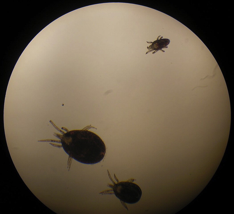
Snake mite (Ophionyssus natricis).
Fig. 5.

Abnormal shell in a leopard tortoise with secondary nutritional hyperparathyroidism.
Amphibians
The thin, relatively unprotected skin of amphibians combined with the significant diversity of amphibian habitats and their biphasic life cycles render them particularly susceptible to a wide range of infectious and noninfectious cutaneous diseases.
Skin Structure and Function
Amphibians belong to three distinct Orders: Anura (frogs); Caudata (salamanders); and Gymnophiona (caecilians). The skin of amphibians is clinically the most important organ system of amphibians and varies depending on the life stage (premetamorphosis or postmetamorphosis); habitat (generally divided into aquatic or terrestrial); and the species.6 The skin functions in osmoregulation, gas respiration, and water absorption.6, 7 Amphibian epidermis is typically thin; keratinized; and consists of the stratum corneum, stratum granulosum, stratum spinosum, and the stratum basale.7, 8, 9, 10, 11 Modifications of amphibian skin include the presence of dermal scales (caecilians); folds and grooves for increased surface area (salamanders); partial ossification of the cranial skin and adherence to the skull (bufonids); a specialized highly vascularized ventral dermal organ for water absorption (“drinking patch” in anurans); and the presence of dermal bones (some anurans).10, 12 The stratum corneum is typically shed in one piece at regular intervals and consumed (dermatophagy) unless the animal is ill.6, 11, 13 The skin of anurans is loosely adhered to the body and can become edematous in disease states.11 Two key features separate adult caecilians and anurans from their larval form: the epidermis is keratinized in adults and the dermis contains a variety of dermal glands.7, 9, 11 Mucus, produced by mucous glands and epithelial cells, aids in respiration, prevents evaporative water loss, contains antibacterial and antifungal properties, can be defensive noxious or contain toxic chemicals, may act as pheromones, and can aid in reproduction.12, 14, 15, 16, 17
Dermatologic Examination and Diagnostic Testing
A thorough history and dermatologic examination are important when evaluating any case of amphibian skin disease. Husbandry-related factors often underlie the development of many skin diseases in amphibians. Important questions to consider include recent introductions into the collection; diet; and tank setup including filtration, aeration, water quality, and temperature. During examination, it is important to always handle amphibians with rinsed gloves to avoid damaging their skin and prevent cutaneous absorption of potentially toxic glandular secretions.18, 19, 20 Many amphibian skin diseases can have a similar appearance with cutaneous hyperemia and discoloration, dermal papules and nodules, ulceration, hemorrhages, edema, and excess mucus being the most common findings.21
Commonly used dermatologic diagnostic tests in amphibians include the following:
-
1.Skin scraping8, 10, 13
-
•Using a coverslip, blunt scalpel blade, or edge of a glass slide, gently scrape over the surface of the skin
-
•Samples taken from lesions may be more diagnostic
-
•Place the sample on a slide
-
•If needed, wet the slide with physiologic saline for a wet mount preparation
-
•Examine immediately using lowest power objective first
-
•Shed skin can also be examined as a wet mount preparation
-
•Samples can also be dried and stained for later examination
-
•
- 2.
-
3.Bacterial culture10, 13, 22, 23
-
•Gentle irrigation of the lesion with sterile physiologic saline or getting a deep sample can reduce contamination of normal surface microflora and environmental bacteria
-
•Dermal glandular secretions and normal microflora may inhibit bacterial growth because of antibacterial properties
-
•Swabs can be moistened with sterile saline or transport media to minimize skin damage and maximize recovery of bacteria
-
•Optimal temperature for sample growth is 35°C/95°F
-
•Most isolates are gram-negative bacteria but gram-positive and mycobacterial infections also occur
-
•
- 4.
-
5.Polymerase chain reaction (PCR) of skin swabs26, 27, 28, 29
-
•Consult laboratory for availability; verification of positive results; type of PCR (conventional, Taqman, real-time, and so forth); use of negative and positive controls; sample collection and swab type; and shipping details
-
•Avoid cross-contamination
-
•Available test for identifying of subclinical carriers of Batrachochytrium dendrobatidis
-
○Test of choice for screening new animals, detection of subclinical infections, and confirmation of positive cytologic examinations
-
○False-negatives can occur with low-level subclinical infections
-
○Skin swabs are preferred sample
-
○Three swabs taken at various times over 14 days increases chance of identification
-
○Tadpole samples are taken from mouthparts (keratinized area)
-
○Can be expensive
-
○
-
•Ranavirus PCR
-
○Frozen tissue, biopsy of skin lesion
-
○
-
•Chlamydophilosis PCR
-
•Flavobacteriosis PCR
-
•Mycobacteriosis PCR
-
○Reliability of results for amphibians is unknown
-
○
-
•
-
6.
Histopathology
Common Differential Diagnoses for Cutaneous Diseases
See Table 2 for a review of common differential diagnoses for dermatologic diseases in amphibians.
Table 2.
Differential diagnoses for cutaneous diseases in amphibians
| Disease/Condition | Causes | Clinical Signs/Properties | Diagnosis | Comments |
|---|---|---|---|---|
| Parasitic | ||||
| Protozoal | Trichodina sp, Epistylis-like ciliates, Piscinoodinum, Ichthyosporidium, Dermocystidium, Tetrahymena, Vorticella, Ichthyobodo | Increased mucus, discoloration, cloudy skin patches, ulcers, secondary skin infection, pruritus | Skin cytology, skin scrapings, histopathology | Trichodinids are typically associated with poor water quality, low numbers may be commensal/nonpathogenic |
| Nematodes | Pseudocapillaroides xenopi; capillarid nematodes that live in tunnels in epidermis of Xenopus laevis | Weight loss, lethargy, skin roughness and ulceration particularly over the dorsum, secondary bacterial and fungal infections | Skin scrapings, histopathology | |
| Trematodes | Clinostomum, Cathaemasia | Cutaneous, yellow nodules | Identification of encysted parasite | Typically not pathogenic |
| Neascus sp | Nodular cysts on lateral line (Xenopus sp) | Identification of encysted parasite | Typically nonpathogenic, but heavy infestation can be fatal | |
| Riberia ondatrae | Limb deformities (usually hind limbs but can affect all) | Histopathology | Damage occurs because of disruption of limb formation in larval stage, usually frogs farmed or housed outdoors with exposure to snails (intermediate hosts) | |
| Arthropods | Argulus sp | Secondary infections, ulcers | Direct observation | Infest aquatic life stages |
| Lernaea sp | Secondary infections, ulcers | Direct observation | Infest aquatic life stages | |
| Leeches | Various species | Secondary infections, open wounds | Direct observation | Can transmit Ichthyophonus sp–like organism |
| Trombiculid mites | Various species | Red-orange vesicular lesions, cutaneous cysts | Microscopic identification | Larval stage only; adults live in the environment also known as “chiggers” |
| Ticks | Various species | Focal irritation, hemorrhage | Direct observation | |
| Fly larvae (myiasis) | Sarcophagidae, Calliphoridae, Chloropidae species larvae | Ulcers, secondary infections, erythema, deep wounds | Direct observation, histopathology | |
| Bacterial | ||||
| Red leg syndrome (bacterial dermatosepticemia) | Bacterial septicemia in amphibians often presents as reddening of skin on ventrum and hindlegs; can be secondary to environmental stressors; most commonly gram-negative pathogens (Aeromonas hydrophila, other) but gram-positive reported | Erythematous hemorrhagic skin, usually ventrally and on extremities, nodules/abscesses, edema, erosions, ulcers, skin sloughing | Clinical signs, culture, histopathology | |
| Flavobacteriosis (“edema syndrome”) | Flavobacterium spp | Generalized edema, hydrocoelom, cutaneous hemorrhages | Bacterial culture, PCR | |
| Mycobacteriosis | Mycobacterium spp | Cutaneous nodules | Stained impression smears, histopathology, culture and identification, PCR | |
| Chlamydophilosis | Chlamydophila sp | Reported in Xenopus laevis Cutaneous petechia and ulceration with edema |
Culture, histopathology | |
| Viral | Ranavirus (an iridovirus) | Edema, red leg syndrome, pale, raised foci, erythema and swelling near gills and hind limbs, cutaneous erosions and ulcers, secondary bacterial infection; thick mucus, cutaneous white polyps and hemorrhage (salamanders); tadpole edema virus infection in larval stages of anurans | Clinical signs, histopathology, PCR, virus isolation, transmission electron microscopy | |
| Fungal | ||||
| Chytrid | Batrochochytrium dendrobatis (chytrid) | Systemic signs (lethargy, anorexia); skin sloughing; color changes; ventral edema and petechiae; mortalities related to osmoregulatory stresses | Cytologic examination of skin scrape, shed skin, PCR, histopathology | Colonizes keratinized skin only, the only keratinized area in larval stages are mouthparts so subclinical infections can occur (can break with clinical disease after metamorphosis); more than 400 amphibian species susceptible; higher incidence in winter months in wild populations |
| Pigmented fungi | Many species including Phialophora, Fonsecaea, Hormodendrum, Cladosporium; fungi found in soil, enter through skin lesions, stress predisposes to infection | Papular and ulcerative skin lesions, nodules, systemic signs | Histopathology, culture | |
| Water molds | Saprolegnia, Aphanomyces; opportunistic, usually secondary to trauma, immunosuppression, severe physical stress, poor water quality | Focal lesions typically, white to tan cottony growth over ulcers or erosions | Stained impression smears, wet mount impression smears or skin scrape, culture, histopathology | |
| Noninfectious diseases | ||||
| Nutritional | ||||
| Metabolic bone disease | Subcutaneous edema, scoliosis, mandibular deformity, postural abnormalities, fractures, tetany, bloating, prolapse | History, clinical signs, radiographs | ||
| Husbandry-related | ||||
| Gas bubble disease | Water supersaturated with oxygen | Gas bubbles in skin especially toe webbing, eyes; erythema and hemorrhage of the skin, mortality | Direct observation of gas bubbles in tissues | |
| Acidic or alkaline environment | Increased or decreased pH (water, soil) | Excess mucus production, skin irritation and ulceration, erythema, respiratory and systemic symptoms | Check pH of environment | |
| Elevated water hardness | Increased water hardness | Skin lesions seen in some species of caecilians | Test water hardness | |
| Ammonia toxicity | Elevated ammonia | Increased mucus production, color changes, erythema, skin sloughing, dyspnea, neurologic signs, secondary infections | Test ammonia levels | Less toxic at lower pH, caution when changing water to prevent overall pH increases (favors more toxic unionized ammonia) |
| Lead toxicity | Lead (plumbing fixtures, décor) | Epidermal sloughing, postural abnormalities, muscular twitching, lethargy, death | Lead levels in tissues | |
| Rostral abrasions | Shipping, jumping in startled animals, iatrogenic handling, cagemate aggression, live prey items, inappropriate cage | Abrasion of the rostrum, color changes, secondary infections, atrophy of rostrum | History, observation | Usually secondary to nervous, easily startled animals. Buffer panels/coating rough surfaces inside enclosure may help reduce incidence |
| Neoplasia | Many including squamous cell carcinoma, adenomas, papillomas, chondromas | Masses (focal or diffuse), color changes, secondary infections | Histopathology | |
Fish
Cutaneous disease is an extremely common presenting complaint to the fish veterinarian. Many owners notice abnormalities in the integumentary system as the first sign of disease in their pet fish. In addition, the skin is an extremely common target for many infectious diseases of ornamental fish. The skin of fish provides a protective barrier against infection, osmotic pressure, and injury. Disruptions of the skin can result in osmotic disturbance, disruption of internal homeostasis, morbidity, and mortality.
Skin Structure and Function
The skin can be divided histologically into the cuticle, epidermis, dermis, and subcutis. The cuticle (outermost layer) is approximately 1 μm thick and contains mucus, sloughed cells, and cellular debris. It has antimicrobial properties mediated by antibodies (IgM), free fatty acids, and lysozymes.30, 31 This layer is commonly referred to as the “slime coat” by aquarium hobbyists because of its high concentration of mucus. This layer is usually lost during routine processing for histopathology. Together with the cuticle, the epidermis produces a waterproof barrier. The epidermis is a nonkeratinizing (most species) stratified squamous epithelium that contains 3 to 20 cell layers.30, 31 It contains many mucus-producing goblet cells and, in some species, club cells that secrete an “alarm substance” when the skin is damaged. Unlike mammals, epidermal cells are not keratinized and are capable of mitotic division in all layers; however, division most commonly occurs in cells adjacent to the basement membrane where the epidermis junctions with the dermis.30, 31 The upper dermis contains collagen and reticulin and forms a supportive network; the deeper dermis contains more compact collagen and provides the main structural strength to the skin.30, 31 Scales are flexible bony plates that develop in scale pockets in the dermis; they are not shed regularly.30, 31 As scales emerge they are covered by a layer of epidermis, and often overlap one another, providing structural support and protection. Two main types (ctenoid and cycloid scales) are described that differ in surface sculpture.30, 31 Ultrastructurally, scales contain collagen fibers interspersed with an organic matrix in which hydroxyapatite crystals are deposited.30, 31 Some fish are scaleless and histologically have a thicker epidermis. Chromatophores (pigment cells) are present in the dermis and include melanophores; xanthophores (yellow); erythrophores (orange-red); leucophores (white); and iridophores (reflective/iridescent/silver). The pigments consist mainly of carotenoids. The subcutis contains connective tissue and fat and is highly vascular; bacterial disease can spread rapidly along this layer.30, 31
Dermatologic Examination and Diagnostic Testing
The diagnostic approach to a fish with dermatologic disease should include a complete history, direct observation of the fish in its aquarium or pond, dermatologic examination, complete water quality, skin scrapings, and a gill biopsy.
As with other species, historical evaluation is extremely important. Because infectious disease is very common in pet fish, questions pertaining to quarantine protocol, most recent fish introduction, and number of fish affected are extremely important. Husbandry-related questions (water changes, filtration, tank or pond setup, water quality testing, and so forth) are extremely important because many diseases in fish are related to poor husbandry and water quality. The owner should be questioned regarding prior treatments because many fish hobbyists attempt numerous over-the-counter remedies before consulting with a veterinarian.
Direct observation is best performed in the home aquarium or pond. Isolation is often an early indication of disease in schooling fish. Other signs that can be seen during direct observation include piping (gasping for air at the surface) and flashing (a sign of pruritus in which the fish rubs against objects in the aquarium or pond). The skin and fins can also be evaluated for abnormalities.
During the dermatologic examination, the skin, fins, and scales should be evaluated thoroughly. Some fish require sedation for this procedure. Latex gloves should be worn to protect the cuticle. Abnormalities that are commonly seen on the dermatologic examination include skin discolorations; erythema; frayed and irregular fins; erosions and ulcerations; petechial and ecchymoses; edema and raised scales; macroparasites (anchor worm, fish lice); papules and nodules; excess mucus production; scale loss; and white-to-gray irregular patches.30
Commonly used dermatologic diagnostic tests in fish include the following:
-
1.Water quality evaluation
-
a.Poor water quality is the most common cause of morbidity and mortality in pet fish
-
b.Poor water quality is the most common underlying cause of immunosuppression and opportunistic infections in pet fish
-
c.Parameters that should be monitored include temperature, pH, ammonia, salinity, nitrite, nitrate, dissolved oxygen, and alkalinity3
-
a.
-
2.Skin scrapings and gill biopsy
-
a.Skin scrapings
-
i.If there are lesions on the skin, a coverslip should be dragged across lesional skin in a head-to-tail direction, collecting mucus on the coverslip. The coverslip is then placed onto a slide with a drop of tank water. Some fish require sedation for this procedure. Sedation may reduce the number of ectoparasites found on skin scrapings.
-
ii.When there are no obvious lesions on the skin, sites commonly sampled include just caudal to the pectoral fin, operculum, and the ventrum. Samples should be taken from two to three different sites; when possible, several fish should be sampled.
-
i.
-
b.Gill biopsy
-
i.Gill is epithelial tissue and many ectoparasites affect the gills and skin. Occasionally, ectoparasites are found only on the gills.
-
ii.Typically requires sedation
-
iii.The operculum is lifted and a small snip of distal gill lamellae is taken (usually with iris scissors) and placed onto a slide with a drop of tank or pond water to examine.
-
i.
-
c.Skin scrapings and gill biopsies are examined under the microscope; superior results are obtained with the condenser down. Most parasites can be seen on ×4 or ×10 magnification. However, with some smaller parasites, such as Ichythobodo, and bacteria, such as Flavobacterium columnare, ×40 magnification is required.
-
a.
-
3.Bacterial culture and sensitivity
-
a.Tissue biopsy for culture sampling is preferred over superficial swabbing of ulcerative lesions
-
a.
-
4.
Histopathology
-
5.
Clinical pathology (complete blood count, biochemistry panel)
-
6.Viral testing
-
a.Koi herpes virus serology and PCR
-
a.
-
7.
Necropsy
Common Differential Diagnoses for Cutaneous Diseases
See Table 3 for a review of common differential diagnoses for dermatologic diseases in fish, including Gyrodactylus and ulcerative bacterial dermatitis (Figs. 6 and 7 ).
Table 3.
Differential diagnoses for cutaneous diseases in fish
| Disease/Condition | Causes | Clinical Signs/Properties | Diagnosis |
|---|---|---|---|
| Ectoparasties | |||
| Ciliated protozoans | |||
| “Ich,” white-spot diseasea | Ichthyophthirius multifiliis (freshwater), Cryptocaryon irritans (marine) | Punctate white nodules (up to 1 mm in size) on the skin/fins caused by the encysted trophont feeding stage, increased mucus, flashing, respiratory symptoms | Skin scrapings, gill biopsy |
| Chilodonella (freshwater), Brookynella (marine)a | Erythema, scale loss, white-to-gray irregular patches, hemorrhages, discolorations, flashing, excessive mucus production, respiratory symptoms | Skin scrapings, gill biopsy | |
| Guppy killer diseasea | Tetrahymena (freshwater), Uronema (marine) | Erythema, scale loss, white-to-gray irregular patches, hemorrhages, discolorations, flashing, excessive mucus production, respiratory symptoms; common in guppies (Poecilia reticulata) | Skin scrapings, gill biopsy |
| Sessile ciliatesa | Epistylis, Ambiphyra (Scyphidia), Apiosoma (Glossatella) | Erythema, scale loss, white-to-gray irregular patches, hemorrhages, discolorations, flashing, excessive mucus production, respiratory symptoms | Skin scrapings, gill biopsy |
| Flagellated protozoans | |||
| Ichthyobodo (Costia)a | Erythema, scale loss, white-to-gray irregular patches, hemorrhages, discolorations, flashing, excessive mucus production, respiratory symptoms | Skin scrapings, gill biopsy | |
| Freshwater and marine velveta | Piscinoodinium (Oodinium) (freshwater), Amyloodinium (marine) | Amber or gold dust–like sheen to the skin, excess mucus, respiratory symptoms | Skin scrapings, gill biopsy |
| Flukesa (see Fig. 6) | Dactylogyrus, Gyrodactylus | Erythema, scale loss, white-to-gray irregular patches, hemorrhages, discolorations, flashing, excessive mucus production, respiratory symptoms | Skin scrapings, gill biopsy |
| Capsalidsa | Benedenia, Neobenedenia | Erythema, scale loss, white-to-gray irregular patches, hemorrhages, discolorations, flashing, excessive mucus production, respiratory symptoms | Skin scrapings, gill biopsy |
| Macroparasites (crustaceans) | |||
| Anchor worma | Laernea | Parasite visible on examination; long, and narrow parasite with anchor at one end and egg sacks at opposite end; erythema and ulceration at site of attachment | Direct observation, microscopic identification |
| Fish licea | Argulus | Parasite visible on examination, erythema, excess mucus production, flashing | Direct observation, microscopic identification |
| Bacterial | |||
| Columnaris diseasea,b | Flavobacterium columnare | Cottony white proliferative lesions on the skin/fins; most commonly affects live bearers | Clinical signs, skin scrapings, bacterial culture |
| Koi ulcer diseasea,b (see Fig. 7) | Multifactorial, often underlying husbandry issues and environmental stressors, secondary bacterial infection | Koi (Cyprinus carpio) with ulcerative skin lesions, often rimmed by annular hemorrhage | Clinical signs, bacterial culture |
| Mycobacteriosisa,b | Mycobacterium spp | Clinical signs include ulcerative skin lesions, reduced appetite, emaciation, lethargy, exophthalmia, swollen abdomen, and fin/tail rot. Mycobacteriosis is zoonotic and can cause “fish tank granuloma” in people | Clinical signs, identification of bacteria on acid-fast stains of histopathology, culture, PCR |
| Bacterial septicemiaa,b (see Fig. 6) | Aeromonas and various other gram-negative isolates | Lethargy, anorexia, abnormal swimming patterns/spinning, hemorrhagic lesions on the skin, abdominal distension/ascites, abnormal position in the water column, exophthalmia, external ulcerative lesions, gill necrosis and mortality | Clinical signs, culture |
| Viral | |||
| Lymphocystisc | Lymphocystivirus | Iridovirus that infects dermal fibroblasts causing them to swell up to 10,000 times results in whitish nodules, typically on the fins Common species of fish affected include freshwater glass fish, marine angelfish and clownfish |
Wet mounts/skin scrapings with classic swollen dermal fibroblasts that appear like a cluster of grapes, histopathology |
| Carp poxc | Cyprinid herpesvirus 1 | Affects koi (Cyprinus carpio), causing epidermal hyperplasia; results in papillomatous “candle-wax” appearing lesions that typically occur on the fins and skin in cooler water temperature (<68°F) during the winter and spring Progression to squamous cell carcinoma reported |
Clinical signs, histopathology |
| Goldfish Herpesvirusc | Cyprinid herpesvirus 2 | Affects goldfish (Carassius auratus), causing mortalities, lethargy, anorexia, and patchy pale areas of gill necrosis and skin lesions including cutaneous ulceration, sloughing of scales, increased mucus production, secondary bacterial/parasitic infections, and petechia/ecchymoses | Clinical signs, histopathology, PCR |
| Koi herpes virusc | Cyprinid herpesvirus 3 | Massive mortality (80%–100%) in koi (Cyprinus carpio) Common cutaneous signs include cutaneous ulceration, sloughing of scales, decreased mucus production, secondary bacterial/parasitic infections, and petechia/ecchymoses All affected fish have gill necrosis and typically show respiratory signs, in addition to lethargy, weight loss, enophthalmos, and occasionally a notched appearance to the head between the eyes and nares |
Clinical signs, histopathology, PCR, virus isolation |
| Neoplasia | |||
| Cutaneous neoplasia | Various types including fibromas; fibrosarcoma; pigment cell tumors (melanoma, erythrophoroma) and tumors of neural origin (neurofibroma, neurofibrosarcoma, schwannoma, peripheral nerve sheath tumor); squamous cell carcinoma | Nodular growths | Histopathology |
| Husbandry-related | |||
| Poor water quality | Various causes including overstocking, overfeeding, inadequate filtration or aeration, infrequent water changes | Skin changes including increased mucus production, erythema, erosions/ulceration, injected fins, flashing; behavioral changes, lethargy, anorexia, poor growth, secondary opportunistic infections, respiratory signs, gill hyperplasia, neurologic abnormalities and mortalities | Water quality evaluation (temperature, pH, ammonia, salinity, nitrite, nitrate, dissolved oxygen, and alkalinity) |
| Gas supersaturation, gas bubble disease | Supersaturation of water caused by faulty equipment, sudden elevations in temperature, Venturi effect | Gas emboli formed in circulation and tissues; gas bubbles may be seen in eyes, on fins, gills, and under skin; behavioral abnormalities, positive buoyancy (small fish), death | Clinical signs, linear gas bubbles can be seen on fin clippings and gill biopsies |
| Idiopathic | |||
| Head and lateral line erosiond | Multifactorial: proposed causes include hexamitid parasites; activated carbon/carbon dust; heavy metals, such as copper; stray electrical voltage; ozone; ultraviolet radiation products; poor nutrition; nutrient deficiencies of vitamins A and C and minerals; internal disease; and various other stressors | Freshwater cichlids (Symphysodon spp, Astronotus ocellatus, other South American cichlids) are commonly affected. Marine fish that are commonly affected include surgeonfishes and tangs (family Acanthuridae) and marine angelfish (family Pomacanthidae). Examination reveals often symmetric, depigmented erosions and ulcerations that coalesce to produce large crateriform lesions and pits on the head; may extend down the lateral line/flanks | Clinical signs, histopathology |
Roberts HR, Palmeiro BS, Weber SW. Bacterial and parasitic diseases of fish. Vet Clin North Am Exot Anim Pract 2009;12(3):609–38.
Palmeiro BS. Bacterial diseases. In: Roberts HR, editor. Fundamentals of ornamental fish health. Ames (IA): Wiley-Blackwell; 2010. p. 125–36.
Palmeiro BS, Weber SW. Viral pathogens of fish. In: Roberts HR, editor. Fundamentals of ornamental fish health. Ames (IA): Wiley-Blackwell; 2010. p. 112–24.
Wildgoose W, Palmeiro BS. Specific syndromes and diseases. In: Roberts HR, editor. Fundamentals of ornamental fish health. Ames (IA): Wiley-Blackwell; 2010. p. 214–23.
Fig. 6.

Gyrodactylus sp (fluke) on a skin scraping from a goldfish (Carassius auratus).
Fig. 7.

Ulcerative bacterial dermatitis in a koi (Cyprinus carpio). Note deep ulcerative lesion with exposed muscle and peripheral annular rim of hemorrhage. This koi also has secondary septicemia and hemorrhages on the skin and fins.
Avian
Cutaneous disease is extremely common in pet birds; assessing the skin can be difficult given the variation in species presenting to the avian practitioner. Avian dermatology cases can be complex and are often multifactorial; nutritional deficiencies, poor management, lack of exercise, and environmental stimulation and behavioral disorders frequently contribute to clinical disease.32
Skin Structure and Function
Avian skin is composed of an epidermis and dermis; the skin is thicker in nonfeathered areas. The layers of the epidermis include the stratum germinativum and the stratum corneum.33 The stratum germinativum (bottom most layer) produces cells that mature to form the keratinized stratum corneum and can be divided into three distinct layers: (1) the stratum basale, (2) the stratum intermedium, and (3) the stratum transitivum. The cells show signs of keratinization in the stratum transitivum.33 Feathers are formed from feather follicles in the dermis. The dermis is thicker than the epidermis and contains structurally supportive collagen, blood vessels, fat, nerves and neuroreceptors, feather follicles, and associated smooth muscle.32, 33 Avian skin is aglandular with the exception of the uropygial (or preen) glands; the pericloacal glands (secrete mucus); and the sebaceous glands of the ear canal.32, 33 The uropygial gland is a holocrine gland found at the base of the tail that secretes a liposebaceous material important in protecting and waterproofing feathers; it is spread through the feathers in a process called preening that is also necessary for interlocking of feather barbules.32, 33
Feathers are arranged into tracts known as pterylae that are separated by featherless areas of skin called apteria.32, 33, 34 Table 4 illustrates the common feather types and their properties. The calamus is the part of the feather that attaches to the follicle.32, 33, 34 The main shaft of the feather is called the rachis; where the rachis meets the calamus is a pulp cap referred to as the superior umbilicus.32, 33, 34 There may be a smaller feather attached to the superior umbilicus that is referred to as the after feather.32, 33, 34 Projections from the rachis are referred to as barbs, which bear projections called barbules.32, 33, 34 Most barbules contain hooks called barbicels that hold the barbs and barbules together.32, 33, 34 Molting occurs when the growth of a new feather in the follicle forces out the older feather; all feathers of adult birds are replaced regularly during molting. Most species of pet birds molt once to twice yearly.32, 33
Table 4.
Feather types
| Feather Type | Feather Properties |
|---|---|
| Natal down | Initial feather covering usually present at time of hatching |
| Juvenile feathers | Smaller and narrower than adult feathers, replace natal down feathers |
| Feather sheath | Cover feathers as they grow from feather follicle. Typically ruptures and releases barbs |
| Contour feathers | Predominant adult feather; main type present on wings and body |
| Remiges | Flight feathers of wings; divided into primary remiges (attach to metacarpus) and secondary remiges (attach to ulna) Typically there are 10 primary feathers and up to 14 secondary feathers per wing |
| Rectrices | Flight feathers on tail |
| Coverts | Feathers that cover the bases of remiges and rectrices |
| Down | Fine feathers that lack barbules on the barbs |
| Filoplume | Close to the follicle of each contour feather, fine hairlike feathers |
| Bristle | Few or no barbs and very stiff rachis; found at base of beak and around eyes |
| Powder down | Specialized down feathers that disintegrate to produce fine granules of keratin that waterproof feathers |
| Semiplume | Large rachis with fluffy vane; present under contour feathers, important in insulation |
| After feathers (hypopenae) | Smaller feather attached to the superior umbilicus |
Dermatologic Examination and Diagnostic Testing
A thorough history and dermatologic examination are important when evaluating any case of avian skin disease. Dermatologic examination in birds should include evaluation of feathers, skin, beak and cere, ears, legs and claws, preen gland, and cloaca. Common findings during clinical examination of birds with skin disease include feather abnormalities (broken or absent feathers, dystrophic and discolored feathers); scaling; crusting; ulceration; redness; and nodules and masses.
Commonly used dermatologic diagnostic tests in pet birds include the following:
-
1.Feather pulp cytology32, 33
-
○Feather pulp cytology is collected from a freshly plucked feather and used to assess for the presence of folliculitis. The calamus can be removed from the feather and contents smeared onto a microscope slide. Possible findings include bacteria, inflammatory cells, viral inclusion bodies, and dermatophytes.
-
○
- 2.
-
3.Feather preparation with potassium hydroxide33
-
○To improve mite identification, the calamus of the feather can be placed into a 10% potassium hydroxide solution, gently heated, and then centrifuged, followed by microscopic examination of the sediment
-
○
-
4.Acetate tape impressions
-
○Used to detect ectoparasites, yeast, and bacterial infections. Feather dander and keratinaceous debris is very abundant on these samples and in some cases can be difficult to differentiate from bacteria and yeast.
-
○
-
5.Impression smear
-
○For moist, exudative, or crusted lesions, direct slide impressions are often used
-
○For drier lesions, direct impressions can be attempted but acetate tape impressions may be preferred. Alternatively, a moistened swab can be used to collect a sample and contents rolled onto a slide.
-
○
-
6.
Skin scrapings
-
7.Culture and sensitivity (bacterial, fungal)
-
○Calamus and feather plucking, sterile tissue biopsy, or superficial swabs
-
○
-
8.Biopsy
-
○Avian skin is much thinner than dogs and cats. In some cases, it is easier to biopsy the skin with a scalpel compared with a punch biopsy. If a punch biopsy is to be performed, a technique has been described where acetate tape is placed over the biopsy site to maintain the structure of the skin.35
-
○
- 9.
-
10.Crop washes33
-
○Can identify Trichomonas or Candida, which can present in birds that feather pluck over the crop area
-
○
- 11.
-
12.Intradermal allergy testing
-
○Codeine phosphate at 1:100,000 wt/vol preferred over histamine as a positive control in birds36
-
○Further research is needed to evaluate appropriate protocols for intradermal allergy testing in pet birds and establish correct allergen dilutions and thresholds
-
○
-
13.
Viral testing including PCR for polyoma virus and psittacine beak and feather disease32, 33
Common Differential Diagnoses for Cutaneous Diseases
See Table 5 for a review of common differential diagnoses for dermatologic diseases in pet birds, including feather picking (Fig. 8 ).
Table 5.
Differential diagnoses for cutaneous diseases in pet birds
| Disease/Condition | Causes | Clinical Signs/Properties | Diagnosis |
|---|---|---|---|
| Parasitic | |||
| Scaly leg/beak mitea,b | Cnemidocoptes spp | Hyperkeratosis and crusting (often honey combed) of the cere/beak, face, legs and feet; common in Budgerigars | Skin scraping |
| Red mitea,b | Dermanyssus gallinae | Some cases asymptomatic, papular eruption, anemia, overpreening | Can be difficult because mite lives off host |
| Ornithonyssus sppa,b | Ornithonyssus spp | Feathers matted with gray-black discoloration, skin thickened and scaly, anemia | Skin scraping |
| Feather mitesa,b | Various species | Usually asymptomatic, large numbers may cause discoloration of the feathers and self-trauma | Direct microscopy of feather |
| Quill mitesa,b | Various species of family Syringophilidae (quill mites), Laminosioptidae and Fainocoptinae (quill wall mites) | Usually asymptomatic, large numbers may cause brittle feathers, hyperkeratosis of quill sheath, pruritus | Direct microscopy of feather or feather preparation with KOH |
| Giardiasisa,b | Giardia spp | Feather plucking over the torso in cockatiels (Nymphicus hollandicus) | Fecal examination |
| Bacterial | |||
| Bumblefoot bacterial/ulcerative pododermatitisa,b | Various bacterial isolates including Staphylococcus and Escherichia coli; hypovitaminosis A; poor perch design (all of same diameter) | Commonly seen in overweight cage birds including buderigars, canaries, and cockatiels; lesions including swelling, hyperkeratosis, and swelling on plantar surface of foot | History, clinical signs, impression smears, and culture/sensitivity |
| Mycobacterial granulomab | Skin lesions most commonly caused by Mycobacterium tuberculosis, less commonly M avium | Most common in Amazons, blue and gold (Ara ararauna) and green wing (Ara chloropterus) macaws; localized lesions often around the head or face; zoonotic risk | Histopathology, microbiology, PCR |
| Fungal | |||
| Aspergillosisa,b | Aspergillus spp (fumigatus most common) | May occur secondary to skin trauma, greenish blue or dark gray ulcerated patches on skin | Clinical signs and fungal culture |
| Candidiasisa,b | Candida albicans | In canaries may cause intense head/neck pruritus, also associated with feather picking | Clinical signs, skin cytology and fungal culture |
| Malasseziac | Malassezia spp | No difference in Malassezia levels were found between feather picking and normal psittacines | Skin cytology |
| Viral | |||
| Psittacine beak and feather diseasea,b | Psittacine circovirus | Chronic form causes feather dystrophy/abnormalities (clubbing and blunting); feather loss; shiny beak; deformed beak and nails; and immunosuppression Acute infections may occur in chicks, with systemic symptoms followed by profound changes in the developing feathers and death (similar to polyoma virus) |
Clinical signs, PCR of blood sample of feather pulp |
| Polyoma virusa,b | Avian polyoma virus | In budgerigars, may cause French moult, which presents as abdominal distention, subcutaneous hemorrhages, lack of down/contour feathers and deformed feathers; other species often subclinical with rare feather abnormalities; subcutaneous and follicle hemorrhages may be seen | Cloacal swab for PCR |
| Papillomasa,b | Considered to be viral induced; herpesvirus or papillomavirus | Papilloma-like hyperplastic/hyperkeratotic lesions most common around palpebrae, commissure of beak or feet (finches), cloaca or choana of psittacines | Clinical signs, histopathology |
| Poxvirusb | Species-specific poxviruses | Dry form causes nodular lesions on nonfeathered areas around face, cere and feet; wet form affects similar areas plus mouth, pharynx, and viscera; canary pox highly infectious with 20%–100% mortality and three forms (cutaneous, diphtheritic, or septicemic) | Histopathology |
| Nutritional | |||
| Hypovitaminosis Aa,b | Most commonly seen in parrots on unsupplemented all seed diets deficient in vitamin A | Skin hyperkeratosis/scaling (worse on feet); white plaques in oral mucosa; rhinitis; blepharitis; sublingual salivary gland abscessation caused by squamous metaplasia | History of inappropriate diet and clinical signs |
| Neoplasia | |||
| Skin neoplasiaa,b | Uropygial adenocarcinoma, lipoma, fibrosarcoma, lymphosarcoma, squamous cell carcinoma, melanoma, hemangiosarcoma | Nodular lesions | Histopathology |
| Idiopathic/multifactorial/miscellaneous | |||
| Feather pickinga,b,d (see Fig. 8) | Many behavioral and nonbehavioral causes Nonbehavioral causes include ectoparasites; endoparasites (Giardia); heavy metal toxicity; hypothyroidism; infectious folliculitis (viral, fungal, bacterial); malnutrition; neoplasia; and other systemic diseases |
Self-induced feather loss, often sparing the head | Rule out nonbehavioral causes of feather plucking before diagnosing as behavioral |
| Chronic ulcerative dermatitisa,b | Unknown; possibly associated with stressful environment | Small Psittaciformes, such as lovebirds, cockatiels, and parakeets ulcerative skin lesions over wing web or patagium and under wing | Ruling out other potential causes |
| Xanthomatosisa,b | Unknown; possibly caused by high-fat diet, trauma, or disorder of lipid metabolism | Nodular lesions caused by accumulation of lipid-containing macrophages Common in smaller Psittaciformes and present as discrete yellow-brown dermal swellings; most common on wing tips |
Histopathology |
| Allergic skin diseasea,b,d | Cutaneous hypersensitivity; IgY seems to be involved in allergic reactions | Presence of true allergic dermatitis is controversial in birds; clinical signs include signs of pruritus (possibly seasonal) including feather plucking and skin mutilation | Rule out other causes of pruritic skin disease, intradermal allergy testing, skin biopsies |
| Feather follicle cystsa,b | Probable hereditary basis, may occur secondary to traumatic damage to feather follicle and nutritional deficiencies | Common in small caged birds, such as budgerigars and canaries; cyst/swelling develops because of inability of growing feather to break through skin, may have caseous exudate or become infected | Ruling out other causes; histopathology |
| Constricted toe syndromea,b | Fibrous band of tissue constricts one or more digits, possibly caused by decreased humidity | Most common in African greys (Psittacus erithacus), macaws, eclectus (Eclectus roratus); swollen toes distal to area of fibrosis | Clinical signs |
| Articular gouta,b | Accumulation of urates in the synovial capsules and tendon sheaths of the joints, most commonly secondary to renal pathology | Most common in psittacines; white gritty swellings around the intertarsal or metatarsal joints | Cytologic demonstration of uric acid crystals, elevated serum uric acid |
| Hypothyroidisma,b | Rare disease of parrots, may be overdiagnosed, causes decreased molting, feather discoloration, hyperkeratosis, alopecia, obesity | Thyroid-stimulating hormone stimulation | |
Girling S. Skin diseases and treatment of caged birds. In: Patterson S, editor. Skin diseases of exotic pets. Ames (IA): Blackwell; 2006. p. 22–47.
Forbes NA. Birds. In: Foster A, Foil C, editors. BSAVA manual of small animal dermatology. Gloucester (England): BSAVA; 2003. p. 256–67.
Preziosi DE, Morris DO, Johnston MS, et al. Distribution of Malassezia organisms on the skin of unaffected psittacine birds and psittacine birds with feather-destructive behavior. J Am Vet Med Assoc 2006;2:216–21.
Nett CS, Tully T. Anatomy, clinical presentation and diagnostic approach to the feather picking pet bird. Comp Cont Educ Pract 2003;25(3):206–19.
Fig. 8.

Feather picking in a Hahns Macaw.
Small mammals
The skin is a common site of disease in small mammals and a very common presenting complaint to the exotic animal practitioner. Parasites, bacterial infections, and husbandry- and environmental-related conditions are most commonly seen.
Skin Structure and Function
The basic structure and function of exotic small mammal skin is very similar to that of the dog and cat. Relevant differences are discussed. The skin is divided into a four-layered avascular epidermis (stratum corneum, stratum granulosum, stratum spinosum, stratum basale) and the underlying, structurally supportive, collagenous, and vascular dermis. The subcutis is below the dermis and consists of connective tissue and fat. In rodents, brown fat is located between the scapulae, in the ventral neck, and in the axillary and inguinal regions; it is more prominent in smaller rodents, rabbits, and ferrets and less so in guinea pigs and chinchillas.37
Hairs can be divided into primary (guard) hairs; secondary (undercoat) hairs; and tactile hairs.37 The number of hairs per follicle varies with the species, breed, age, and other external factors; chinchillas have as many as 60 hairs per follicle, producing the characteristic dense soft coat.37 The keratinized hair consists of the innermost medulla, pigmented cortex, and outermost cuticle. Primary hairs are associated with sebaceous glands, apocrine sweat glands, and an arrector pili muscle. Rodents and ferrets have no epitrichial (apocrine) sweat glands.37 Secondary hairs are typically only accompanied by sebaceous glands.37 The rat and mouse tail is very sparsely haired. In interfollicular regions, there is surface parakeratosis and no stratum granulosum, whereas follicular ostia contain the typical orthokeratosis and stratum granulosum; these changes give the tail its characteristic scaly appearance.37
The footpads are areas of specialized thickened epidermis with underlying shock-absorbing fat deposits. Atrichial (eccrine) sweat glands are located only in the footpad. Rabbits lack foot pads, but instead have coarse fur on their distal limbs. Sebaceous scent glands are a common feature in many small mammal species and are important in scent marking and communication. Hamsters have large darkly pigmented glands on their flanks, more prominent in males.37 Gerbils have large oval-shaped yellowish hairless scent glands on the ventrum.37 Guinea pigs have a large gland over the rump that can secrete an oily substance, especially in boars.37 Rabbits have sebaceous scent glands on the chin (mental gland) that is used for territorial marking, anal glands, and androgen-dependent inguinal scent glands.37 Ferrets have active sebaceous glands throughout their skin that results in their typical musky odor and greasy coat; they also have two prominent perianal scent glands.37
Dermatologic Examination and Diagnostic Testing
As with other exotic species, a thorough questioning and evaluation of the husbandry is critical for successful diagnosis and treatment of small mammal dermatoses. Important questions include those pertaining to the environment and husbandry (type of housing, indoor or outdoor, substrate or bedding, diet, and so forth), and more targeted questions pertaining to skin disease. In small mammals, it is important to know whether the condition is pruritic and whether any other animals are affected.
A thorough dermatologic examination is necessary in all patients with skin disease. Common lesions include hairloss, erythema, scaling, crusting, excoriations, erosions, and ulcers. Restraint to obtain quality diagnostic samples from small mammal skin can be challenging in some cases, so anesthesia or sedation may be needed.
Commonly used dermatologic diagnostic tests in small mammals include the following:
-
1.Impression smear
-
○For moist, exudative, or crusted lesions, direct slide impressions are often used
-
○For drier lesions, direct impressions can be attempted but acetate tape impressions may be preferred. Alternatively, a moistened swab can be used to collect a sample and contents rolled onto a slide.
-
○
-
2.Skin scrapings
-
a.Very useful for detection of ectoparasites
-
b.Given the thin skin of many exotic patients, some practitioners prefer to use scraping spatulas to perform skin scrapings
-
a.
-
3.
Bacterial culture and sensitivity
-
4.
Fungal culture
-
5.Wood lamp
-
a.Limited usefulness in small mammals given that Trichophyton mentagrophytes is the most common dermatophyte isolate in clinical cases
-
a.
-
6.Trichogram
-
a.Useful to evaluate hair structure
-
b.Evaluation for broken or fractured hair ends that would help determine whether hair loss is traumatic. Evaluate for ectoparasites.
-
c.Evaluation for evidence of dermatophytosis (fungal hyphae/ectothrix spores)
-
a.
-
7.Acetate tape impression
-
a.Useful for collection of surface-dwelling mites, such as Cheyletiella and Myobia
-
a.
-
8.
Skin biopsies for histopathology
-
9.
Clinical pathology testing including complete blood count and biochemistry panel
-
10.
Testing for adrenal disease in ferrets
Common Differential Diagnoses for Cutaneous Diseases
See Table 6 for a review of common differential diagnoses for dermatologic diseases in rabbits, including Cheyletiella parasitivorax (Fig. 9 ). See Table 7 for a review of common differential diagnoses for dermatologic diseases in guinea pigs, including Gyropus ovalis and noninflammatory flank alopecia (Figs. 10 and 11 ). See Table 8 for a review of common differential diagnoses for dermatologic diseases in gerbils and hamsters, including demodicosis, (Demodex aurati and Demodex criceti, [Fig. 12 ]). See Table 9 for a review of common differential diagnoses for dermatologic diseases in mice and rats, including Myobia musculi and Polyplax spinulosa (Figs. 13 and 14 ). See Table 10 for a review of common differential diagnoses for dermatologic diseases in ferrets, including alopecia (Fig. 15 ). See Table 11 for a review of common differential diagnoses for dermatologic diseases in chinchillas.
Table 6.
Differential diagnoses for cutaneous diseases in rabbits
| Disease/Condition | Causes | Clinical Signs/Properties | Diagnosis |
|---|---|---|---|
| Ectoparasites | |||
| Ear mitesa, b, c, d | Psoroptes cuniculi | Pruritic otitis, pinnal crusting, head shaking, canal erythema, thick ceruminous debris in canals, otitis externa and secondary otitis media; lesions rarely reported on face, neck, trunk extremities, and perineum; life cycle 3 wk and adults can live in environment for up to 3 wk | Otoscopic examination, microscopy of aural debris |
| Scabiesc | Notoedres cati var cuniculi, Sarcoptes scabei var cuniculi | Crusting, pruritic dermatitis, most often affecting the head | Skin scrapings, trichogram, acetate tape impression |
| Cheyletiellosisa, b, c, d (see Fig. 9) | Cheyletiella spp (parasitivorax most common) | Scaling, walking dandruff, pruritus, alopecia, some cases asymptomatic, lacks host specificity and is zoonotic; life cycle 3 wk and can live off host for up to 10 d | Skin scrapings, trichogram, acetate tape impression |
| Fur-clasping mitea, b, c, d | Listrophorus (Leporacarus) gibbus | Often asymptomatic, scaling, alopecia; coinfestation with Cheyletiella common | Skin scrapings, trichogram, acetate tape impression |
| Demodicosis | |||
| Demodex cuniculib,c | Most often aclinical, alopecia | Skin scrapings, trichograms | |
| Fleasa | Numerous species including Spilopsyllus cuniculi (rabbit stick-tight flea), Ctenocephalides felis (cat flea), Cediopsylla simplex (Eastern rabbit flea), Odontopsyllus multispinous (giant Eastern rabbit flea), Echidnophaga gallinacea (stick-tight flea) | Often asymptomatic, may have pruritus or poor coat; S cuniculi: flea life cycle tied to reproductive cycle, transmits myxomatosis; C felis most commonly found on pet rabbits | Removal and microscopic identification |
| Licea | Haemodipsus ventricosus | Anemia, pruritus | Trichograms, scrapings, microscopic identification |
| Ticksa,c | Numerous species including Haemaphysalis leporis-palustris | Removal and microscopic identification | |
| Myiasisa,c,d | Various fly species including Wohlfahrtia vigil, Lucilia, and Calliphora spp | Fly strike common in outdoor environments in warm summer months, typically seen in rabbit with soiled perineum, inguinal/perineal skin most commonly affected | Removal and microscopic identification |
| Cuterebraa, b, c, d | Cuterebra spp larvae | Subcutaneous nodular swelling with small breathing hole; neurologic and respiratory signs rare; rabbits housed outdoors most commonly affected in summer months | Surgical removal, identification |
| Fungal | |||
| Dermatophytosisa, b, c, d | Trichophyton mentagrophytes (most common), Microsporsum gypseum, Microsporum canis | Crusting, scaling alopecic lesions most common on the face and feet | Trichogram, fungal culture |
| Viral | |||
| Myxomatosisa, b, c, d | Myxoma virus (poxvirus) | Swelling of eyelids, genitals, and pinna; fever; lethargy; anorexia; nodular swellings of the face and ears; death typically within 14 d; more mild form of the disease with widespread cutaneous nodules reported in vaccinated rabbits Insect vectors, such as mosquitoes and rabbit flea; vaccines developed, availability depending on country |
Histopathology, virus isolation |
| Shope papilloma virusa, b, c, d | Shope papilloma virus (papovavirus) | Multifocal hyperkeratotic papillomas typically around ears and eyelids; can become neoplastic (squamous cell carcinoma) and metastasize to axillary lymph node or resolve over several months; insect vector | Histopathology, virus isolation |
| Shope fibroma virusa | Shope fibroma virus (poxvirus) | Fibroma lesion; single or multiple flat subcutaneous nodules especially on genitals, perineum, ventral abdomen, legs, nose, pinna, eyelid; up to 7 cm in diameter, tumors typically regress over a period of months | Histopathology, virus isolation |
| Bacterial | |||
| Rabbit syphilis, venereal spirochaetosisa, b, c, d | Treponema paraluiscuniculi | Venereal transmission and by direct contact Lesions (redness, edema, vesicles, ulcers, hemorrhagic crusts) often limited to mucocutaneous junctions of nares, philtrum, vulva, perineum, eyes Can be subclinical |
Dark field microscopic visualization of organism or silver stains on histopathology, serology |
| Subcutaneous abscessesc,d | Dental disease, bite wounds, other injuries; isolates include various anaerobic bacteria, Pasturella multocida (may be less common than previously reported), Staphylococcus spp, Streptococcus spp | Rabbit heterophils cannot liquefy pus so abscesses are caseous with thick capsule; facial abscesses most commonly caused by dental disease | Clinical signs, fine-needle aspirate/cytology, culture/sensitivity, imaging for dental-associated abscesses |
| Moist dermatitis “blue fur disease”a,c,d | Severe chronic dental disease and excess salivation (slobbers), Overweight animals with large dewlap; constant wetting prediposes to colonization with Pseudomonas spp | Moist erythematous dermatitis of chin, neck, and dewlap, blue-green discoloration to fur (from pyocyanin pigment produced by Pseudomonas) | Clinical signs, impression cytology, culture/sensitivity |
| Neoplasia | |||
| Neoplasiae | Reported types (in decreasing frequency) trichoblastoma, collagenous hamartoma, shope fibroma, lipoma, squamous cell carcinoma, myxosarcoma, peripheral nerve sheath tumor, malignant melanoma, fibrosarcoma, carcinoma, squamous papilloma, liposarcoma, leiomyosarcoma, trichoepithelioma, apocrine carcinoma, shope papilloma | Cutaneous growths | Histopathology |
| Husbandry-related/multifactorial/miscellaneous | |||
| Urine scalding | Urinary tract disease (hypercalciuria, urinary calculi, urinary tract infection), wet bedding, obesity, inactivity, neuromuscular disease, and so forth | Moist erythematous dermatitis perineal region, plantar hind limbs | Clinical signs |
| Frostbitea | Cold environmental temperatures | Necrosis of pinnal margins | Clinical signs; histopathology |
| Ulcerative pododermatitisa,d | Loss of thick fur on plantar/palmar limbs leads to pressure induced necrosis of skin Overweight, inactive rabbits, wet/soiled bedding, grid wire floors, hereditary factors with Rex rabbits being commonly affected because of lack of protective guard hairs; secondary infection with Staphylococcus aureus common |
Alopecia, erythematous, painful ulcerative dermatitis of the metatarsal (less commonly metacarpal) regions; can progress to osteomyelitis | Clinical signs, impression cytology, culture/sensitivity |
| Barberinga, b, c, d | Dominant animals in collection; occasionally self-barbering during estrus or with low-fiber diet | Broken hairs, alopecia | History, clinical signs, trichograms showing broken hairs |
| Sebaceous adenitisf | Unknown; immune-mediated attack on sebaceous glands | Nonpruritic scaling and alopecia, follicular casting | Histopathology |
| Telogen defluxiona, b, c, d | Systemic stress/illness or after parturition | Widespread hairloss 4–6 wk after systemic stress, nonpruritic, hair easily epilated, patchy alopecia | History, clinical signs, histopathology |
| Cutaneous asthenia | Heritable collagen defect | Hyperextensible skin, thin atrophic scars, wounds | Electron microscopy, histopathology may be supportive |
| Thymoma-associated exfoliative dermatitisg | Thymoma | Generalized scaling, alopecia | Histopathology, thoracic radiographs |
Meredith A. Dermatology of mammals. In: Patterson S, editor. Skin diseases of exotic pets. Ames (IA): Blackwell; 2006. p. 175–312.
Scarff D. Rabbits and rodents. In: Foster A, Foil C, editors. BSAVA manual of small animal dermatology. Gloucester (England): BSAVA; 2003. p. 242–51.
Jenkins JR. Skin disorders of the rabbit. Vet Clin North Am Exotic Anim Pract 2001;4:543–63.
Johnston MS. Small, cute, fluffy and itchy: clinical approach to rabbit and rodent skin diseases. In: Proceedings North American Veterinary Dermatology Forum. Denver (CO): 2008. p. 74–8.
von Bomhard W, Goldschmidt MH, Shofer FS, et al. Cutaneous neoplasms in pet rabbits: a retrospective study. Vet Pathol 2007; 44(5):579–88.
White SD, Linder KE, Schultheiss P, et al. Sebaceous adenitis in four domestic rabbits (Oryctalagus cuniculus). Vet Dermatol 2000;11:53–60.
Florizoone K. Thymoma-associated exfoliative dermatitis in a rabbit. Vet Dermatol 2005;16(4):281–4.
Fig. 9.

(A, B) Note moderate scaling and self-induced alopecia. (C) Cheyletiella parasitivorax.
Table 7.
Differential diagnoses for cutaneous diseases in guinea pigs
| Disease/Condition | Causes | Clinical Signs/Properties | Diagnosis | Comments |
|---|---|---|---|---|
| Infectious | ||||
| Bacterial | ||||
| Cervical lymphadenitis | Bacterial infection of cervical lymph nodes, coarse feed causes oral trauma | Fluctuant to firm swelling in cervical lymph nodes | History of consumption of coarse feed causing oral trauma, typical clinical signs, culture | Node may rupture, Streptococcus zooepidemicus most commonly isolated, stress increases predisposition |
| Staphylococcal pyoderma | Staphylococcus aureus, Staphylococcus epidermidis, other; secondary to bites or wounds, self-trauma | Alopecia, erythema, crusts, abscessation, ulcers, folliculitis | Clinical signs, cytology, culture/sensitivity | |
| Otitis media/interna | Multiple bacterial etiologies | Head tilt, head shaking, circling, purulent discharge, ataxia | Clinical signs, diagnostic imaging of bulla, culture exudate | |
| Abscesses | Bite wounds, environmental trauma | Fluctuant to firm subcutaneous swelling, drainage | Clinical signs, culture | |
| Ectoparasites | ||||
| Lice (see Fig. 10) | Trixacarus caviae | Pruritus, alopecia, crusts/scales, erythema, excoriations, secondary pyoderma, Pruritus can be intense, resembling seizures | Skin scraping, acetate tape impression, trichogram | Zoonotic but self limiting |
| Glirocola porcelli, Gyropus ovalis | Often subclinical, rough coat, scale, alopecia, pruritus in heavy infestations | Skin scraping, acetate tape impression, trichogram, direct visualization | Biting lice; environmental cleaning essential part of treatment | |
| Chirodiscoides caviae | Subclinical, pruritus, self-induced alopecia | Skin scraping, acetate tape impression, trichogram | ||
| Demodex caviae | Alopecia, erythema, crusts, affected animals immunosuppressed | Skin scraping | ||
| Fungal | ||||
| Dermatophytosis | Trichophyton mentagrophytes | Scaling alopecia on face, legs, ears; occasional pruritus; crusts; papules; pustules; secondary bacterial pyoderma | Trichogram, fungal culture, biopsy | |
| Noninfectious/husbandry-related/miscellaneous | ||||
| Hypovitaminosis C | Vitamin C deficiency | Poor wound healing, depression, rough hair coat, pinnal scaling, swollen joints, abnormal gait, petechiae of mucous membranes, lameness, secondary infections | Guinea pigs cannot synthesize vitamin C, condition can be seen in cavies fed rabbit pellets or other ascorbic acid–deficient diet | |
| Cystic ovarian disease (see Fig. 11) | Cause unknown; estrogenic substances in hay have been implicated | Bilateral, symmetric alopecia (back, flanks, ventrum), nonpruritic | Clinical signs in a female cavy, palpation, diagnostic imaging | |
| Pregnancy-associated alopecia | Sow with nonpruritic bilateral flank alopecia during late pregnancy | History, ruling out other causes | ||
| Pododermatitis | Poor cage hygiene, wire cage flooring, obesity, sedentary cavy, hypovitaminosis C; Staphylococcus aureus most commonly isolated | Mild swelling of plantar surface of foot progressing to ulcerations and osteomyelitis | Clinical signs, culture lesions, history | Multimodal approach to treatment is required Prognosis is poor after deep ulceration present |
| Cheilitis | Oral trauma; feeding acidic and abrasive food stuffs, hypovitaminosis C; Staphylococcus commonly isolated; possible pox virus etiology | Perioral ulceration erythema and crusting | History, clinical signs, impression cytology culture | |
| Scent gland impaction | Scent glands on rump become impacted | Malodorous dermatitis, matted hairs, secondary infection | History, clinical signs | |
| Barbering | Barbering in group of animals or self-barbering | Incomplete/traumatic alopecia, chewed whiskers | History, trichogram showing broken hair shafts | |
| Neoplasia | Trichofolliculoma most common cutaneous neoplasm, others include sebaceous adenoma, lipoma, fibromas, fibrosarcomas, schwannoma, vascular anomaly | Nodules, masses and lumps; trichofolliculomas often have central pore through which keratinaceous debris is discharged | Biopsy, fine-needle aspirate/cytology | |
Fig. 10.

Gyropus ovalis from a guinea pig.
Fig. 11.

Cystic ovarian disease resulting in symmetric noninflammatory flank alopecia.
Table 8.
Differential diagnoses for cutaneous diseases in gerbils and hamsters
| Disease/Condition | Causes | Clinical Signs/Properties | Diagnosis | Comments |
|---|---|---|---|---|
| Infectious causes | ||||
| Bacterial | ||||
| Bacterial pyoderma | Secondary to trauma, ectoparasites, or accumulated hardenian gland secretions in nasal dermatitis (gerbils) or dental disease (hamsters) Staphylococcus spp most commonly isolated | Erythema, crusting, alopecia | Impression smears, cytology, culture | |
| Viral | ||||
| Hamster polyomavirus (papovavirus) | HaPV | Associated with cutaneous epithelioma/trichoepithelioma; verrucous mass near eyes, mouth, and perianal region in young hamsters; transmitted by urine | Histopathology | |
| Parasitic | Pruritus, scales, crusts, secondary infections | Skin scraping, impression smear, biopsy | ||
| Demodex aurati (hamsters) (see Fig. 12) | Alopecia, scaling, erythema | Skin scraping | Cigar-shaped, inhabits hair follicles; evaluate for underlying immunosuppressive disease | |
| Demodex criceti (hamsters) (see Fig. 12) | Alopecia, scaling, erythema | Skin scraping | Short- and fat-bodied, superficial, inhabits keratin; evaluation for underlying immunosuppressive disease | |
| Notoedres notoedres (hamster), N cati (hamster) | Yellow crusts, pinnae, tail, paws, muzzle | Skin scraping | ||
| Demodex meroni (gerbils) | Alopecia, scaling, ulceration, secondary bacterial infection; most commonly affects face, thorax, abdomen, and limbs | Trichogram, skin scrapings | ||
| Acarus farris (fur mite, gerbils) | Alopecia, scaling, thickening of skin over tail, head, hind end | Trichogram, skin scrapings | ||
| Trixacarus caviae (hamster) | Pruritus, alopecia | Skin scraping | Transmissible to other animals including humans | |
| Fungal | ||||
| Dermatophytosis | Trichophyton mentagrophytes, Microsporum canis, M gypseum | Pruritus, alopecia, crusts, scales, erythema, dry skin, secondary bacterial infections | Fungal culture, trichogram | Asymptomatic carriers possible, environmental cleaning essential, can be zoonotic and also spread to other susceptible species |
| Noninfectious | ||||
| Hyperadrenocorticism (hamster) | Primary-neoplasia of adrenal gland, secondary-pituitary tumor, iatrogenic | Symmetric alopecia, hyperpigmentation, thin skin, comedones, polyuria/polydipsia, polyphagia, pot-bellied, secondary demodicosis | Clinical signs, adrenal ultrasound Dynamic function tests like ACTH stimulation test or dexamethasone suppression test, urine cortisol creatinine ratio not well described and difficult because of required blood and urine volumes |
Can resemble demodicosis and cutaneous lymphoma Hyperadrenocorticism with secondary demodicosis is common |
| Hair coat roughness (hamster, gerbils) | Aging, fighting, high humidity (gerbils, >50%), overall bad health, stress | Rough appearing, greasy coat | History, clinical signs, ruling out other causes | |
| Facial dermatitis, nasal dermatitis, “sore nose” (gerbils) | Gerbils stressed by overcrowding and high humidity, hypersecretion of gland results in accumulation of porphyrin pigment around nares; may lead to self-trauma and secondary staphylococcus infection | Alopecia, erythema and crusting around the nares, can progress to face, paws, and ventral abdomen, alopecia, secondary moist dermatitis | Clinical signs, impression smears, bacterial culture porphyrins fluoresce under ultraviolet light | |
| Bald nose | Rubbing on wire cage or feeders or burrowing | Traumatic alopecia on dorsum of nose and muzzle | Clinical signs, history, trichogram | |
| Barbering | Dominant individual chews hair off of other animals | Traumatic alopecia on dorsal head and tail base | Clinical signs, history, trichogram | |
| Tail slip (gerbils) | Improper handling of tail | Skin lost from tail exposing muscle and bone | History and clinical signs | |
| Neoplasia | ||||
| Hamsters | Epitheliotrophic lymphoma | Alopecia, erythema, scaling, pruritus, secondary infections, ulceration, crusts, plaques, or nodules | Histopathology | Rule outs include demodicosis or hyperadrenocorticism; demodicosis can be secondary to epithelioptrophic lymphoma |
| Melanoma, melanocytoma, epithelioma, trichoepithelioma, squamous cell carcinoma, fibrosaroma, basal cell carcinoma, papilloma | Fine-needle aspirate, histopathology | |||
| Gerbils | Melanoma, melanocytoma, neoplasia of ventral scent gland (scent gland carcinoma), squamous cell carcinoma, basal cell carcinoma | Fine-needle aspirate, histopathology | ||
Fig. 12.

(A) Demodicosis resulting in alopecia and mild crusting in a long-haired golden hamster (Mesocricetus auratus). (B) Demodex aurati: note long cigar shape. (C) Demodex criceti: note short stubby appearance.
Table 9.
Differential diagnoses for cutaneous diseases in mice and rats
| Disease/Condition | Causes | Clinical Signs/Properties | Diagnosis | Comments |
|---|---|---|---|---|
| Infectious causes | ||||
| Bacterial | ||||
| Pyoderma |
Staphylococcus aureus, Streptococcus, other Can be secondary to ectoparasites, trauma, or salivary gland infection |
Pruritus, hairloss, abscessation | Impression smears, culture | |
| Viral | ||||
| Sialodacryoadenitis (rats; rat coronavirus) | Coronavirus | Sneezing, oculonasal discharge, swelling near eyes, cervical edema, cervical lymphadenopathy, corneal ulceration/hyphema, secondary infections | Clinical signs, serology, histopathology | |
| Ectoparasites | ||||
| Fur mite (mice, rats) | Radfordia spp (fur mite, mice and rats) | Alopecia, pruritus, ulceration, scaling, secondary bacterial dermatitis; asymptomatic | Skin scraping, acetate tape impression, trichograms | |
| Fur mite (mice, rats) (see Fig. 13) | Myobia musculi | Alopecia, pruritus, ulceration, scaling, secondary bacterial dermatitis; asymptomatic | Skin scraping, acetate tape impression, trichograms | |
| Psorergates muricola (mice) | Small white nodules, especially on the pinnae | Skin scraping, acetate tape impression, trichograms | Burrowing mite, found in stratum corneum | |
| Demodex musculi (mice), Demodex ratticola (rats) | Rare, follicular mite, localized alopecia, secondary infection | |||
| Rat mange mite | Notoedres muris (rat) | Most common on pinnae and nose, hyperkeratotic, papules, yellow crusts | Skin scraping, acetate tape impression | |
| Myocoptes musculinus | Alopecia, pruritus, ulceration, scaling, secondary bacterial dermatitis; asymptomatic | Skin scraping, acetate tape impression, trichograms | ||
| Lice (see Fig. 14) | Polyplax serrata (mouse), Polyplax spinulosa (rats) | Pruritus, hairloss, restlessness, anemia | Possible vector of tularemia | |
| Pinworms | Syphacia sp | Perianal pruritus | Acetate tape impression from perineal region | |
| Fungal | ||||
| Dermatophytosis | Trichophyton mentagrophytes, Microsporum canis, M. gypseum | Alopecia, crusts, scales, erythema, dry skin, secondary bacterial infections; asymptomatic carriers common | Fungal culture, trichogram | Environmental cleaning essential, can be zoonotic and also spread to other susceptible species |
| Noninfectious | ||||
| Neoplasia | Mammary gland fibroadenoma (rats); adenocarcinoma; fibrosarcoma (mice); squamous cell carcinoma (mice, rats); fibroma; papillomas; basal cell carcinomas | Clinical signs and typical location, fine-needle aspirate/cytology, biopsy | Histopathology | |
| Mammary masses in rats can get very large | ||||
| Husbandry-related | ||||
| Barbering (mice) | Hair and whiskers of subordinates are chewed by dominant mouse | Incomplete/traumatic alopecia, chewed whiskers, dominant mouse has intact whiskers | History, clinical signs | Typical in group housing, especially males Reducing numbers may help |
| Ring tail (mice, rats) | Low environmental humidity | Annular constriction at base of tail, secondary edema and necrosis develop | History, clinical signs | Usually young mice/rats, not common in pet rats |
Fig. 13.

Myobia musculi from a mouse.
Fig. 14.

Polyplax spinulosa from a rat.
Table 10.
Differential diagnoses for cutaneous diseases in ferrets
| Disease/Condition | Causes | Clinical Signs/Properties | Diagnosis | Comments |
|---|---|---|---|---|
| Infectious | ||||
| Viral | Canine distemper virus (paramyxovirus) | Brown crusted lesions on chin, nose, inguinal, and perianal region Hyperkeratosis and swelling of footpads; pyrexia, nasal, and ocular discharge, coughing, anorexia, neurologic signs and death | Clinical signs, fluorescent antibody of conjunctival smears, peripheral blood smear, serum antibody titers, histopathology | Vaccine available |
| Bacterial | ||||
| Bacterial pyoderma | Secondary to trauma, bite wounds, rough playing, ectoparasites; most commonly caused by Staphylococcus or Streptococcus | Superficial to deep pyoderma, abscesses, cellulitis | Cytology, culture | |
| Fungal | ||||
| Dermatophytosis | Trichophyton mentagrophytes, Microsporum canis; uncommon, may be secondary to underlying immunosuppression | Circular alopecia, erythema, scaling, secondary pyoderma | Trichogram, fungal culture | |
| Parasitic | ||||
| Ectoparasites | Fleas (Ctenocephalides felis) | Pruritus, scaling, crusting, alopecia, excoriations | Observation, clinical signs, flea “dirt” or live fleas on flea combing | |
| Ear mites (Otodectes cynotis) | Otic pruritus, excess dark brown ceruminous debris, head shaking, ectopic sites include feet and tail tip | Otoscopic examination, microscopy of aural debris | ||
| Sarcoptic mange mite (Sarcoptes scabei) | General form: focal to diffuse alopecia, pruritus, scaling; Localized form: only toes/feet affected inflammation, swelling, crusts, and pruritus of paws; nails may become deformed and slough | Skin scraping, mites may be difficult to find | ||
| Noninfectious | ||||
| Endocrine | ||||
| Hyperadrenocorticism (see Fig. 15) | Adrenocortical hyperplasia, adenoma or adenocarcinoma; neutering may play role in pathogenesis | Bilateral, symmetric alopecia, pruritus, vulvar enlargement, comedones, prostatic hyperplasia, stranguria, and urinary obstruction in males | Clinical signs, abdominal palpation, elevations of one or more levels of circulating sex hormones, ultrasonography, pancytopenia may be present | |
| Hyperestrogenism | Unmated females not stimulated to ovulate may result in prolonged estrus | Swollen vulva, alopecia, bone marrow suppression, anemia | Clinical signs, history, CBC | |
| Hypersensitivity | Atopic dermatitis, food allergy | Pruritus | Rule out more common causes of pruritus, intradermal allergy testing, food trial | |
| Neoplasia | ||||
| Mast cell tumors | Small, round, slightly raised, dermal mass, Occasional yellow crusty surface or pruritic | Fine-needle aspirate, histopathology | Usually benign Can occur anywhere but common head, neck, shoulders, or trunk | |
| Apocrine scent tumors | Adenocarcinoma, adenoma | Located in areas of high concentration of scent glands; head, neck, prepuce, vulva, perineum | Fine-needle aspirate, histopathology | Can exhibit rapid growth and be locally aggressive and metastatic |
| Basal cell tumor | Discrete, solitary, often pedunculated or ulcerated | Fine-needle aspirate, histopathology | ||
| Cutaneous lymphoma | Nodules, ulcerated masses, swelling, pruritus, alopecia, erythema, scaling; most commonly affects feet and extremities | Cytology, histopathology | ||
| Sebaceous adenomas/epitheliomas | Mass may be ulcerated, have necrotic centers | Fine-needle aspirate, histopathology | ||
| Environmental | ||||
| Seasonal alopecia | Seasonal molting | Bilaterally symmetric alopecia of tail, inguinal region, and perineum during breeding season | Clinical signs, history, season, ruling out other etiologies | |
| Telogen defluxion | 2–3 mo after stressful event | Thinning of coat | History, ruling out other causes | |
| Nutritional | ||||
| Biotin deficiency | Raw eggs in diet | Bilaterally, symmetric alopecia | Dietary history, clinical signs | Compound in egg whites, avidin, binds dietary biotin |
Fig. 15.

Alopecia in a ferret with hyperadrenocorticism.
Table 11.
Differential diagnoses for cutaneous diseases in chinchillas
| Disease/Condition | Causes | Clinical Signs/Properties | Diagnosis |
|---|---|---|---|
| Bacterial | |||
| Abscessesa | Bite wounds, dental disease; Staphylococcus and Streptococcus are common isolates | Soft fluctuant swelling | Fine-needle aspirate, culture/sensitivity, imaging |
| Moist dermatitisa | Staphylococcal infection caused by excessive salivation from dental disease | Moist erythematous dermatitis, ventral chin and neck | Clinical signs, culture/sensitivity |
| Fungal | |||
| Dermatophytosisa | Trichophyton mentagrophytes most common, Microsporum canis and Microsporum gypseum less common | Alopecia; scaling; crusting and erythema around eyes, nose, mouth, legs, and feet | Trichogram, fungal culture |
| Husbandry-related | |||
| Dietary deficiencies of fatty acids, zinc, and panthothenic acida | Unbalanced diet | Patchy alopecia, scaly skin | History, clinical signs, and response to supplementation |
| Yellow ears, yellow fata | Diet deficient in choline, methionine, or vitamin E; impaired metabolism of plant pigments leads to concentration of yellow-orange pigment in skin and fat | Yellowish discoloration of skin worse on the ventral abdomen and perineal, painful swellings on ventral abdomen | Clinical signs and history |
| Cotton fur syndromea | High protein diet (crude protein >28%) | Wavy, weak hair that appears like cotton | Dietary analysis for protein levels and clinical signs |
| Fur chewinga | Barbering, may be related to overcrowding or other stressor | Traumatic alopecia | Trichogram |
| Matted fura | Lack of dust baths, high relative humidity | Matted fur | Clinical signs and history |
| Miscellaneous | |||
| Fur-slipa | Rough handling, frightened, trauma, fighting causes rapid shedding of patch of fur; natural defense mechanism | Well-circumscribed alopecia | History, clinical signs, and ruling out other differentials, especially dermatophytosis |
Meredith A. Dermatology of mammals. In: Patterson S, editor. Skin diseases of exotic pets. Ames (IA): Blackwell; 2006. p. 175–312.
Summary
Skin disease is an extremely common presenting complaint to the exotic animal practitioner. These cases may be challenging because dermatologic diseases are often multifactorial and many have underlying husbandry or environmental deficiencies that must be identified. A thorough diagnostic evaluation is critical for successful management of exotic animal cutaneous disease.
Footnotes
Disclosures: The authors have nothing to disclose.
References
- 1.White S.D., Bourdeau P., Bruet V. Reptiles with dermatological lesions: a retrospective study of 301 cases at two university veterinary teaching hospitals (1992-2008) Vet Dermatol. 2011;22(2):150–161. doi: 10.1111/j.1365-3164.2010.00926.x. [DOI] [PubMed] [Google Scholar]
- 2.Hoppmann E., Barron H.W. Dermatology in reptiles. J Exot Pet Med. 2007;16(4):210–224. [Google Scholar]
- 3.Goodman G. Dermatology of reptiles. In: Patterson S., editor. Skin diseases of exotic pets. Blackwell: Ames (IA); 2006. pp. 73–118. [Google Scholar]
- 4.Johnston MS. Scales and sheds: the ins and outs of reptile skin disease. In: Proceedings North American Veterinary Dermatology Forum. Denver (CO): 2008. p. 62–6.
- 5.Mitchell M., Colombini S. Reptiles. In: Foster A., Foil C., editors. BSAVA manual of small animal dermatology. BSAVA; Gloucester (England): 2003. pp. 269–275. [Google Scholar]
- 6.Gentz E.J. Medicine and surgery of amphibians. ILAR J. 2007;48(3):255–259. doi: 10.1093/ilar.48.3.255. [DOI] [PubMed] [Google Scholar]
- 7.Wright K.M. Pathology of amphibia. In: Wright K.M., Whitaker B.R., editors. Amphibian medicine and captive husbandry. Krieger; Malabar (FL): 2001. pp. 401–485. [Google Scholar]
- 8.Pessier A.P. Cytologic diagnosis of disease in amphibians. Vet Clin North Am Exot Anim Pract. 2007;10:187–206. doi: 10.1016/j.cvex.2006.10.006. [DOI] [PubMed] [Google Scholar]
- 9.Campbell C.R., Voyles J., Cook D.L. Frog skin epithelium: electrolyte transport and chytridiomycosis. Int J Biochem Cell Biol. 2012;44:431–434. doi: 10.1016/j.biocel.2011.12.002. [DOI] [PMC free article] [PubMed] [Google Scholar]
- 10.Clayton L.A., Gore S.R. Amphibian emergency medicine. Vet Clin North Am Exot Anim Pract. 2007;10:587–620. doi: 10.1016/j.cvex.2007.02.004. [DOI] [PubMed] [Google Scholar]
- 11.Wright K.M. Anatomy for the clinician. In: Wright K.M., Whitaker B.R., editors. Amphibian medicine and captive husbandry. Krieger; Malabar (FL): 2001. pp. 15–30. [Google Scholar]
- 12.Wright K.M. Applied physiology. In: Wright K.M., Whitaker B.R., editors. Amphibian medicine and captive husbandry. Krieger; Malabar (FL): 2001. pp. 31–34. [Google Scholar]
- 13.Wright K.M. Clinical techniques. In: Wright K.M., Whitaker B.R., editors. Amphibian medicine and captive husbandry. Krieger; Malabar (FL): 2001. pp. 89–110. [Google Scholar]
- 14.Conlon J.M., Mechkarska M., King J.D. Host-defense peptides in skin secretions of African clawed frogs (Xenopodinae, Pipidae) Gen Comp Endocrinol. 2012;176:513–518. doi: 10.1016/j.ygcen.2011.10.010. [DOI] [PubMed] [Google Scholar]
- 15.McKenzie V.J., Bowers R.M., Fierer N. Co-habiting amphibian species harbor unique skin bacterial communities in wild populations. ISME J. 2012;6:588–596. doi: 10.1038/ismej.2011.129. [DOI] [PMC free article] [PubMed] [Google Scholar]
- 16.Prates I., Antoniazzi M.M., Sciani J.M. Skin glands, poison and mimicry in dendrobatid and leptodactylid amphibians. J Morphol. 2012;273:279–290. doi: 10.1002/jmor.11021. [DOI] [PubMed] [Google Scholar]
- 17.Raspotnig G., Norton R.A., Heethoff M. Oribatid mites and skin alkaloids in poison frogs. Biol Lett. 2011;7(4):555–556. doi: 10.1098/rsbl.2010.1113. [DOI] [PMC free article] [PubMed] [Google Scholar]
- 18.Bennett T.D. Frogs and toads. In: Meredith A., Johnson-Delaney C., editors. BSAVA manual of exotic pets. British Small Animal Veterinary Association; Gloucester (United Kingdom): 2010. pp. 316–330. [Google Scholar]
- 19.de la Navarre B.J. Common procedures in reptiles and amphibians. Vet Clin North Am Exot Anim Pract. 2006;9:237–267. doi: 10.1016/j.cvex.2006.04.002. [DOI] [PubMed] [Google Scholar]
- 20.Wright K.M. Restraint techniques and euthanasia. In: Wright K.M., Whitaker B.R., editors. Amphibian medicine and captive husbandry. Krieger; Malabar (FL): 2001. pp. 111–122. [Google Scholar]
- 21.Pessier A.P. An overview of amphibian skin disease. Semin Avian Exot Pet. 2002;11(3):162–174. [Google Scholar]
- 22.Klaphake E. Bacterial and parasitic diseases of amphibians. Vet Clin North Am Exot Anim Pract. 2009;12:597–608. doi: 10.1016/j.cvex.2009.06.005. [DOI] [PubMed] [Google Scholar]
- 23.McCampbell S. Clinical microbiology of amphibians for the exotic practice. In: Wright K.M., Whitaker B.R., editors. Amphibian medicine and captive husbandry. Krieger; Malabar (FL): 2001. pp. 123–128. [Google Scholar]
- 24.Taylor S. Mycoses. In: Wright K.M., Whitaker B.R., editors. Amphibian medicine and captive husbandry. Krieger; Malabar (FL): 2001. pp. 188–192. [Google Scholar]
- 25.Wright K.M. Bacterial diseases. In: Wright K.M., Whitaker B.R., editors. Amphibian medicine and captive husbandry. Krieger; Malabar (FL): 2001. pp. 160–179. [Google Scholar]
- 26.Johnson A.J., Wellehan J.F. Amphibian virology. Vet Clin North Am Exot Anim Pract. 2005;8:53–65. doi: 10.1016/j.cvex.2004.09.001. [DOI] [PubMed] [Google Scholar]
- 27.Pessier AP, Mendelson JR III. Quarantine. In: Proceedings from Workshop on Infectious Diseases in Amphibian Survival Assurance Colonies and Reintroduction Programs. San Diego (CA): 2009. p. 69–101.
- 28.Pessier AP, Mendelson JR III. Diagnostic testing. In: Proceedings from Workshop on Infectious Diseases in Amphibian Survival Assurance Colonies and Reintroduction Programs. San Diego (CA): 2009. p. 102–58.
- 29.Searle C.M., Gervasi S.S., Hua J. Differential host susceptibility to Batrachochytrium dendrobatidis, an emerging amphibian pathogen. Conserv Biol. 2011;25(5):965–974. doi: 10.1111/j.1523-1739.2011.01708.x. [DOI] [PubMed] [Google Scholar]
- 30.Palmeiro BS. Skin to fins: diving into pet fish dermatology. In: Proceedings North American Veterinary Dermatology Forum. Denver (CO): 2008. p. 55–9.
- 31.Wildgoose W.H. Skin diseases. In: Wildgoose W.H., editor. BSAVA manual of ornamental fish. 2nd edition. BSAVA; Gloucester (England): 2001. pp. 269–275. [Google Scholar]
- 32.Forbes N.A. Birds. In: Foster A., Foil C., editors. BSAVA manual of small animal dermatology. BSAVA; Gloucester (England): 2003. pp. 256–267. [Google Scholar]
- 33.Fraser M. Dermatology of birds. In: Patterson S., editor. Skin diseases of exotic pets. Blackwell; Ames (IA): 2006. pp. 3–14. [Google Scholar]
- 34.Nett C.S., Tully T. Anatomy, clinical presentation and diagnostic approach to the feather picking pet bird. Comp Cont Educ Pract. 2003;25(3):206–219. [Google Scholar]
- 35.Nett C.S., Hodgin E.C., Foil C.S. A modified biopsy technique to improve histopathological evaluation of avian skin. Vet Dermatol. 2003;14:147–151. doi: 10.1046/j.1365-3164.2003.00333.x. [DOI] [PubMed] [Google Scholar]
- 36.Columbini S., Foil C., Hosgood G. Intradermal skin testing in Hispaniolan parrots (Amazonia ventralis) Vet Dermatol. 2000;11:271–276. [Google Scholar]
- 37.Meredith A. Dermatology of mammals. In: Patterson S., editor. Skin diseases of exotic pets. Blackwell; Ames (IA): 2006. pp. 175–312. [Google Scholar]
- 38.Williams D. Reptiles. In: Foster A., Foil C., editors. BSAVA manual of small animal dermatology. BSAVA; Gloucester (England): 2003. pp. 281–287. [Google Scholar]
- 39.Wright K.M. Trauma. In: Wright K.M., Whitaker B.R., editors. Amphibian medicine and captive husbandry. Krieger; Malabar (FL): 2001. pp. 233–238. [Google Scholar]
- 40.Johnson-Delaney C. Guinea pigs, chinchillas, degus and duprasi. In: Meredith A., Johnson-Delaney C., editors. BSAVA manual of exotic pets. British Small Animal Veterinary Association; Gloucester (United Kingdom): 2010. pp. 28–62. [Google Scholar]
- 41.O'Rourke D.P. Disease problems of guinea pigs. In: Quesenberry K.E., Carpenter J.W., editors. Ferrets, rabbits, and rodents: clinical medicine and surgery. 2nd edition. Saunders; St Louis (MO): 2003. pp. 245–254. [Google Scholar]
- 42.Donnelly T.M. Disease problems of small rodents. In: Quesenberry K.E., Carpenter J.W., editors. Ferrets, rabbits, and rodents: clinical medicine and surgery. 2nd edition. Saunders; St Louis (MO): 2003. pp. 299–315. [Google Scholar]
- 43.Sayers I., Smith S.A. Mice, rats, hamsters, and gerbils. In: Meredith A., Johnson-Delaney C., editors. BSAVA manual of exotic pets. British Small Animal Veterinary Association; Gloucester (United Kingdom): 2010. pp. 1–27. [Google Scholar]
- 44.Garner M. Cytologic diagnosis of diseases of rabbits, guinea pigs, and rodents. Vet Clin North Am Exot Anim Pract. 2007;10:25–49. doi: 10.1016/j.cvex.2006.10.002. [DOI] [PubMed] [Google Scholar]
- 45.Antinoff N., Hahn K. Ferret oncology: disease, diagnostics, and therapeutics. Vet Clin North Am Exot Anim Pract. 2004;7:579–625. doi: 10.1016/j.cvex.2004.05.001. [DOI] [PubMed] [Google Scholar]
- 46.Orcutt C. Dermatologic diseases. In: Quesenberry K.E., Carpenter J.W., editors. Ferrets, rabbits, and rodents: clinical medicine and surgery. 2nd edition. Saunders; St Louis (MO): 2003. pp. 107–114. [Google Scholar]
- 47.Pollock C. Emergency medicine of the ferret. Vet Clin North Am Exot Anim Pract. 2004;10:463–500. doi: 10.1016/j.cvex.2007.02.002. [DOI] [PMC free article] [PubMed] [Google Scholar]
- 48.Quesenberry K.E., Rosenthal K.E. Endocrine diseases. In: Quesenberry K.E., Carpenter J.W., editors. Ferrets, rabbits, and rodents: clinical medicine and surgery. 2nd edition. Saunders; St Louis (MO): 2003. pp. 79–90. [Google Scholar]
- 49.Schoemaker N.J. Ferrets, skunks, and otters. In: Meredith A., Johnson-Delaney C., editors. BSAVA manual of exotic pets. British Small Animal Veterinary Association; Gloucester (United Kingdom): 2010. pp. 127–138. [Google Scholar]
- 50.Williams B.H. Neoplasia. In: Quesenberry K.E., Carpenter J.W., editors. Ferrets, rabbits, and rodents: clinical medicine and surgery. 2nd edition. Saunders; St Louis (MO): 2003. pp. 91–106. [Google Scholar]