

Abstract
Feline calicivirus (FCV) is a common pathogen of cats that is particularly widespread in multi-cat environments such as shelters and catteries. FCV infections are usually associated with acute, mild and self-limiting upper respiratory tract disease characterized by oral vesicles/ulcers. Recently, virulent systemic disease (VSD) associated with FCV infection has been reported in the USA and Europe. This paper describes a case of VSD affecting one adult, FIV infected cat (“Oscar”) living in a shelter located in Northern Italy; the clinical, post-mortem and laboratory findings indicate that this is the first case of suspected FCV-VSD in this country. Similar to a previous report (Meyer et al., 2011), the disease affected only one cat, while others remained asymptomatic, despite their direct contact with “Oscar”. Phylogenetic analysis identified unique features in the “Oscar” FCV isolate. The FIV infection of the patient might have favoured the generation of the virulent FCV strains in this cat.
Keywords: Feline calicivirus, Virulent systemic disease, VS-FCV, Cats, Phylogenetic analysis
1. Introduction
Feline calicivirus (FCV) is a highly prevalent pathogen of cats, with a widespread distribution in the feline population. FCV infections are associated with a range of clinical syndromes. They can be clinically inapparent or be seen as relatively mild oral and upper respiratory tract disease, with or without acute polyarthritis. Less commonly, sudden death, ulcerative dermatitis, limping, abortion, jaundice and severe pneumonia can occur (Greene, 2006). FCV has also been implicated in the pathogenesis of the feline chronic gingivo-stomatitis complex (FCGS), but its role remains uncertain (Knowles et al., 1991).
FCV is a RNA virus with strong genetic and antigenic variability. The genomic adaptability and strain variability is believed to generate viral strains of variable virulence that may be responsible for the wide range of clinical manifestations (Pedersen et al., 2000).
Vaccination is widely practiced and provides moderate protection against acute disease but does not prevent FCV infection and both vaccinated and unvaccinated cats can become persistently infected carriers (Wardley, 1976, Wardley et al., 1974).
More recently, virulent systemic disease (VSD) associated with FCV infection has been reported in the USA and Europe (Pedersen et al., 2000, Schorr-Evans et al., 2003, Hurley et al., 2004, Coyne et al., 2006, Reynolds et al., 2009). Outbreaks have been associated with a high mortality rate (up to 50%); besides upper respiratory tract disease, affected cats showed a range of clinical signs of variable severity, including fever, cutaneous oedema, ulcerative dermatitis, anorexia and jaundice. VSD, initially termed haemorrhagic-like fever, shares some features with rabbit haemorrhagic disease caused by another calicivirus, the Rabbit Haemorrhagic Disease Virus (RHDV). FCV-VSD strains are described as highly contagious, with marked tropism for endothelial and epithelial cells of the skin and parenchymal organs which explains the clinical manifestation of the disease. Adult cats are often more severely affected than kittens, and vaccination does not prevent VSD.
A recent report of FCV-VSD in a cat in Germany which was not associated with an epizootic course, demonstrated that FCV-VSD can also occur as a disease of individual animals (Meyer et al., 2011). This has also been indicated by earlier clinical reports of FCV infected cats which mentioned jaundice and sudden death (Love and Baker, 1972, Ellis, 1981).
This paper describes a systemic caliciviral disease affecting an adult cat living in a shelter in Northern Italy. Clinical, laboratory and post-mortem findings indicate that this is the first reported case of FCV-VSD in this country.
2. Materials and methods
2.1. Case description
A 10-years-old neutered male Domestic Shorthair cat, named “Oscar”, which had been living in a rescue shelter in a residential area of Bologna (Emilia-Romagna region, Northern Italy) for two months, was presented to and treated by one of the authors (MSC), for a 7-day period of disease. No other cats living in the rescue shelter which practices strict annual vaccination with Feligen CRP (Virbac), developed similar clinical signs while “Oscar” was ill or afterwards. Clinical signs worsened during the following days and the cat died 7 days after initial presentation despite intensive care treatment.
2.2. Sample collection
Venous blood was collected from “Oscar” upon admission for a minimum database and FIV-FeLV testing (SNAP® FIV/FeLV Combo Test, IDEXX, Italy). Blood samples were taken by jugular venipuncture using a vacuum collection system (Sarstedt®) with tubes containing K3EDTA or clot activator. Samples were immediately analysed or stored at −20 °C. Complete blood count (CBC) and biochemistry were performed, using an automated blood cell counter (Cell-Dyn 3500R) and a biochemistry analyzer (Olympus AU 400), respectively. Dry cotton swabs were rolled over the caudal pharynx and were then placed in 1.0 ml of sterile 0.01 M phosphate buffered saline and stored at −80 °C for virus culture or RNA isolation. In addition, oropharyngeal samples were collected from three other healthy cats, aged between one and two years (cat #3, cat #6, cat #7) that had lived in direct contact with “Oscar”. These specimens were submitted for a virological examination at the Department of Veterinary Medical Sciences, University of Bologna (DIMEVET).
2.3. Post mortem, histopathological and immunohistological examination
The carcase was submitted to DIMEVET for a full post mortem examination. Tissue samples (tongue, tonsils, heart, lungs, liver, intestine, bone marrow, spleen, kidney, brain) were fixed in 4% buffered formalin and paraffin wax-embedded. Sections (3–5 μm) were prepared and stained with haematoxylin and eosin (HE) for light microscopical examination.
Immunohistology for the demonstration of FCV antigen was carried out on sections from the lung, liver, kidney, intestine and tongue, using a rabbit anti-peptide antibody directed against a conserved epitope (antigenic site 4) of the FCV capsid following a previously published protocol (Coyne et al., 2006). Briefly, the peroxidase anti-peroxidase method was applied after blocking of endogenous peroxidase, antigen retrieval with citrate buffer (pH 6.0) at 96 °C and incubation with the primary antibody at 4 °C for 15–18 h and using diaminobenzidine for visualisation. A formalin-fixed and paraffin-embedded cell pellet of FCV strain F9-infected feline embryo cells and sections from a tongue ulcer with marked FCV antigen expression from previous diagnostic case were used as positive controls. Consecutive sections from the tested organs of the present case, in which the primary antibody was replaced by an irrelevant isotype-matched antibody served as negative controls.
2.4. Virological investigations
2.4.1. Virus isolation
Virus culture was performed for FCV and Feline Herpesvirus (FHV) isolation from the oropharyngeal swabs from cat #3, cat #6, cat #7 and several tissue specimens, collected at the time of necropsy from the cat “Oscar”, such as tongue, liver, lungs, intestine, brain, kidney, bladder and bone marrow.
Specimens were cultured on a confluent monolayer of Crandell-Reese Feline Kidney (CrFK) cells at 37 °C in a 5% CO2 atmosphere in Dulbecco’s modified Eagle’s medium (DMEM; Gibco®, Life Technologies Corporation, Grand Island, NY, USA) supplemented with 1% antibiotic-antimycotic solution (10 000 units/mL of penicillin, 10000 μg/mL of streptomycin, and 25 μg/mL of Fungizone® (amphotericin B; Gibco®, Life Technologies Corporation), 10% fetal calf serum (FCS), 2 mmol/L of l-glutamine, 1 mmol/L of sodium pyruvate and 7.5% sodium bicarbonate. For virus isolation, CRFK monolayers were incubated for 2 h with a 0.1 ml aliquot of supernatant derived from oropharyngeal swabs to which antibiotic-antimycotic solution (0.02 mg/ml) had been added. The inoculum was then removed and replaced by culture medium with 2% FCS.
Tissue samples were grinded with a sterile pestle in a mortar with sterile sand to a 10% suspension in DMEM supplemented with antibiotic-antimycotic solution. After cold centrifugation at 3000g for 15 min, the supernatants were filtered through a 0.45-μm filter, and 0.1 ml of each was added to a well of a 24-well tissue culture plate. The culture plates were incubated at 37 °C in a 5% CO2 humid atmosphere.
Infection was confirmed by the presence of the characteristic cytopathic effects within 12–48 h post infection; three blank cell passages were carried out for each sample (Povey and Johnson, 1971), followed by FCV-FHV specific molecular techniques described below.
2.4.2. Molecular diagnostic assay
The third cell passage supernatant was used to detect FHV DNA and FCV RNA by molecular techniques.
The FHV polymerase chain reaction (PCR) was performed as previously described (Vögtlin et al., 2002, Vögtlin et al., 2002) on DNA extracted from the freeze-thawed cell lysate of each specimen using a QIAamp DNA mini kit (Qiagen, Hilden, Germany) according to the manufacturer’s instructions.
To detect FCV RNA and to quantify viral loads, a two step specific real-time reverse transcriptase PCR (RT-PCR) was performed as previously described (Helps et al., 2002).
Total RNA was isolated from 140 μl of freeze-thawed cell lysate, using a QIAamp Viral RNA Kit (Qiagen) according to the manufacturer’s instructions.
Complementary DNA (cDNA) was generated, using the Multiscribe Reverse Transcriptase (Applied Biosystem®, Life Technologies Corporation, Grand Island, NY, USA) and random hexamers in a final volume of 20 μl. The cycling parameters were the following: 10 min at 25 °C, 15 min at 45 °C and 2 min at 95 °C. Quantitative PCR was performed, using a Rotor Gene 3000 (Corbett Research, Sydney, Australia) and Sybr Green technology. Reactions were carried out in a final volume of 25 μl with 12.5 μl of Sybr Premix Ex Taq (Takara Bio Inc., Otsu, Japan), 100 nM of forward and reverse primers (qFCV_ for 5′-TAA TTC GGT GTT TGA TTT GGC CTG GGC T-3′; qFCV_rev 5′ CAT ATG CGG CTC TGA TGG CTT GAA AC TG 3′), 9.5 μl of water and 2 μl of cDNA. Samples were subjected to the following thermal cycling conditions: 10 min at 95 °C, followed by 40 cycles of 10 s at 95 °C, 10 s at 60 °C and 20 s at 72 °C. Immediately following the PCR a melting curve was performed, raising the incubation temperature from 72 °C to 95 °C in 0.1 °C increments, with a hold of 10 s at each increment. In order to generate a standard curve, FCV RNA was amplified by RT-PCR and cloned into a pCR 4-TOPO Vector (TOPO TA Cloning Kit, Life Technologies Corporation). The plasmid was used as standard in serial 10-fold dilutions to precisely quantify the viral load. A limit of detection of 2–4 copies of viral cDNA (detected in 95% of real time RT-PCR runs) was achieved.
2.4.3. Capsid gene amplification and sequencing
To investigate the relationships of the FCV isolates obtained in this study, the ORF2 encoding the precursor of the major structural capsid protein was amplified and sequenced from each sample.
For appropriate primer design, sequences of FCV strains with low and high virulence were retrieved from the database GenBank (accession numbers: M86379, D31836, DQ424892, AY560118, AF479590) and aligned.
Two primers pairs overlapping the hypervariable region (HVR) E of the gene ORF2 were used: FW3–5′ CTT ATG TCC GAC ACT GAT AC 3′ and RW3–5′ GTG GTG ATG AAA GCA GT 3′ predicted to anneal to bp 6145–6164 and 6763–6785 of the FCV genomes aligned, respectively; FW4–5′ CCT GAT GGT TGG CCA GAC AC 3′ and RW4–5′ GTA CCC TTT GCT CAA GAA TTT TGT 3′ predicted to anneal to bp 6562–6582 and 7477–7500 respectively. Reverse trascription of RNA into cDNA was carried out as described above and PCRs were performed using the Taq DNA polymerase (Qiagen). Cycling condition for the amplification of the cDNA using the primers FW3 and RW3 were the following: initial denaturation of 5 min at 94 °C; 45 cycles of 30 s at 94 °C, 30 s at 53 °C and 45 s at 72 °C; a final extension of 5 min at 72 °C. Thermal-cycling conditions using the primers FW4 and RW4 consisted of 94 °C for 2 min, 50 cycles of 94 °C for 45 s, 58 °C for 45 s and 72 °C for 1 min, with a final elongation step of 72 °C for 5 min.
The PCR products were electrophoresed in TAE buffer on a 2% agarose gel stained with ethidium bromid and visualised using UV light.
Amplicons were purified using the commercial kit High Pure PCR Product Purification Kit (Roche, Mannheim, Germany) according to the manufacturer’s instructions and were directly sequenced by automated sequencing (ABI 3730 DNA Analyzer; Applied Biosystems®, Life Technologies Corporation) using both forward and reverse primers.
2.5. Phylogenetic analysis
Forward and reverse sequences obtained from each amplicon were assembled and a consensus sequence was generated using the BIOEDIT software version 7.0.9 (Hall, 1999). Nucleotide sequence alignments of the partial ORF2 gene were carried out in CLUSTAL W web interface (Larkin et al., 2007) and the nucleotide alignments for the codon positions were subsequently corrected with the DAMBE software version 4.1.19 (Xia and Xie, 2001). Final alignments were manually edited and translated into amino acid sequences; the degree of similarity among the sequences at both the nucleotide and the amino acid levels was determined using the BIOEDIT sequence alignment editor.
The NetNGlyc 1.0 web-server (http://www.cbs.dtu.dk/services/NetNGlyc) was used to predict N-glycosylation sites (Asn-Xaa-Ser/Thr, where Xaa is any amino acid except Pro) in all FCV HVR_E protein sequences.
Phylogenetic relationships among FCV sequences were estimated using the software PHYLIP 3.69 package (Felsenstein, 1989). A maximum-likelihood tree was constructed by PROML with JTT amino acid substitution model. For bootstrap analyses, 1000 data sets were generated by SEQBOOT; the consensus bootstrap tree was obtained with the CONSENSE programme.
Phylograms were drawn using the tree drawing software Tree View (Page, 1996). Reference FCV strains were obtained from Genbank and included in the phylogenetic analysis (Table 1 ).
Table 1.
Calicivirus strains with known gene sequences that were used as a basis for the sequence analysis of the Feline calicivirus (FCV) isolates in the present study. Rabbit Haemorrhagic Disease Virus (RHDV) was used as outgroup.
| Virus strain group | Virus strains | Genbank accession no. |
|---|---|---|
| VS-FCV strains | FCV-Deuce | DQ910789 |
| FCV-5 | DQ910790 | |
| FCV-Georgie | DQ910791 | |
| FCV-George Walder | DQ910792 | |
| FCV-Jengo | DQ910793 | |
| FCV-Ari | DQ910794 | |
| FCV-Kaos | DQ910795 | |
| UTCVM-H1 | AY560116 | |
| UTCVM-H2 | AY560117 | |
| FCV-1 | EU202911 | |
| FCV-2 | EU202912 | |
| FCV-3 | EU202913 | |
| FCV-4a | EU202914 | |
| FCV-4b | EU202915 | |
| FCV-5a | EU202916 | |
| FCV-5b | EU202917 | |
| FCV-6a | EU202918 | |
| FCV-6b | EU202919 | |
| FCV-6c | EU202920 | |
| FCV-6d | EU202921 | |
| FCV-7 | EU202922 | |
| FCV-8 | EU202923 | |
| Classical FCV strains | FCV-796 | DQ910788 |
| FCV-131 | DQ910787 | |
| FCV-127 | DQ910786 | |
| UTCVM-NH1 | AY560113 | |
| UTCVM-NH2 | AY560114 | |
| UTCVM-NH3 | AY560115 | |
| USDA | AY560118 | |
| V276 | AF032106 | |
| FCV/DD/2006/GE | DQ424892 | |
| V77 | AF038126 | |
| V274 | AF031877 | |
| V83 | AF031876 | |
| 182cvs5A | AF031875 | |
| 2280 | X99445 | |
| KS109 | X99446 | |
| FCV-KS20 | X99447 | |
| KS40 | X99448 | |
| KS8 | X99449 | |
| LS015 | AF109464 | |
| F65 | AF109465 | |
| JOK63 | AF109466 | |
| LS012 | AF109467 | |
| A4 | AF109468 | |
| 213/95 | AF283778 | |
| NADC | L09718 | |
| CFI/68 | M32819 | |
| KCD | L09719 | |
| FCV-255 | U07130 | |
| LLK | U07131 | |
| FCV/DD/2006/GE | DQ424892 | |
| Cranleigh | AY299541 | |
| FCV-U2 | AY053460 | |
| FCV-V66/97 | AJ009721 | |
| AF486286 | AF486286 | |
| FCV-U1 | AF357010 | |
| Urbana | NC001481 | |
| Vaccine strains | F9 | M86379 |
| FCV-2024 | AF479590 | |
| F4 | D90357 | |
| RHDV strain | RHDV-FRG | NC001543 |
VS-FCV: virulent systemic feline calicivirus; RHDV: rabbit haemorrhagic disease virus.
3. Results
3.1. Clinical, pathological and immunohistological findings in the clinically affected cat “Oscar”
Upon physical examination, “Oscar” was depressed and hyperthermic (40.2 °C) and exhibited oral ulcers and generalised lameness.
CBC and serum biochemistry panels showed moderate normocytic, normochromic anaemia (Red Blood Cells RBCs 3.85 × 10⁶ /μl; reference interval 5.0–10 × 106 /μl; haematocrit value HCT% 17.9%; reference interval 24.0–45.0%) associated with thrombocytopenia (platelet count PLT 70.0 × 10³ /μl; reference interval 300–700 × 103 /μl).
A mild to moderate increase in transaminase concentrations (aspartate aminotransferase AST 356 U/l; reference interval 14–41; alanine transaminase ALT 445 U/l; reference interval 22–45), hyperprotidemia (10.9 g/dl, reference interval 6.0–8.0) with reduced albumin to globulin ratio (A/G 0.43, reference interval 0.6–1.20) and mild hypernatraemia (159 m Eq/l, reference interval 141–155) were also detected.
The gross post mortem examination revealed moderate anaemia and a multifocal chronic ulcerative glossitis. Haemoperitoneum (100 ml) due to liver ruptures was observed. The liver was small and showed an enhanced lobular pattern and two subcapsular haematomas (approximately 2 × 2 cm and 1 × 2 cm in size). The stomach exhibited multifocal petechial haemorrhage. A focal area of parenchymal haemorrhage (6–7 mm in diameter) was found in the right cerebral hemisphere. No other organ or parenchyma showed significant gross changes.
The histological examination of the tongue confirmed the gross findings and immunohistology identified FCV antigen within macrophages in the inflammatory infiltrates underlying the epithelial defects (data not shown). The liver exhibited diffuse acute hyperaemia with hepatocyte atrophy, multifocal random necrosis of individual or clustered hepatocytes and periportal and parenchymal mixed cellular inflammatory cell infiltration (Fig. 1 a). Viral antigen was detected in Kupffer cells and hepatocytes that exhibited signs of degeneration/necrosis (Fig. 1b). The brain showed diffuse perivascular oedema and a spotted focal perivascular infiltration by lymphocytes and a few macrophages (Fig 1c). Some vessels in the cerebellum exhibited weak FCV antigen expression in the cytoplasm of endothelial cells and a more intense reaction in pericytes (Fig. 1d). Further histological changes were restricted to erosions in the stomach and severe enteritis, with shortening of villi and diffuse mucosal infiltration by lymphoid cells, and to mild multifocal desquamation of alveolar macrophages in the lung. The latter also showed variable FCV antigen expression (data not shown).
Fig. 1.

Cat “Oscar”. (a) Liver. Portal and parenchymal infiltration by mononuclear cells and a few neutrophils. HE stain. Bar = 50 μm. (b) Liver. FCV antigen is expressed by Kupffer cells, necrotic hepatocytes (short arrows) and endothelial cells (long arrow). Peroxidase anti-peroxidase method, Papanicolaou’s haematoxylin counterstain. Bar = 10 μm. (c) Brain, frontal cortex. Mild perivascular lymphocyte-dominated infiltration. HE stain. Bar = 50 μm. (d) Brain, cerebellum. Viral antigen is expressed by activated endothelial cells (long arrow). A strong reaction is also seen in some pericytes (short arrows). Peroxidase anti-peroxidase method, Papanicolaou’s haematoxylin counterstain. Bar = 10 μm.
3.2. Virological findings in the clinically affected cat “Oscar”
“Oscar” was tested positive for FCV by molecular assays and virus isolation in oropharyngeal swab and all tested organs and tissues (tongue, liver, brain, lungs, kidney, urinary bladder, intestine and bone marrow). He was negative for FeLV, but positive for FIV antibodies.
Viral cultures from the oropharyngeal swab as well as from the tissue specimens of the tongue, liver, intestine, brain and kidney developed a typical cytopathic effect (Povey and Johnson, 1971), from the first passage on CrFK cell cultures. Lungs, bladder and bone marrow cultures did not develop an evident cytopathic effect.
The third cell passage of all samples examined in cultured cells, independent of the results, were screened for FHV and FCV by conventional PCR and quantitative PCR, respectively, for viral molecular identification. None of the specimens examined yielded a positive result for FHV. FCV infection of cell cultures was confirmed for all samples by quantitative PCR. High viral loads were detected in the oropharyngeal swab as well as in tongue, liver, intestine, brain and kidney, with viral loads ranging from 105 to 107 cDNA copies/μl. Low viral loads were detected in bladder, bone marrow and lungs, with values ranging from 100 to 102 cDNA copies/μl.
3.3. Virological screening of cats in the shelter
All tested cats (cats #3, #6, #7) that lived in the shelter in direct contact with “Oscar” were found positive for FCV by molecular assay and viral isolation from oropharyngeal swabs. Viral loads ranged from 105 to 106 cDNA copies/μl. Serological and virological testing did not identify any FeLV, FHV or FIV infected animals.
3.4. Sequence and phylogenetic analyses
The ORF2 1014-nt sequences were obtained from FCV isolates detected in oral swabs of all examined cats (“Oscar”, cat #3, cat #6, cat #7) and from liver, intestine, kidney, tongue and brain of cat “Oscar”. Amplicons from the remaining tissues could not be sequenced due to poor product amplification.
Comparison of the nucleotide sequences obtained from the FCV isolates of cat “Oscar” showed complete identity between isolates from intestine, tongue and kidney. The nucleotide sequence similarity between the isolate from the oropharyngeal swab and those from the tissues ranged from 75.3% to 76% (Table 2 ). However, there was a high level of sequence identity (98.6–99.3%) between the tissue isolates themselves (Table 2). A similarly high degree of nucleotide sequence identity (99.3–99.6%) was observed between FCV isolates from the asymptomatic cats (cat #3, #6, and #7) (Table 2). However, lower sequence similarity was observed when the isolates from the asymptomatic cats were compared to the isolates from the oropharyngeal swab (82.2–82.9%) and tissues (63.8–64.9%) of “Oscar” (Table 2). A comparison with the sequences of VS-FCV reference strains (GenBank DQ910792 and AY560117) showed nucleotide sequence similarities between 60.7–62.8% in the tissue isolates and 77.7–82.6% in the isolate from the oropharyngeal swab of Oscar. The amino acid identity between the FCV isolates in this study (isolates from oropharyngeal swabs of cat #3, #6, and #7 and from oropharyngeal swab and tissues of “Oscar”) ranged from 62.5% to 98.9% (Table 2).
Table 2.
Nucleotide and amino acid percentage similarities between the FCV isolates from the present study and reference VSD-FCV strains (including their Genbank accession number). To simplify the presentation of results, identical sequences have not been included in the table. The percentages of amino acid similarities are shown in brackets.
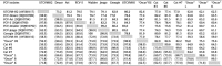 |
OS: oral swab; L: liver; B: brain; K: kidney.
For better insight into the variability of the hypervariable region E, the deduced amino acids sequences were compared. Accumulation of the amino acid substitutions occurred between residues 427–481 (Fig. 2 ), i.e. positions 427, 430, 436, 439, 440, 441, 443, 448, 449, 450, 456, 460 and 471, were variable among viruses, with distinct mutations allowing distinction between the FCV isolated from the asymptomatic cats #3, #6, #7 and the FCVs detected in the clinically affected cat “Oscar”. Prediction of the glycosylation pattern identified one glycosylation site at residues Asn-459 in all isolates (Fig. 2).
Fig. 2.

Protein alignment of the amino acid residues 427–481 of the hypervariable region E of the capsid protein from different VS-FCV strains from the Genbank database and from the current study, compared to the vaccine strain F9. M86379 vaccine strain F9; AY560116: UTCVM-H1; AY560117: UTCVM-H2; DQ910789: FCV-Deuce; DQ910790: FCV-5; DQ910791: Georgie DQ910792 Georgie Walder; DQ910793: Jengo; DQ910794: Ari; DQ910795: Kaos. OS:oral swab; L: liver; B: brain; K: kidney; T: tongue; I: Intestine. Amino acids that differed from VS-FCV and FCV detected in healthy cats are marked. The predicted glycosylation site is underlined.
To better understand the evolutionary relationships between FCV strains isolated in our outbreak and those isolated in other parts of the world, a phylogenetic analysis was carried out. A robust, well-supported phylogenetic tree was identified (Fig. 3 ). This showed that all FCV isolates from the tissues of the cat “Oscar” segregate between them, forming a distinct sub-group. These viruses are different from the FCV isolated from “Oscar’s” oropharyngeal swab which forms a separate lineage within the subgroup. Furthermore, the FCV strains recovered from asymptomatic cats in the same shelter fall into a distinct subgroup with other FCV reference strains (Fig. 3). A comparison with published sequences from different hypervirulent FCV strains did not identify any relationship between viruses detected in “Oscar” and FCV strains isolated during other VSD outbreaks.
Fig. 3.

Phylogenetic tree constructed with the partial ORF2 amino acid sequences generated in this study and FCV sequences from the GenBank database. GenBank accession numbers of the reference strains are shown in brackets. Only one arbitrary sequence from each group of identical sequences was included. Bootstrap (1000 replicates) values >70 are indicated at the internal nodes. In bold: VSD-FCV reference strains and sequences obtained from “Oscar”. Rabbit haemorrhagic disease virus (RHDV) was used as outgroup to root the tree. The scale bar to the left of the tree indicates the estimated number of amino acid substitution per site.
4. Discussion and conclusion
The present case represents the first FCV-associated virulent systemic disease (FCV-VSD) reported in Italy. The disease observed in “Oscar” closely resembles that seen in outbreaks of haemorrhagic systemic fever caused by VS-FCV reported in several parts of the world (Pedersen et al., 2000, Schorr-Evans et al., 2003, Hurley et al., 2004, Coyne et al., 2006, Reynolds et al., 2009, Meyer et al., 2011). The severe clinical course is typical and the pathological findings are within the reported scope. Immunohistology confirmed the viral tropism for parenchymal epithelial cells and for endothelial cells. Damage to these cells in various organs can be regarded as the cause for the range of changes observed in the present case: ulcerative glossitis, liver necrosis with haemoperitoneum and multifocal haemorrhage. Calicivirus infection was definitively confirmed by virus isolation and real-time PCR not only in the diseased cat “Oscar”, but also in asymptomatic cats that lived in direct contact with him, confirming the presence of FCV in the shelter and its maintenance in the environment by carriers cats (Wardley, 1976).
FCV-VSD predominantly occurs as a highly contagious disease, affecting a large number of animals living in direct contacts (Pesavento et al., 2004, Reynolds et al., 2009), but a recent report showed that FCV-VSD is not necessarily associated with high morbidity (Meyer et al., 2011). At present, however, the cause for such variability in the clinical courses in different outbreaks is unknown. We also report a single case of FCV-VSD in a cat kept in a highly crowded environment, in which the diseased animal was found to be carrier of a different strain of FCV.
It is generally speculated that genetic differences between FCV strains of low and high virulence account for the presence of local mild FCV infection or systemic infection in affected cats. Mutations within the viral genome may be responsible for the highly virulent phenotype. Since the FCV strains isolated in each outbreak of VSD were genetically distinct from each other, if viral mutations are required to cause the hyper-virulent phenotype, they must evolve independently in each outbreak. To our knowledge, no specific genetic markers for an increase in virulence have been identified, although previous reports showed that sequence differences in HVR_E lead to the acquisition of an additional glycosylation site in some VSD isolates (Foley et al., 2006, Abd-Eldaim et al., 2005). Changes in the glycosylation pattern of viral proteins could affect the host-virus interactions and represent a mechanism for immune evasion, as reported for other viruses, such as influenza virus (Vigerust and Sheperd, 2007). In the present study, we could identify only one additional glycosylation site in the 5′ HVR_E sequences, in the Asn-459 residue, in all FCV isolates, but observed a high diversity in the 5′ HVR_E region, which allowed the distinction between FCV isolated from the cat with VSD and asymptomatic cats in the present study. Although these mutations do not affect the viral glycosylation pattern, they might modify the antigenic profile of the HVR_E. The capsid protein of FCV is an important target for the immune response and molecular studies have mapped antigenic epitopes located in the HVR_E (Radford et al., 1999, Geissler et al., 2002). Since these amino acid differences occurred in a putatively neutralising region of HVR_E, the underlying mutations might represent a mechanism for FCV immune evasion and possibly also differences in viral pathogenicity (Henzel et al., 2012). The results of our phylogenetic analyses showed that clustering of reference viruses occurs independent of clinical signs; in particular VS-FCV associated with haemorrhagic disease are spread over different lineages of the phylogenetic tree. This result support the hypothesis of Hurley et al. (2004) who suggested that VSD can be associated with a range of FCV genotypes.
The present study identified a clear genetic divergence between FCV from asymptomatic cats, viruses isolated from the tissues of the diseased cat and the virus isolated from an oropharyngeal swab of the same cat. This finding suggests that FCV of variable virulence were circulating simultaneously in the cattery. The results of our study also confirm that each VS-FCV outbreak could be caused by a distinct, individual FCV strain (Radford et al., 2007).
Cats suffer from a range of infections with viruses that have acquired a particularly opportunistic character and are associated with persistent, chronic and/or asymptomatic infections (Pedersen et al., 2004; Bannasch and Foley, 2005). The risk of infections is increased in multi-cat households, catteries and shelters, due to the generally close contact between animals; furthermore, immunodepression, i.e. due to chronic FIV infection, may support the generation and selection of virulent mutants and impair the host’s ability to combat the mutant viruses. A similar mechanism was observed in FIV-infected cats, since they developed FIP more frequently upon experimental infection with Feline Coronavirus (FCoV; Poland et al., 1996). In the case described here, the cat was also FIV infected and therefore immunosuppressed to some degree. This might have supported the development of the disease by allowing newly formed virulent FCV to replicate and spread systemically.
In conclusion, in analogy to FCoV infection and the development of FIP, FCV-VSD may preferentially develop in individual, persistently FCV infected cats due to host factors that allow the generation of FCV mutants with enhanced virulence.
Acknowledgements
The authors wish to thank Drs. Alan Radford and Carol Porter and Prof. Rosalind Gaskell, University of Liverpool, for providing the anti-FCV peptide antibody for diagnostic purposes.
References
- Abd-Eldaim M., Potgieter L., Kennedy M. Genetic analysis of feline caliciviruses associated with a hemorrhagic-like disease. Journal of Veterinary Diagnostic Investigation. 2005;17:420–429. doi: 10.1177/104063870501700503. [DOI] [PubMed] [Google Scholar]
- Bannasch M.J., Foley J.E. Epidemiological evaluation of multiple respiratory pathogens in cats in animal shelters. Journal of Feline Medicine and Surgery. 2005;7:109–119. doi: 10.1016/j.jfms.2004.07.004. [DOI] [PMC free article] [PubMed] [Google Scholar]
- Coyne K.P., Jones B.R., Kipar A., Chantrey J., Porter C.J., Barber P.J., Dawson S., Gaskell R.M., Radford A.D. Lethal outbreak of disease associated with feline calicivirus infection in cats. The Veterinary Record. 2006;158:544–550. doi: 10.1136/vr.158.16.544. [DOI] [PubMed] [Google Scholar]
- Ellis T.M. Jaundice in a siamese cat with in utero feline calicivirus infection. Australian Veterinary Journal. 1981;57:383–385. doi: 10.1111/j.1751-0813.1981.tb00527.x. [DOI] [PubMed] [Google Scholar]
- Felsenstein J. PHYLIP – Phylogeny Inference Package (Version 3.2) Cladistics. 1989;5:164–166. [Google Scholar]
- Foley J., Hurley K., Pesavento P.A., Poland A., Pedersen N.C. Virulent systemic feline calicivirus infection: local cytokine modulation and contribution of viral mutants. Journal of Feline Medicine and Surgery. 2006;8:55–61. doi: 10.1016/j.jfms.2005.08.002. [DOI] [PMC free article] [PubMed] [Google Scholar]
- Geissler K., Schneider K., Truyen U. Mapping neutralizing and non-neutralizing epitopes on the capsid protein of feline calicivirus. Journal of Veterinary Medicine Series B: Infectious Disease and Veterinary Public Health. 2002;49:55–60. doi: 10.1046/j.1439-0450.2002.00529.x. [DOI] [PubMed] [Google Scholar]
- Greene G.E., editor. Infectious Diseases of the Dog and Cat. Saunders Elsevier; 2006. pp. 145–154. [Google Scholar]
- Hall T.A. BIOEDIT: a user-friendly biological sequence alignment editor and analysis program for window 95/98/NT. Nucleic Acids Symposium Series. 1999;41:95–98. [Google Scholar]
- Henzel A., Sá e Silva M., Luo S., Lovato L.T., Weiblen R. Genetic and phylogenetic analyses of capsid protein gene in feline calo virus isolates from Rio Grande do Sul in southern brazil. Virus Research. 2012;163:667–671. doi: 10.1016/j.virusres.2011.12.008. [DOI] [PubMed] [Google Scholar]
- Helps C., Lait P., Tasker S., Harbour D. Melting curve analysis of feline calicivirus isolates detected by real-time reverse transcription PCR. Journal of Virological Methods. 2002;106:241–244. doi: 10.1016/s0166-0934(02)00167-2. [DOI] [PubMed] [Google Scholar]
- Hurley K.F., Pesavento P.A., Pedersen N.C., Poland A.M., Wilson E., Foley J. An outbreak of virulent systemic feline calicivirus disease. Journal of the American Veterinary Medical Association. 2004;224:241–249. doi: 10.2460/javma.2004.224.241. [DOI] [PubMed] [Google Scholar]
- Knowles J.O., McArdle F., Dawson S., Carter S.D., Gaskell C.J., Gaskell R.M. Studies on the role of feline calicivirus in chronic stomatitis in cats. Veterinary Microbiology. 1991;27:205–219. doi: 10.1016/0378-1135(91)90148-9. [DOI] [PubMed] [Google Scholar]
- Larkin M.A., Blackshields G., Brown N.P., Chenna R., McGettigan1 P.A., McWilliam H., Valentin F., Wallace I.M., Wilm A., Lopez R., Thompson J.D., Gibson T.J., Higgins D.G. Clustal W and clustal X version 2.0. Bioinformatics. 2007;23:2947–2948. doi: 10.1093/bioinformatics/btm404. [DOI] [PubMed] [Google Scholar]
- Love D.N., Baker K.D. Sudden death in kittens associated with a feline picornavirus. Australian Veterinary Journal. 1972;48:643. doi: 10.1111/j.1751-0813.1972.tb05105.x. [DOI] [PubMed] [Google Scholar]
- Meyer A., Kershaw O., Klopfleisch R. Feline calicivirus-associated virulent systemic disease: not necessarily a local epizootic problem. The Veterinary Record. 2011;168:589a. doi: 10.1136/vr.d160. [DOI] [PubMed] [Google Scholar]
- Page R.D. TreeView: an application to display phylogenetic trees on personal computers. Molecular Biology and Evolution. 1996;4:406–425. doi: 10.1093/bioinformatics/12.4.357. [DOI] [PubMed] [Google Scholar]
- Pedersen N.C., Elliott J.B., Glasgow A., Poland A., Keel K. An isolated epizootic of hemorrhagic-like fever in cats caused by a novel and highly virulent strain of feline calicivirus. Veterinary Microbiology. 2000;73:281–300. doi: 10.1016/S0378-1135(00)00183-8. [DOI] [PMC free article] [PubMed] [Google Scholar]
- Pedersen N.C., Sato R., Foley J.E., Poland A.M. Common virus infections in cats, before and after being placed in shelters, with emphasis on feline enteric coronavirus. Journal of Feline Medicine and Surgery. 2004;7:14–18. doi: 10.1016/j.jfms.2003.08.008. [DOI] [PMC free article] [PubMed] [Google Scholar]
- Pesavento P.A., Maclachlan N.J., Dillard-Telm L., Grant C.K., Hurley K.E. Pathologic, immunohistochemical, and electron microscopic findings in naturally occurring virulent systemic feline calicivirus infections in cats. Veterinary Pathology. 2004;41:257–263. doi: 10.1354/vp.41-3-257. [DOI] [PubMed] [Google Scholar]
- Poland A.M., Vennema H., Foley J.E., Pedersen N.C. Two related strains of feline infectious peritonitis virus isolated from immunocompromised cats infected with a feline enteric coronavirus. Journal of Clinical Microbiology. 1996;34:3180–3184. doi: 10.1128/jcm.34.12.3180-3184.1996. [DOI] [PMC free article] [PubMed] [Google Scholar]
- Povey R.C., Johnson R.H. A survey of feline viral rhinotracheitis and feline picornavirus infection in britain. Journal of Small Animal Practice. 1971;12:233–247. doi: 10.1111/j.1748-5827.1971.tb06226.x. [DOI] [PubMed] [Google Scholar]
- Radford A.D., Willoughby K., Dawson S., McCracken C., Gaskell R.M. The capsid gene of feline calicivirus contains linear B-cll epitopes in both variable and conserved regions. Journal of Virology. 1999;73:8496–8502. doi: 10.1128/jvi.73.10.8496-8502.1999. [DOI] [PMC free article] [PubMed] [Google Scholar]
- Radford A.D., Coyne K.P., Dawson S., Porter C.J., Gaskell R.M. Feline calicivirus. Veterinary Research. 2007;38(2):319–335. doi: 10.1051/vetres:2006056. [DOI] [PubMed] [Google Scholar]
- Reynolds B.S., Poulet H., Pingret J.-L., Jas D., Brunet S., Lemeter C., Etievant M., Boucraut-Baralon C. A nosocomial outbreak of feline calicivirus associated virulent systemic disease in france. Journal of Feline Medicine and Surgery. 2009;11:633–644. doi: 10.1016/j.jfms.2008.12.005. [DOI] [PMC free article] [PubMed] [Google Scholar]
- Schorr-Evans E.M., Poland A., Johnson W.E., Pedersen N.C. An epizootic of highly virulent feline calicivirus disease in a hospital setting in New England. Journal of Feline Medicine and Surgery. 2003;5:217–226. doi: 10.1016/S1098-612X(03)00008-1. [DOI] [PMC free article] [PubMed] [Google Scholar]
- Vigerust D.J., Sheperd V.L. Virus glycosylation: role in virulence and immune interqactions. Trends in Microbiology. 2007;15(5):211–218. doi: 10.1016/j.tim.2007.03.003. [DOI] [PMC free article] [PubMed] [Google Scholar]
- Vögtlin C., Fraefel S., Albini S., Leutenegger C.M., Schraner E., Spiess B., Lutz H., Ackermann M. Quantification of feline herpesvirus 1 DNA in ocular fluid samples of clinically diseased cats by real-time TaqMan PCR. Journal of Clinical Microbiology. 2002;40:519–523. doi: 10.1128/JCM.40.2.519-523.2002. [DOI] [PMC free article] [PubMed] [Google Scholar]
- Wardley R.C. Feline calicivirus carrier state: a study of the host/virus relationship. Archives of Virology. 1976;52:243–249. doi: 10.1007/BF01348021. [DOI] [PubMed] [Google Scholar]
- Wardley R.C., Gaskell R.M., Povey R.C. Feline respiratory viruses-their prevalence in clinically healthy cats. Journal of Small Animal Practice. 1974;15:579–586. doi: 10.1111/j.1748-5827.1974.tb06538.x. [DOI] [PubMed] [Google Scholar]
- Xia X., Xie Z. DAMBE: software package for data analysis in molecular biology and evolution. Journal of Heredity. 2001;92:371–373. doi: 10.1093/jhered/92.4.371. [DOI] [PubMed] [Google Scholar]