

Abstract
Background
The patterns of corticosteroids usage in severe acute respiratory syndrome (SARS) and associated treatment outcomes in Hong Kong were studied.
Method
Patients ≥ 18 years old who either had not received corticosteroid or had taken corticosteroids within 14 days from symptom onset were included. Patients receiving corticosteroids beyond 15 days or other investigational treatment within 21 days from symptom onset were excluded. Of 1313 eligible patients, 1287 with major corticosteroid dosage-type combinations were analysed.
Results
Crude death rate was lower among 1188 steroid-treated patients compared to 99 patients in Group No Steroid (17.0% vs. 28.3%). Among four corticosteroid groups studied, mortality was lowest in the low-dose oral prednisolone (Group P) and high-dose methylprednisolone (Group MP) groups. On multivariate analysis of the corticosteroid groups, independent factors related to death were: corticosteroid group, older age, co-morbidity, worse chest X-ray score, worse respiratory status at Days 8–10 and higher admission white cell count. Again Groups P and MP had significantly lower adjusted odds ratios for death and lower bacterial and fungal culture rates. Despite worse chest X-ray scores and higher cumulative corticosteroid dosages in Group MP compared to Group P, fewer patients required rescue pulsed corticosteroid. Patients on hydrocortisone (Group HC) had the highest positive culture rates.
Conclusion
We speculate that corticosteroid with higher in-vitro inflammatory potency administered at timing and dosages commensurate with disease severity may be conducive to better outcome from SARS as a consequence of more effective control of immunopathological lung damage.
Keywords: Corticosteroids, Severe acute respiratory syndrome, Treatment
Introduction
The role of corticosteroids in the treatment of severe acute respiratory syndrome (SARS) is controversial. In Hong Kong, corticosteroids were widely used because of its reported efficacy in mainland China,1 and because four of five initial SARS patients died with best supportive treatment alone without corticosteroid. Survival rates in Hong Kong had improved with combined corticosteroids and ribavirin treatment in subsequent patients.2, 3, 4, 5 Methylprednisolone in “pulsed” dosages (at ≥500 mg per dose) was also frequently added to the baseline corticosteroid regimen on further deterioration or recrudescence of symptoms and signs.1, 2, 3, 4, 5, 6
SARS has been proposed to be a triphasic disease, commencing with viral replication, followed by immunopathological damage and finally pulmonary destruction.6 Advocates of corticosteroids aimed at controlling the immunopathological phase of SARS5, 7 and suppressing elevated cytokines levels.8 Opponents contended that a self-limiting course had been observed in SARS even with significant initial chest radiograph involvement,9, 10 and that corticosteroids only increased susceptibility to secondary infections.11, 12
The Hong Kong Hospital Authority SARS Collaborative Group (HASCOG) tracked information of all probable and confirmed Hong Kong SARS patients as defined by the World Health Organisation (WHO) criteria.13 In the following study, we aimed to study the patterns of corticosteroids usage and the associated treatment outcomes.
Materials and methods
Study design and subjects
Clinical information of all SARS patients was retrospectively collected from May to October 2003, with enhancement by the HA Clinical Management System (CMS) which provided on-line laboratory and medication information. The following patients were included: (i) ≥18 years of age and still hospitalised for SARS after 22 March 2003, when discovery of the novel coronavirus (CoV) was announced in Hong Kong; and either (ii) had not been given corticosteroids throughout the course of hospitalisation; or (iii) had received corticosteroid within 0–14 days of symptom onset. All included patients had to fulfil the latest case definition for SARS.13 Patients were excluded if they: (i) had been discharged or had died before 22 March 2003, when lack of knowledge had resulted in variations in management; (ii) had corticosteroids started beyond 15 days after symptom onset; or (iii) had received other investigational treatment within 0 to 21 days of symptom onset, including lopinavir/ritonavir combination, immunoglobulin, convalescent plasma, anti-tumour necrosis factor-α and traditional Chinese medicine.
Data collection
Demographic data, serial laboratory test results, intensive care unit admission, requirement for pulsed methylprednisolone rescue and assisted ventilation, microbiological findings including bacterial, viral and fungal cultures, antibiotics usage and final outcomes were recorded. Presence of co-morbidities were defined by illnesses with possible adverse impacts on patient outcome, namely, (i) diabetes with documented end-organ complications (vascular, cardiac, neurological, renal); (ii) chronic obstructive pulmonary disease (excluding chronic asthma); and (iii) other chronic diseases requiring long-term treatment (cardiac, pulmonary, neurological, renal, liver). Because of laboratory variations in reference ranges of lactate dehydrogenase (LDH), an LDH ratio, obtained by dividing measured values by the respective upper limits of normal range, was used for comparison.
Microbiology
Positive cultures of microorganisms including bacteria and fungi were documented. Positive laboratory confirmation of SARS-CoV infection was defined as two or more positive real-time reverse transcriptase–polymerase chain reaction (RT–PCR) for SARS-CoV in nasopharyngeal aspirate, nasopharyngeal swab or stool, or positive RT–PCR of one specimen confirmed by two laboratories, or four-fold rise in IgG against SARS-CoV in paired acute-convalescent sera or four-fold of threshold IgG in single sera. The WHO criteria in identifying laboratory-confirmed cases were adopted.13
Corticosteroid regimens
To study the effect of initial dosages on the subsequent disease course, predominant types of corticosteroid employed were defined as those that contributed to at least two-thirds of the total dosage during the first four days of treatment, excluding dosages due to pulsed MP rescue. Subsequent analysis was based on the following corticosteroid types: intravenous hydrocortisone (Group HC), intravenous methylprednisolone (Group MP), oral prednisolone (Group P), or intravenous pulsed corticosteroid (Group Pulse). “Pulsed corticosteroid” is defined as intravenous methylprednisolone administered at 500 mg or more per dose for at least 1 day from the day of corticosteroid commencement. “Rescue pulse” is defined as intravenous methylprednisolone administered at 500 mg or more per dose for at least 1 day started after at least 1 day of corticosteroid treatment. Patients who had not received any corticosteroids were designated ‘Group No Steroid’, within which patients who had and had not received ribavirin were identified. Patients with no major types of corticosteroids were excluded from all analyses, and were defined as receiving more than one type of corticosteroid (excluding pulsed methylprednisolone), with total dosage of any type not reaching at least two-thirds of the total corticosteroid dosage in the first 4 days after steroid commencement. To reflect anti-inflammatory effects, all steroid types were converted to hydrocortisone-equivalent based on relative glucocorticoid activities in the following ratios: 4 mg methylprednisolone = 5 mg prednisolone = 20 mg hydrocortisone.
Scoring of chest radiographs
Chest radiographs were scored using a “0–4” scoring system to indicate the geographical extent of radiographical opacities: score 0 = 0%, 1 = 1–25%, 2 = 26–50%, 3 = 51–75%, 4 = 76–100%. Total score ranged from 0 to 24 for all six lung zones (lower/middle/upper in both left and right lungs), with higher scores denoting more extensive chest X-ray (CXR) involvement. The scoring method was standardised and performed by radiologists from individual hospitals.
Respiratory status
The PaO2/FiO2 (P/F) ratio, defined in this study as the worst arterial partial pressure of oxygen (PaO2) divided by the fractional inspired oxygen concentration (FiO2) on any particular day was used as a reflection of the patients' respiratory status. Where PaO2 values were not available, they were estimated from oxygen saturation values obtained by pulse oximetry (SpO2) using the oxygen dissociation curve, with the help of an equation which relates SpO2 to PaO2 at standard temperature (37 °C).14 Respiratory status was categorised according to the worst P/F ratio on any day: 0–26.5 kPa (0–199 mmHg) being “acute respiratory distress syndrome (ARDS)” and 26.6–39.9 kPa (200–299 mmHg) being “acute lung injury (ALI)”.15 Higher values were arbitrarily divided into “mild respiratory impairment” (RI, 40.0–53.2 kPa or 300–399 mmHg) and normal (53.3–62.0 kPa or 400–465 mmHg). Since SARS-related acute respiratory failure (ARF) usually peaks by Day 8 after symptom onset,6 patient outcome was studied according to their respiratory status on Days 8–10 from symptom onset.16, 17
Outcomes
The primary outcome studied was the survival benefits of different treatment groups. To study the potentially life-threatening infectious complications of corticosteroid with possible impact on outcome, results of bacterial and fungal cultures together with treatment for fungal infection and tuberculosis were recorded as secondary outcomes. Since ribavirin was used in 1219 SARS patients in this study (93.9%), the final analysis was performed only on patients who had ever received ribavirin in order to remove confounding effect of ribavirin, if any.
Ethics
Ethical committee approval was considered not necessary because direct patient intervention was not involved and individual patient data were not presented.
Statistical analysis
Data are presented in mean ± SD or median (interquartile range [IQR]). Two-sided Kruskal–Wallis test or chi-squared test was used for univariate comparisons of demographic and clinical parameters among the groups where appropriate. Multivariate logistic regression analyses were performed by backward selection of variables showing a P value of <0.20 in the univariate analysis by Wilcoxon test (with normal approximation) or chi-squared test to predict the effect of various corticosteroid groups on survival after adjusting for possible confounding factors. The selected model gave the adjusted ratios of odds of death and their 95% confidence intervals for individual corticosteroid groups relative to a chosen reference group after controlling for all identified confounding factors. Using demographic variables and clinical parameters on admission for model entry, the first multivariate model studied the survival outcome of various corticosteroid groups over the Group No Steroid. Further analyses were performed using clinical parameters at steroid commencement rather than on admission to compare among the steroid groups their survival rates. In these further models, the corticosteroid group showing the highest survival benefit over the Group No Steroid in the first multivariate model was selected as the reference group. In all analyses a P value of <0.05 was considered statistically significant. All statistical analyses were done using SAS version 8.02.
Results
During the 2003 outbreak in Hong Kong, a total of 1755 probable SARS patients (WHO criteria)13 had been treated in 14 acute general hospitals under HA. As per defined exclusion criteria, 468 patients were excluded for reasons shown in Table 1, resulting in a final cohort of major corticosteroid groups consisting of 1287 patients.
Table 1.
Description of inclusions and exclusions of patients in the SARS database in analyses on the effectiveness of corticosteroid treatment of SARS
 |
TCM, traditional Chinese medicine; CS, convalescent serum; PG, Pentaglobin (IgM enriched immunoglobulin).
The demographic and clinical parameters of these 1287 patients from 14 acute hospitals in Hong Kong are shown in Table 2. The majority of patients, totalling 1188 (92.3%), had received corticosteroid. Two hospitals had used centre-specific treatment protocols3, 5 on most of their patients throughout the outbreak, in which the dosages and types of corticosteroids used were clearly defined, while the other 12 hospitals had utilised two or more corticosteroid regimens. Patients were grouped based on the commonest corticosteroid types used in 1188 patients, in order of frequency, as follows: hydrocortisone (Group HC), methylprednisolone (Group MP), pulsed methylprednisolone (Group Pulse), and prednisolone (Group P). Only 99 patients received no steroid (Group No Steroid) and were distributed in 13 hospitals. A step-down strategy in corticosteroid dosages was observed in all steroid treatment groups unless deterioration occurred which called for the use of pulsed methylprednisolone rescue. Details of individual patients in the Group No Steriod were not retrieved, but to our knowledge some patients had minor disease diagnosed only on or after discharge (in particular in the early part of the outbreak before the virus was identified and diagnostic tests developed), some presented late in the course of the outbreak with atypical symptoms (such as diarrhoea), while some physicians did not believe in administering corticosteroid in this unknown disease. All baseline characteristics and clinical parameters on admission were statistically different among the groups (Table 2). In particular, Group P consisted of the youngest patients with the largest number of health care workers (47%) and the mildest disease on admission, as evidenced by the lowest median CXR score, normal respiratory status and absence of ARDS. Group MP had the highest median CXR score on admission. The two groups with the highest prevalence of ARDS on admission (8.3% and 8.2% for Groups Pulse and No Steroid, respectively) also had the highest mortality rates (30% and 28.3%, respectively). Overall, crude death rates were lowest for Group P and Group MP.
Table 2.
Demographic and clinical parameters of all major corticosteroid groups (N = 1287) at hospital admission, bacteriology and final outcomes
| Group |
||||||
|---|---|---|---|---|---|---|
| No steroid (N = 99) | P (N = 170) | HC (N = 621) | MP (N = 177) | Pulse (N = 220) | P value | |
| Demographic and clinical parameters | ||||||
| Median age (IQR) (years) | 61 (40–81) | 34 (26–47) | 40 (31–52) | 39 (29–48) | 46.5 (32–70) | <0.0001 |
| Gender female, N (%) | 48 (48.5) | 108 (63.5) | 369 (59.4) | 102 (57.6) | 110 (50.0) | 0.0191 |
| Comorbidity, N (%) | 43 (43.4) | 16 (9.4) | 72(11.6) | 18 (10.2) | 61 (27.7) | <0.0001 |
| Positive lab confirmation, N (%) | 53 (53.5) | 156 (91.8) | 538 (86.6) | 147 (83.1) | 168 (76.4) | <0.0001 |
| Health care workers, N (%) | 6 (6.1) | 80 (47.1) | 99 (15.9) | 42 (23.7) | 44 (20.0) | <0.0001 |
| At hospital admission | ||||||
| Respiratory status on admission | 85 | 163 | 591 | 172 | 206 | |
| Normal, N (%) | 30 (35.3) | 81 (49.7) | 208 (35.2) | 80 (46.5) | 62 (30.1) | <0.0001 |
| RI, N (%) | 35 (41.2) | 73 (44.8) | 313 (53.0) | 73 (42.4) | 108(52.4) | |
| ALI, N (%) | 13 (15.3) | 9 (5.5) | 44 (7.5) | 13 (7.6) | 19 (9.2) | |
| ARDS, N (%) | 7 (8.2) | 0 (0) | 26 (4.4) | 6 (3.5) | 17 (8.3) | |
| WBC (109/L) | 7.15 | 5.21 | 5.55 | 5.35 | 5.90 | <0.0001 |
| median (IQR) | (5.43–10.35) | (3.90–6.30) | (4.20–6.90) | (4.25–7.10) | (4.40–7.69) | |
| N | 96 | 166 | 604 | 176 | 210 | |
| Neutrophil (109/L) | 5.60 | 3.50 | 4.07 | 3.85 | 4.35 | <0.0001 |
| Median (IQR) | (3.53–8.61) | (2.60–4.84) | (2.90–5.50) | (2.97–5.40) | (3.10–6.32) | |
| N | 90 | 165 | 595 | 170 | 204 | |
| LDH ratio | 0.93 | 0.92 | 0.97 | 1.03 | 1.22 | <0.0001 |
| Median (IQR) | (0.77–1.23) | (0.78–1.17) | (0.80–1.29) | (0.76–1.43) | (0.92–1.66) | |
| N | 69 | 115 | 481 | 138 | 137 | |
| CXR scorea | 2 | 1 | 2 | 3 | 2 | <0.0001 |
| Median (IQR) | (1–7) | (0–2) | (1–4) | (1–6) | (1–5) | |
| N | 90 | 162 | 569 | 151 | 196 | |
| Bacteriology | ||||||
| Positive fungal culture and ever received antifungal treatment, N (%) | 0 (0.0) | 0 (0) | 32 (5.2) | 3 (1.7) | 7(3.2) | <0.0001 |
| New cases of tuberculosis, N (%) | 2 (2) | 2 (1.2) | 4 (0.6) | 1 (0.6) | 1 (0.5) | 0.4675 |
| Final outcomes | ||||||
| ICU care, N (%) | 4 (4.0) | 6 (3.5) | 159 (25.6) | 30 (17.0) | 48 (21.8) | <0.0001 |
| Intubation, N (%) | 4 (4.0) | 0 (0) | 117 (18.8) | 12 (6.8) | 32 (14.6) | <0.0001 |
| Death, N (%) | 28 (28.3) | 8 (4.7) | 111 (17.9) | 17 (9.6) | 66 (30.0) | <0.0001 |
IQR, Interquartile range.
CXR score (range 0 to 24); a higher score denotes worse CXR.
Table 3a–c shows the details of 1188 patients who had received corticosteroids. Table 3a shows that general characteristics were significantly different amongst the groups. Steroid commencement was earliest in Group HC and latest in Group Pulse [median Days 4 (IQR 3–6) and 6 (IQR 4–8), respectively, after symptom onset]. Among the groups, Group MP had the lowest proportion of patients requiring pulsed corticosteroid rescue (41% compared to 53–100% in the other groups), which was also given at the latest time point (median Day 9 after symptom onset, compared to Days 6–8 in the other groups). The total cumulative corticosteroid dosage was highest in Group Pulse, while duration of steroid treatment was shortest in Group P (median 15 days) and longest in Group MP (median 21 days), the latter of which was dictated by protocol.3 Table 3b shows the patients' clinical parameters at corticosteroid commencement. Group P still had the mildest disease. Comparatively, Group MP and Group Pulse had the worst clinical status as evidenced by the highest percentages of patients with ARDS (9.4% and 18.1%, respectively) and the highest median CXR score [4 (IQR 2–7) and 5 (IQR 2–9), respectively]. Table 3c shows the clinical parameters at pulsed corticosteroid commencement. Group P consistently had the mildest disease, with the lowest median CXR score and only 12.4% of patients in either ALI or ARDS. In contrast, patients in Group MP had the worst clinical status with the highest median CXR score, with 45.9% having either ALI or ARDS.
Table 3.
Details of corticosteroid usage (N = 1188)
| Characteristics | Group |
P value | |||
|---|---|---|---|---|---|
| P (N = 170) | HC (N = 621) | MP (N = 177) | Pulse (N = 220) | ||
| (a) General characteristics | |||||
| Days from symptom onset median (IQR) | 5 (3–7) | 4 (3–6) | 5 (3–7) | 6 (4–8) | <0.0001 |
| Days of steroid treatment | 15 | 19 | 21 | 19 | <0.0001 |
| Median (IQR) | (13–18) | (14–24) | (18–24) | (12–23) | |
| Cumulative dosages in hospital | 6400 | 12150 | 11040 | 16430 | <0.0001 |
| Median (IQR) | (2300–9640) | (5100–22800) | (8040–21440) | (10305–24310) | |
| Total cumulative steroid dosage | 7020 | 13200 | 11350 | 17560 | |
| Median (IQR) | (2820–12380) | (5280–23880) | (8420–22140) | (10650–25380) | <0.0001 |
| Pulsed steroid given, N (%) | 90 (53) | 372 (60) | 72 (41) | 220 (100) | <0.0001 |
| Days of pulsed steroid initiation from symptom onset | 8 | 8 | 9 | 6 | <0.0001 |
| Median (IQR) | (7–10) | (6–11) | (7–12) | (4–8) | |
| (b) Clinical parameters at corticosteroid commencement | |||||
| Respiratory status on steroid start, N | 165 | 599 | 170 | 216 | |
| Normal, N (%) | 70 (42.4) | 177 (29.6) | 60 (35.3) | 46 (21.3) | <0.0001 |
| RI, N (%) | 83 (50.3) | 319 (53.3) | 79 (46.5) | 98 (45.4) | |
| ALI, N (%) | 10 (6.1) | 59 (9.9) | 15 (8.8) | 33 (15.3) | |
| ARDS, N (%) | 2 (1.2) | 44 (7.4) | 16 (9.4) | 39 (18.1) | |
| WBC (109/L) | 4.55 | 5.30 | 5.13 | 5.51 | 0.0004 |
| Median (IQR) | (3.33–5.99) | (4.00–6.80) | (4.06–6.80) | (4.00–8.10) | |
| N | 164 | 595 | 166 | 215 | |
| Neutrophil (109/L) | 3.10 | 4.00 | 3.70 | 4.26 | <0.0001 |
| Median (IQR) | (2.25–4.34) | (2.77–5.40) | (2.70–5.47) | (2.63–6.97) | |
| N | 164 | 592 | 161 | 212 | |
| LDH ratio | 0.94 | 1.03 | 1.14 | 1.49 | <0.0001 |
| Median (IQR) | (0.82–1.20) | (0.83–1.46) | (0.77–1.55) | (1.08–2.04) | |
| N | 90 | 421 | 131 | 137 | |
| CXR score | 2 | 2 | 4 | 5 | <0.0001 |
| Median (IQR) | (1–3) | (1–5) | (2–7) | (2–9) | |
| N | 154 | 564 | 159 | 188 | |
| (c) Clinical parameters on pulsed corticosteroid commencement | |||||
| Number of patients with Pulsed steroid given | 90 | 372 | 72 | 220 | |
| Respiratory status on pulsed steroid start, N | 89 | 363 | 72 | 216 | |
| Normal, N (%) | 34 (38.2) | 90 (24.8) | 13 (18.1) | 46 (21.3) | 0.0002 |
| RI, N (%) | 44 (49.4) | 154 (42.4) | 26 (36.1) | 98 (45.4) | |
| ALI, N (%) | 9 (10.1) | 58 (16.0) | 11 (15.3) | 33 (15.3) | |
| ARDS, N (%) | 2 (2.3) | 61 (16.8) | 22 (30.6) | 39 (18.1) | |
| WBC (109/L) | 5.30 | 8.67 | 9.26 | 5.51 | <0.0001 |
| Median (IQR) | (3.70–7.60) | (6.10–12.10) | (6.50–14.00) | (4.00–8.10) | |
| N | 89 | 350 | 70 | 215 | |
| Neutrophil (109/L) | 4.20 | 7.42 | 7.91 | 4.26 | <0.0001 |
| Median (IQR) | (2.70–6.17) | (5.00–11.10) | (5.60–13.00) | (2.63–6.97) | |
| N | 89 | 349 | 70 | 212 | |
| LDH ratio | 1.01 | 1.16 | 1.64 | 1.49 | <0.0001 |
| Median (IQR) | (0.85–1.38) | (0.90–1.63) | (1.17–2.46) | (1.08–2.04) | |
| N | 41 | 151 | 58 | 137 | |
| CXR score | 2 | 5.5 | 7.5 | 5 | <0.0001 |
| Median (IQR) | (1–4) | (3–10) | (3–14) | (2–9) | |
| N | 89 | 328 | 62 | 188 | |
To remove possible confounding effects due to ribavirin, 68 patients not having been treated with ribavirin (50 from Group No Steroid, 11 from Group HC, 1 from Group MP, 5 from Group P and 1 from Group Pulse) were excluded from further analyses of the final outcomes. Among the 99 patients in Group No Steroid, comparison between the 50 patients who had not and the 49 who had received ribavirin by univariate analysis failed to show any significant difference in demographics and all other characteristics including co-morbidity, percentage with laboratory confirmation and percentage of health care workers, admission parameters (respiratory status, white cell count, neutrophils, LDH, CXR scores), as well as major outcomes (data not shown). Univariate comparisons of the characteristics between survivors and fatal cases among 1219 patients ( Table 4) indicated that age, sex, co-morbidity, worst respiratory status at Days 8 to 10, worst X-ray score, LDH ratio, WBC and neutrophil counts on admission and corticosteroid treatment reached statistical significance (P < 0.05).
Table 4.
Univariate analysis of the characteristics between alive and dead patients who had had been treated with both corticosteroid and ribavirin (N = 1219)
| Variable | No. of patient data available for analysis | Survivor, N (Mean ± SD or %) | Death, N (Mean ± SD or %) | P value |
|---|---|---|---|---|
| Age | 1219 | 1006 | 213 | <0.0001 |
| (% 18–44) | 69.0% | 16.4% | ||
| (% 45–64) | 23.3% | 21.1% | ||
| (% 65+) | 7.7% | 62.5% | ||
| Sex (% female) | 1219 | 1006 | 213 | <0.0001 |
| 60.5% | 42.7% | |||
| Comorbidity (% yes) | 1219 | 1006 | 213 | <0.0001 |
| 8.2% | 47.9% | |||
| Worst respiratory status at day 8–10 (%RF)a | 1155 | 959 | 196 | <0.0001 |
| 23.7% | 79.1% | |||
| LDH ratiob | 887 | 743 | 144 | <0.0001 |
| 1.13 ± 0.57 | 1.49 ± 0.92 | |||
| WBCb (×109/L) | 1186 | 995 | 191 | <0.0001 |
| 5.73 ± 2.61 | 7.86 ± 4.22 | |||
| Lymphocyteb (×109/L) | 1162 | 979 | 183 | 0.2103 |
| 0.90 ± 0.48 | 1.04 ± 0.73 | |||
| Plateletb (×109/L) | 1186 | 995 | 191 | 0.2118 |
| 178.6 ± 68.3 | 188.5 ± 81.9 | |||
| Neutrophilb (×109/L) | 1162 | 979 | 183 | <0.0001 |
| 4.32 ± 2.50 | 6.04 ± 3.30 | |||
| Worst X-ray (% 7–24) | 1115 | 918 | 197 | <0.0001 |
| 44.2% | 92.9% | |||
| Ever treated with steroid (% yes) | 1219 | 1006 | 213 | 0.0012 |
| 96.8% | 92.0% |
RF, respiratory failure (ALI or ARDS).
On admission.
In 1032 patients with complete data for multivariate analysis, the aggregate effect of corticosteroid usage compared with Group No steroid on survival was insignificant after controlling for age, sex, co-morbidity, highest CXR score, worst respiratory status at Days 8–10 and WBC on admission.
Table 5a,b shows the multivariate models for the ratios of odds of deaths for individual corticosteroid groups after controlling for identified confounding factors. Table 5a shows that, when individual corticosteroid groups were studied, only Group MP demonstrated survival benefit compared with Group No steroid (Adjusted OR 0.25, 95% CI 0.07–0.90, P = 0.0345). Table 5b shows that Group MP as the reference group demonstrated survival benefits over Group HC (Adjusted OR 3.77, 95% CI 1.73–8.23, P = 0.0009) and Group Pulse (adjusted OR 2.76, 95% CI 1.18–6.41, P = 0.0186) but not Group P (adjusted OR 0.67, 95% CI 0.15–2.93, P = 0.5904).
Table 5.
Multiple logistic regression analysis for the odds of death in relation to different corticosteroid groups among patients who had been treated with ribavirin
| Effect | Reference | OR | 95% CI | P value | |
|---|---|---|---|---|---|
| (a) Logistic model for odds of Death in 1032 subjects with breakdown into different steroid types (163 deaths and 869 survivors) | |||||
| Major steroid group | HC vs. No Steroid | 1.00 | 0.33 | 3.07 | 0.9984 |
| MP vs. No Steroid | 0.25 | 0.07 | 0.90 | 0.0345 | |
| P vs. No Steroid | 0.17 | 0.03 | 1.05 | 0.0560 | |
| Pulse vs. No Steroid | 0.70 | 0.22 | 2.22 | 0.5431 | |
| Age | 45–64 vs. 18–44 | 1.72 | 0.98 | 3.02 | 0.0578 |
| 65+ vs. 18–44 | 8.83 | 4.80 | 16.24 | <0.0001 | |
| Sex | Male vs. Female | 1.60 | 1.01 | 2.54 | 0.0451 |
| Comorbidity | Yes vs. No | 4.52 | 2.47 | 8.27 | <0.0001 |
| Worst X-ray score | 7–24 vs. 0–6 | 14.17 | 6.08 | 33.00 | <0.0001 |
| Worst respiratory status at day 8–10 | RF vs. Normal/RI | 4.21 | 2.56 | 6.93 | <0.0001 |
| WBC on admission | per 109/L increase | 1.09 | 1.02 | 1.17 | 0.0127 |
| (b) Logistic model for odds of Death in 1044 subjects using Group MP as the reference group in steroid groupings (176 deaths and 868 survivors) | |||||
| Major steroid group | HC vs. MP | 3.77 | 1.73 | 8.23 | 0.0009 |
| P vs. MP | 0.67 | 0.15 | 2.93 | 0.5904 | |
| Pulse vs. MP | 2.76 | 1.18 | 6.41 | 0.0186 | |
| Age | 45–64 vs. 18–44 | 2.08 | 1.21 | 3.57 | 0.0077 |
| 65+ vs. 18–44 | 10.85 | 6.02 | 19.56 | <0.0001 | |
| Sex | Male vs. Female | 1.57 | 1.01 | 2.46 | 0.0471 |
| Comorbidity | Yes vs. No | 4.91 | 2.71 | 8.90 | <0.0001 |
| Worst X-ray score | 7–24 vs. 0–6 | 23.03 | 10.02 | 52.95 | <0.0001 |
| Respiratory status on steroid start | RF vs. Normal/RI | 2.04 | 1.27 | 3.28 | 0.0033 |
Tabe 5(a). c-index = 0.923; Hosmer and Lemeshow Goodness-of-Fit test P value = 0.7850. Table 5(b). c-index = 0.923; Hosmer and Lemeshow Goodness-of-Fit test P value = 0.8960.
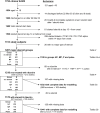
Fig. 1 shows that Group P had the lowest percentage of survivors who had positive bacterial cultures throughout hospitalisation. Group MP, which had received the highest total dosage of corticosteroid (Table 3a), showed slightly higher rates after Days 1–7, and both these groups also had the highest survival rates. Groups HC, No Steroid and Pulse had comparatively higher culture rates after Day 22 and higher mortality rates. Group HC had the highest positive culture and antifungal treatment rates while Groups P and MP had similar low rates. New cases of tuberculosis were uncommon, and again the rate was highest for Group HC.
Figure 1.

Percentage of survivors with positive bacterial cultures in all corticosteroid groups at different time points during hospitalisation.
Discussion
The present study is the largest comprehensive review to date of the use of corticosteroids in SARS treatment. On multivariate analysis, corticosteroid use as a whole did not show survival benefit compared with no steroid use. However, when individual corticosteroid types were analysed, Group MP (intravenous methylprednisolone) conferred lower mortality compared with Group No Steroid, which is statistically significant. Among the corticosteroid groups, Group MP and Group P (oral prednisolone) showed similar survival outcome.
These results should be interpreted with caution in the light of its limitations of being a retrospective study without control groups. Disparities of baseline characteristics could have been secondary to selection bias, i.e. the choice of corticosteroid use and dosage might have been related to severity of illness or the treatment protocols adopted by individual hospitals. We thus observed that steroid types and dosages had varied from protocol-driven low-dose prednisolone,4 high-dose MP,3 and even pulsed corticosteroid on commencement of treatment.5 Although corticosteroids and ribavirin were usually given together, the effect of the latter was likely small, if any.18, 19, 20 Predominant corticosteroid type was classified according to their use in the first four days, but subsequent use including rescue pulsed doses and the total dose and duration administered could also have confounded the final conclusion. We believe that the cytokine storm in the initial days of the SARS illness has the most critical effect on the manifestations of the disease8, 21 and that an appropriate level of immunomodulation at this stage may have the most impact on the subsequent course of the illness.8, 22 We have therefore arbitrarily classified our patients as described, which was also the most reasonable since the majority of patients given corticosteroid (1188, or 97.9%) could be classified. Also, PaO2 and FiO2 could only be estimated in most instances based on assumptions, nevertheless, the same rules were applied to every patient. Finally, while we have already excluded patients with experimental therapy (especially antivirals like lopinavir/ritonavir which may have beneficial effects23) and those with ‘do not resuscitate’ orders, it is still possible for the outcome to be affected by different modes of subsequent management, including the choice of ventilatory strategy.
Our results suggest that SARS patients given prednisolone and methylprednisolone had fared better than those given hydrocortisone. We speculate that this may be related to the comparatively higher anti-inflammatory potency of the former two drugs as demonstrated in in vitro studies.24, 25 We observed that corticosteroid dosages employed for SARS in Hong Kong had been diverse, ranging from low dose regimens4, 26 through dosages commonly employed in treating acute asthmatic attacks1, 6, 27 and higher dosages triggered by surrogate markers of clinical deterioration,3, 28 to pulsed MP dosages for treatment of acute autoimmune pulmonary vasculitis.5 At first sight, it does appear intriguing that both low-dose (P) and high-dose (MP) groups should result in similarly low mortality rates in our study. On detailed analysis, however, it appeared that dosages had been tailored in accordance with disease severity: Group P (prednisolone) with the mildest clinical and radiographic parameters had received lower dosages of corticosteroid on steroid commencement, whereas Group MP with worse clinical status received higher initial dosages. The relative good outcomes of MP and P groups could be related to their lower cumulative doses compared to Groups HC and Pulse and hence more modest immunosuppressive effects. However, it must be pointed out that Group P had the mildest disease throughout hospital stay while Group MP had much more severe illness. Hence low cumulative dosages should not be the major reason for the better outcomes of these two groups. We speculate, therefore, that corticosteroid administered at dosages commensurate to disease severity may be conducive to effectively controlling immunopathological lung damage. In addition, our data do not support the contention that higher corticosteroids dosages as treatment for SARS would necessarily lead to an infection-prone immunocompromised state, because the high-dose protocol-driven3 Group MP (methylprednisolone) had higher (worse) median CXR score on corticosteroid commencement but displayed low mortality rate and the lowest proportionate requirement for pulsed MP rescue. It also turned out to have consistently lower rates of positive bacterial and fungal cultures as well as anti-fungal treatment. That high dose methylprednisolone was more beneficial than no or lower dosages of corticosteroid has also been shown in a comparative study from mainland China.29 As only two hospitals had used specific treatment protocols throughout the outbreak, while the other 12 had applied different regimens to different patients at different phases of the outbreak, definite conclusions about the effects of corticosteroids are difficult even though we have adjusted for most baseline differences using multivariate analysis.
In most reported series in the literature, corticosteroids had been started to treat SARS at variable but usually much earlier time points. These included: as soon as the diagnosis of SARS was made and without response to antibiotics treatment within 48 h;1 on confirming an epidemiological link or contact history;5, 30 or on finding lung involvement on high-resolution CT thorax alone despite clear chest radiographs.5, 6, 30 Such early corticosteroid administration strategies might have led to the drug being given too early in the viral replicative phase, which may result in aggravation and/or prolongation of viral replication and thus less satisfactory outcome.5, 6, 30, 31, 32 We therefore submit that the low mortality and morbidity in Group MP may have resulted from effective control of severe disease by appropriately high dosages of corticosteroid with high anti-inflammatory activity given at a strategic timing as per protocol3 to coincide with the period of high SARS-related immunopathological activity. This strategy would allow the smooth tailing down of corticosteroid dosages to avoid disease rebound as well as the risk of secondary infections during the subsequent phase of immunoparesis.3, 28 In contrast, Group HC, which also received high cumulative corticosteroid dosages and had high proportions of patients requiring pulsed MP rescue, displayed the highest positivity for bacterial, fungal and tuberculosis infections. We postulate that comparatively weaker anti-inflammatory effects of hydrocortisone24, 25 in these groups might have resulted in incomplete control of SARS and thus persistent elevation of cytokines at an earlier stage, which in turn may promote bacterial proliferation.7, 33 In the same context, high cumulative corticosteroid dosages in the later stages of hospitalisation resulting from increased need for rescue dosages could also have contributed to excessive secondary infections among patients receiving hydrocortisone. Cytokine assays have not been specifically performed for the purpose of this study.
Although definitive answers to the best timing, dosage and duration of corticosteroid administration can only be obtained in large-scale randomised placebo-controlled trial, in which a potentially effective antiviral agent is supplemented with corticosteroids as secondary or rescue treatment, our analyses do suggest that appropriate severity-adjusted corticosteroid dosages, especially high-dose methylprednisolone commenced only on overt respiratory deterioration in the more severe cases, were associated with better outcome in the management of severe acute respiratory syndrome.
Acknowledgements
The authors acknowledge the funding support for the HA SARS Database on data collection and management from the Research Fund for the Control of Infectious Diseases (RFCID) of Health, Welfare and Food Bureau of the Government of the Hong Kong Special Administrative Region, People's Republic of China. We thank members of the Hong Kong Hospital Authority SARS Collaborative Group (HASCOG) for contribution to data collection and permission to perform this study. Contributions from the following radiologists to chest radiograph scoring are also acknowledged: Dr Greg Antonio, Dr C.C. Chan, Dr Eddie Chan, Dr James Griffith, Dr C.M. Lee, Dr Lilian Leong, Dr T.H. Siu, Dr K.W. Tang, Dr Wong Yick, and Dr M.K. Yuen. We would also like to thank Ms Vivian Man for secretarial assistance and our health care workers for their dedication to the care for SARS patients in Hong Kong.
References
- 1.Zhong N.S., Zeng G.Q. Our strategies for fighting severe acute respiratory syndrome (SARS) Am J Respir Crit Care Med. 2003;168:7–9. doi: 10.1164/rccm.200305-707OE. [DOI] [PubMed] [Google Scholar]
- 2.Choi K.W., Chau T.N., Tsang O., Tso E., Chiu M.C., Tong W.L. Outcomes and prognostic factors in 267 patients with severe acute respiratory syndrome in Hong Kong. Ann Intern Med. 2003;139:715–723. doi: 10.7326/0003-4819-139-9-200311040-00005. [DOI] [PubMed] [Google Scholar]
- 3.So L.K.Y., Lau A.C.W., Yam L.Y.C., Cheung T.M., Poon E., Yung R.W. Development of a standard treatment protocol for severe acute respiratory syndrome. Lancet. 2003;361:1615–1617. doi: 10.1016/S0140-6736(03)13265-5. [DOI] [PMC free article] [PubMed] [Google Scholar]
- 4.Lee N., Hui D., Wu A., Chan P., Cameron P., Joynt G.M. A major outbreak of severe acute respiratory syndrome in Hong Kong. N Engl J Med. 2003;348:1986–1994. doi: 10.1056/NEJMoa030685. [DOI] [PubMed] [Google Scholar]
- 5.Ho J.C., Ooi G.C., Mok T.Y., Chan J.W., Hung I., Lam B. High dose pulse versus non-pulse corticosteroid regimens in severe acute respiratory syndrome. Am J Respir Crit Care Med. 2003;168:1449–1456. doi: 10.1164/rccm.200306-766OC. [DOI] [PubMed] [Google Scholar]
- 6.Peiris J.S.M., Chu C.M., Cheng V.C., Chan K.S., Hung I.F., Poon L.L. Clinical progression and viral load in a community outbreak of coronavirus-associated SARS pneumonia: a prospective study. Lancet. 2003;361:1767–1772. doi: 10.1016/S0140-6736(03)13412-5. [DOI] [PMC free article] [PubMed] [Google Scholar]
- 7.Lau A.C., So L.K. Severe acute respiratory syndrome treatment: present status and future strategy. Curr Opin Investig Drugs. 2003;4:918–920. [PubMed] [Google Scholar]
- 8.Wong C.K., Lam C.W., Wu A.K., Ip W.K., Lee N.L., Chan I.H. Plasma inflammatory cytokines and chemokines in severe acute respiratory syndrome. Clin Exp Immunol. 2004;136:95–103. doi: 10.1111/j.1365-2249.2004.02415.x. [DOI] [PMC free article] [PubMed] [Google Scholar]
- 9.Wong R.S., Hui D.S. Index patient and SARS outbreak in Hong Kong. Emerg Infect Dis. 2004;10:339–341. doi: 10.3201/eid1002.030645. [DOI] [PMC free article] [PubMed] [Google Scholar]
- 10.Hsu L.Y., Lee C.C., Green J.A., Ang B., Paton N.I., Lee L. Severe acute respiratory syndrome (SARS) in Singapore: clinical features of index patient and initial contacts. Emerg Infect Dis. 2003;9:713–717. doi: 10.3201/eid0906.030264. [DOI] [PMC free article] [PubMed] [Google Scholar]
- 11.Li X.W., Jiang R.M., Guo J.Z. Glucocorticoid in the treatment of severe acute respiratory syndrome patients: a preliminary report. Chin J Intern Med. 2003;42:378–381. [PubMed] [Google Scholar]
- 12.Wang H., Ding Y., Li X., Yang L., Zhang W., Kang W. Fatal aspergillosis in a patient with SARS who was treated with corticosteroids. N Engl J Med. 2003;349:507–508. doi: 10.1056/NEJM200307313490519. [DOI] [PubMed] [Google Scholar]
- 13.World Health Organisation Case definitions for surveillance of severe acute respiratory syndrome (SARS) 2003. http://www.who.int/csr/sars/casedefinition revised May 1. [accessed July 8, 2004]
- 14.Severinghaus J.W., Stafford M., Thunstrom A.M. Estimation of skin metabolism and blood flow with tcPO2 and tcPO2 electrodes by cuff occlusion of the circulation. Acta Anaesthesiol Scand Suppl. 1978;68:9–15. doi: 10.1111/j.1399-6576.1978.tb01386.x. [DOI] [PubMed] [Google Scholar]
- 15.Bernard G.R., Artigas A., Brigham K.L., Carlet J., Falke K., Hudson L. The American–European Consensus Conference on ARDS. Definitions, mechanisms, relevant outcomes, and clinical trial coordination. Am J Respir Crit Care Med. 1994;149(3 Pt 1):818–824. doi: 10.1164/ajrccm.149.3.7509706. [DOI] [PubMed] [Google Scholar]
- 16.Fowler R.A., Lapinsky S.E., Hallet D., Detsky A.S., Sibbald W.J., Slutsky A.S. Critically ill patients with severe acute respiratory distress syndrome. JAMA. 2003;290:363–373. doi: 10.1001/jama.290.3.367. [DOI] [PubMed] [Google Scholar]
- 17.Lew T.W., Kwek T.K., Tai D., Earnest A., Loo S., Singh K. Acute respiratory distress syndrome in critically ill patients with severe acute respiratory distress syndrome. JAMA. 2003;290:374–380. doi: 10.1001/jama.290.3.374. [DOI] [PubMed] [Google Scholar]
- 18.Cinatl J., Morgenstern B., Bauer G., Chandra P., Rabenau H., Doerr H.W. Glycyrrhizin, an active component of liquorice roots, and replication of SARS-associated coronavirus. Lancet. 2003;361:2045–2046. doi: 10.1016/S0140-6736(03)13615-X. [DOI] [PMC free article] [PubMed] [Google Scholar]
- 19.Chen F., Chan K.H., Jiang Y., Kao R.Y., Lu H.T., Fan K.W. In vitro susceptibility of 10 clinical isolates of SARS coronavirus to selected antiviral compound. J Clin Virol. 2004;31:69–75. doi: 10.1016/j.jcv.2004.03.003. [DOI] [PMC free article] [PubMed] [Google Scholar]
- 20.Morgenstern B., Michaelis M., Baer P.C., Doerr H.W., Cinatl J., Jr. Ribavirin and interferon-beta synergistically inhibit SARS-associated coronavirus replication in animal and human cell lines. Biochem Biophys Res Commun. 2005;326:905–908. doi: 10.1016/j.bbrc.2004.11.128. [DOI] [PMC free article] [PubMed] [Google Scholar]
- 21.Jiang Y., Xu J., Zhou C., Wu Z., Zhong S., Liu J. Characterization of cytokine/chemokine profiles of severe acute respiratory syndrome. Am J Respir Crit Care Med. 2005;17:850–857. doi: 10.1164/rccm.200407-857OC. [DOI] [PubMed] [Google Scholar]
- 22.Beijing Group of National Research Project of SARS Dynamic changes in blood cytokine levels as clinical indicators in severe actue respiratory syndrome. Chin Med J (Engl) 2003;116:1283–1287. [PubMed] [Google Scholar]
- 23.Chan K.S., Lai S.T., Chu C.M., Tsui E., Tam C.Y., Wong M.M. Treatment of severe acute respiratory syndrome with lopinavir/ritonavir: a multicentre retrospective matched cohort study. Hong Kong Med J. December 2003;9(6):399–406. [PubMed] [Google Scholar]
- 24.Langhoff E., Ladefoged J., Dickmeiss E. The immunosuppressive potency of various steroids on peripheral blood lymphocytes, T cells, NK and K cells. Int J Immunopharmacol. 1985;7:483–489. doi: 10.1016/0192-0561(85)90067-0. [DOI] [PubMed] [Google Scholar]
- 25.Langhoff E., Ladefoged J. Relative immunosuppressive potency of various corticosteroids measured in vitro. Eur J Clin Pharmacol. 1983;25:459–462. doi: 10.1007/BF00542111. [DOI] [PubMed] [Google Scholar]
- 26.Sung J.J., Wu A., Joynt G.M., Yuen K.Y., Lee N., Chan P.K. Severe acute respiratory syndrome: report of treatment and outcome after a major outbreak. Thorax. 2004;59:414–420. doi: 10.1136/thx.2003.014076. [DOI] [PMC free article] [PubMed] [Google Scholar]
- 27.Chan J.W., Ng C.K., Chan Y.H., Mok T.Y., Lee S., Chu S.Y. Short term outcome and risk factors for adverse clinical outcomes in adults with severe acute respiratory syndrome (SARS) Thorax. 2003;58:686–689. doi: 10.1136/thorax.58.8.686. [DOI] [PMC free article] [PubMed] [Google Scholar]
- 28.Lau A.C., So L.K., Miu F.P., Yung R.W., Poon E., Cheung T.M. Outcome of coronavirus-associated severe acute respiratory syndrome using a standard treatment protocol. Respirology. 2004;9:173–183. doi: 10.1111/j.1440-1843.2004.00588.x. [DOI] [PMC free article] [PubMed] [Google Scholar]
- 29.Zhao Z., Zhang F., Xu M., Huang K., Zhong W., Cai W. Description and clinical treatment of an early outbreak of severe acute respiratory syndrome (SARS) in Guangzhou, PR China. J Med Microbiol. 2003;52(Pt 8):715–720. doi: 10.1099/jmm.0.05320-0. [DOI] [PubMed] [Google Scholar]
- 30.Tsui P.T., Kwok M.L., Yuen H., Lai S.T. Severe acute respiratory syndrome: clinical outcome and prognostic correlates. Emerg Infect Dis. 2003;9:1064–1069. doi: 10.3201/eid0909.030362. [DOI] [PMC free article] [PubMed] [Google Scholar]
- 31.Tsang O.T., Chau T.N., Choi K.W., Tso E.Y., Lim W., Chiu M.C. Coronavirus-positive nasopharyngeal aspirate as predictor for severe acute respiratory syndrome mortality. Emerg Infect Dis. 2003;9:1381–1387. doi: 10.3201/eid0911.030400. [DOI] [PMC free article] [PubMed] [Google Scholar]
- 32.Lee N., Allen Chan K.C., Hui D.S., Ng E.K., Wu A., Chiu R.W. Effects of early corticosteroid treatment on plasma SARS-associated coronavirus RNA concentration in adult patients. J Clin Virol. 2004;31:304–309. doi: 10.1016/j.jcv.2004.07.006. [DOI] [PMC free article] [PubMed] [Google Scholar]
- 33.Meduri G.U., Kanangat S., Stefan J., Tolley E., Schaberg D. Cytokines IL-1beta, IL-6, and TNF-alpha enhance in vitro growth of bacteria. Am J Respir Crit Care Med. 1999;160:961–967. doi: 10.1164/ajrccm.160.3.9807080. [DOI] [PubMed] [Google Scholar]