

Highlights
► A one-step real time quantitative RT-PCR assay was developed to detect published Dugbe virus genomes of the Nairovirus genus. ► Primers and probes were designed to detect specific sequences on the most conserved regions of the S segment. ► The limit of detection was 10 copies per reaction improving of 3 log10 FFU/mL the conventional RT-PCR. ► The specificity of the primers and probe was confirmed with the closely related Nairoviruses CCHFV and Hazara virus. ► 498 ticks collected in the Republic of Chad were screened: one sample was found positive.
Keywords: Nairovirus, Dugbe virus, One step real-time RT-PCR, Screening tool, Tick vector, Epidemiology
Abstract
A one-step real time quantitative RT-PCR (qRT-PCR) assay was developed to detect all published Dugbe virus (DUGV) genomes of the Nairovirus genus. Primers and probes were designed to detect specific sequences on the most conserved regions of the S segment. The limit of detection of the assay was 10 copies per reaction which is an improvement of 3 log10 FFU/mL over the sensitivity of conventional RT-PCR. The specificity of the primers and probe was confirmed with the closely related Nairoviruses CCHFV and Hazara virus, and on the non-related viruses Coronavirus and Influenza A virus. This qRT-PCR assay was used to screen nucleic acids extracted from 498 ticks collected in the Republic of Chad. One sample was found positive suggesting that DUGV is present in this part of the world. The molecular assay developed in this study is sensitive, specific and rapid and can be used for research and epidemiological studies.
1. Introduction
Dugbe virus (DUGV), a member of the genus Nairovirus of the Bunyaviridae family, was first isolated in 1964 from the Amblyomma variegatum tick in Nigeria (Causey, 1970). Frequently detected in tick-borne virus surveys in Africa (Guilherme et al., 1996), DUGV is a tri-segmented single-stranded negative RNA enveloped virus and is considered endemic in arid regions (Burt et al., 1996). DUGV is also one of the most common tick-borne viruses found throughout Africa (David-West and Porterfield, 1974). For example, a study in Kenya documented frequent isolation of DUGV from ticks infesting market livestock (Sang et al., 2006). Among the 7 serogroups characterized in the Nairovirus genus (Elliott et al., 2000), DUGV belongs to the Nairobi sheep disease group which includes the Nairobi sheep disease virus, pathogenic to sheep and goats (Davies, 1997) and Kupe virus, the most closely related virus (Crabtree et al., 2009). The genetically and serologically related Crimean-Congo hemorrhagic fever (CCHF) group, which also includes Hazara virus (Begum et al., 1970), is the other group of importance because of the very high pathogenicity of CCHF virus in humans, leading to fatal hemorrhagic disease in 30% of patients (Swanepoel et al., 1987). In contrast, the pathogenicity of DUGV is relatively low in humans – a thrombocytopenia case was once described (Burt et al., 1996) – but in newborn mice the virus is neuroinvasive and lethal (Boyd et al., 2006).
Detection of most Nairovirus is currently dependant on the time consuming virus isolation in cell culture or intra-cerebral inoculation in newborn mice from infected samples. An ELISA has also been established (Burt et al., 1996) as well as a conventional RT-PCR (Ward et al., 1990) for the detection of DUGV. To date, this conventional RT-PCR method alone has been used for the study of DUGV replication in supernatants, clinical samples or for epidemiological studies (Bridgen et al., 2002, Marriott et al., 1992, Marriott and Nuttall, 1996, Sang et al., 2006).
The aim of this study was to develop a sensitive, specific and rapid one-step quantitative real time RT-PCR assay (qRT-PCR) to detect DUGV in infected cell supernatants, ticks or serum samples.
2. Materials and methods
2.1. qRT-PCR development
2.1.1. Virus preparation, titration and RNA extraction
DUGV (IbH 11480), CCHF (IbAr 10200), Hazara (JC280), Coronavirus (ATCC VR-740), Influenza A (ATCC VR-547), were used in this study. All infectious work was carried out in a biosafety-level 2 and 4. The batches of viral inocula used in this study were prepared by 2 passages in BHK21 cells (ATCC CCL-10) grown at +37 °C, 5% CO2 in DMEM medium (Invitrogen, Karlsruhe, Germany), supplemented with 10% fetal calf serum (FCS) (PAN, Biotech, GmbH, Aidenbach, Germany), 5 × 10⁴ U Penicillin and 50 mg Streptomycin (Invitrogen, Karlsruhe, Germany). Briefly, cells were incubated for 1 h at +37 °C, 5% CO2 in the presence of the initial virus inoculum at a multiplicity of infection (MOI) of 0,1 in DMEM medium without FCS. Medium containing 5% of FCS was then added and cells were maintained for 3 days. Absence of mycoplasma was confirmed using the Mycoalert kit (Lonza, Verviers, Belgium).
Cell culture supernatants were centrifuged (5 min, 3000 × g, +4 °C), aliquoted and frozen (−80 °C) until used for viral RNA extraction. Viral titers, expressed in focus forming units per ml (FFU/mL) were estimated by the immunoperoxydase assay on Vero cells seeded in a flat bottom 12-well plate (BD, Franklin Lakes, NJ, USA), according to the dilution methods reported previously using an in-house hyperimmunized mouse ascitic fluid specific to DUGV (Peyrefitte et al., 2010). All viral RNAs were extracted from 200 μL of cell supernatants using the QIAmp Viral RNA Mini Kit (Qiagen, Courtaboeuf, France) according to the manufacturers’ instructions. To evaluate the influence of the serum, virus from infected cells supernatant was serially diluted in human serum (Invitrogen, Karlsruhe, Germany).
2.1.2. DUGV primers and probe
Sequences retrieved from GenBank database (accession numbers: AF434165, AF434164, AF434163, AF434162, and AF434161) were aligned using Clustal W 1.7. Software (Thompson et al., 1994) to select the most genetically conserved regions. Primers and probe (Table 1 ) were chosen in the highest conserved S segment region and according to the specifications needed for a real-time RT-PCR detection system (Bustin, 2000). The aligned sequences were identical in the region used for the primers and probe design. The specific DUGV probe was labeled at the 5′-end with the reporter 6-carboxy-fluorescein (FAM) and at the 3′-end with the quencher 6-carboxytetrametyl-rhodamine (TAMRA) (Eurogentec S.A., Herstal, France).
Table 1.
Primers and probe used for qRT-PCR of DUGV RNA.
| Name | 5′ → 3′ sequence | Genome positiona |
|---|---|---|
| F-DUGV | CTGGCTCAAGCAGTGGAACT | 904–923 |
| S-DUGV | bCACAAGGAGCACAAATAGACAc | 950–970 |
| R-DUGV | AGAGGAATTGAGACAAAGTGA | 1028–1048 |
According to the S fragment of the DUGV isolate IbH 11480, Genbank accession number AF434163.
Reporter dye (FAM).
Quencher dye (TAMRA).
2.1.3. qRT-PCR conditions
The qRT-PCR was performed in a final volume of 25 μL, containing 5 μL of extracted RNA, 5 μL of 5× one step RT-PCR buffer (Qiagen), 1 μL of dNTP 10 mM, 3 pmol of the probe, 15 pmol of each primer (Eurogentec S.A., Herstal, France), 8 U of RNase inhibitor (RNasin, Promega) and 2 μL of the one step hot start enzyme mix (Qiagen). Assays were carried out in a Biorad® CFX96 instrument with the following steps: reverse transcription at +50 °C for 30 min, hot start and denaturation at +95 °C for 15 min, and 45 cycles with +94 °C for 30 s, +53 °C for 30 s, +72 °C for 30 s, followed by a +30 °C for 30 s cooling step.
2.1.4. Generation of DUGV RNA synthetic transcripts
In the conditions described in Section 2.1.3, except for the thermocycler used (GeneAmp PCR System 9700, Applied Biosystems, Courtaboeuf, Villebon sur Yvette, France) and the absence of probe, a RT-PCR product was obtained using DUGV isolate IbH 11480 (Genbank accession number AF434163). The 145 bp amplicon was cloned into the T3 polymerase expression vector pBluescript II SK (±) (Stratagene, CA, USA). The plasmid was digested with the XhoI endonuclease (Roche, Heidelberg, Germany), then purified and in vitro transcribed using the Kit-Ribomax™ Large Scale RNA Production System T3 (Promega, WI, USA). Residual DNA was removed by DNAse I treatments (Roche). The RNA was quantified using the Genesys 10 Bio spectrophotometer (Thermo Electron Corporation, OH, USA). Absence of residual DNA was assessed by PCR using the Expand High Fidelity Enzyme mix (Roche).
The quality of the synthetic DUGV transcripts was evaluated by controlling the absence of residual DNA. No amplification was observed when synthetic RNA was subjected to PCR, showing no DNA contamination. The RNA was quantified on the spectrophotometer, then converted to copy number based on base composition and length of the RNA fragment.
2.2. Sensitivity, specificity and reproducibility of the qRT-PCR DUGV assay
The sensitivity and reproducibility of the qRT-PCR DUGV assay was determined using 10-fold dilutions of in vitro transcribed DUGV (105 to 100 copies per reaction) in triplicate. After a 10-fold serial dilution of titrated virus supernatant, RNA was extracted and tested in triplicate (2.6 × 10⁴ to 2.6 × 10⁻² FFU/mL). The specificity was evaluated by using RNA extracted from supernatants from CCHFV, Hazara virus, Coronavirus and Influenza A virus infected cells.
To mimic infected human sera, viral supernatants were 10-fold diluted in human serum (Sigma–Aldrich, Saint Quentin Fallavier, France).
To compare the qRT-PCR we used the conventional RT-PCR described by Ward et al. (1990).
2.3. Field study
Total RNA was extracted from 498 ticks collected from zebu and sheep, in the slaughter house of N’Djamena, in Chad in 2006. The attached ticks were pulled off manually and placed in sterile plastic tubes containing 70% camphorated ethanol, then stored at +4 °C, until identification. After a washing step in sterile PBS, ticks were identified to the species level by using the identification key (Walker et al., 2003). Each tick was individually triturated by using a Fast-Prep FP 120 cell disrupter (QBiogene, Illkirch, France) in 400 μL of sterile PBS. The mixture was centrifuged (5 min, 3000 × g, +4 °C), aliquoted and frozen (−80 °C) until used for viral RNA extraction.
3. Results
3.1. Development of the qRT-PCR DUGV assay
3.1.1. Assay sensitivity and specificity
The sequence of the DUGV transcript was confirmed by sequencing (data not shown).
The primers-probe set selected to detect potentially all DUGV known strains was able to detect the tested DUGV RNA. Optimal qRT-PCR conditions were determined using virus supernatant. The standard curve was obtained using the qRT-PCR amplification of the quantified RNA transcript. Results of the sensitivity and specificity of the qRT-PCR assay are summarized in Table 2 . The correlation coefficient of the standard curve ranged from 0.97 to 0.99. The limit of detection of the assay was 10 synthetic copies per reaction and 2.6 × 10⁻² FFU/mL of viral supernatant.
Table 2.
Sensitivity and specificity of the DUGV real-time RT-PCR assay.
| Sample | Quantity |
Assay |
||
|---|---|---|---|---|
| FFU/mL | RNA copies/assay | Cta | Result | |
| DUGV RNA transcriptb | ||||
| Dilution | ||||
| 10–8 | 105 | 22.49 ± 0.34 | Pos | |
| 10–9 | 104 | 26.18 ± 0.44 | Pos | |
| 10–10 | 103 | 29.79 ± 0.58 | Pos | |
| 10–11 | 102 | 33.24 ± 0.37 | Pos | |
| 10–12 | 101 | 36.84 ± 0.42 | Pos | |
| 10–13 | >41 | Neg | ||
| Titrated stock virusc | 2.6 × 10⁵ | |||
| Dilution | ||||
| 10–2 | 2.6 × 10³ | 1.2 × 10⁵ | 22.49 ± 0.46 | Pos |
| 10–3 | 2.6 × 10² | 1.6 × 10⁴ | 25.29 ± 0.52 | Pos |
| 10–4 | 2.6 × 101 | 1.2 × 10³ | 29.50 ± 0.32 | Pos |
| 10–5 | 2.6 × 100 | 9.6 × 101 | 33.11 ± 0.15 | Pos |
| 10–6 | 2.6 × 10⁻¹ | 1.0 × 101 | 36.78 ± 0.36 | Pos |
| 10–7 | 2.6 × 10⁻² | NDd | 40.02 ± 0.62 | Pos |
| 10–8 | >41 | Neg | ||
| Hazara virus | >107 | – | Neg | |
| CCHF virus | >107 | – | Neg | |
| Coronavirus | >107 | – | Neg | |
| Influenza A virus | >107 | – | Neg | |
Pos: Positive, Neg: Negative, and ND: not determined.
Cycle threshold (Ct) value obtained from 3 independent assays performed in duplicate are represented as mean ± standard deviation if greater than zero.
Ten-fold dilutions of the DUGV RNA transcripts.
Ten-fold dilutions of the DUGV stock (isolate IbH 11480, Genebank accession number AF434163).
Calculation of the initial RNA titre was not performed because the obtained Ct value was out of the standard curve range.
The specificity of the assay was evaluated by testing the closely related CCHFV and Hazara virus which were not detected. Kupe virus was not assayed because it was not in our virus collection. Despite the closest phylogenetical relationship between Kupe and Dugbe viruses, a nucleotide difference was observed when the primers and probe were aligned (GeneBank accession number EU257626). No cross reactivity was detected with the unrelated viruses Coronavirus and Influenza A virus (Table 2).
3.2. Evaluation of the qRT-PCR DUGV assay
The qRT-PCR assay was first compared with the conventional RT-PCR (Ward et al., 1990) using extracted viral RNA. The presented DUGV assay was 3 log10 FFU/mL more sensitive than the conventional RT-PCR (Fig. 1 ). The qRT-PCR assay detected as low as 10 RNA copies/assay while the conventional RT-PCR detected 5.5 × 10⁴ copies/assay, in our hands.
Fig. 1.
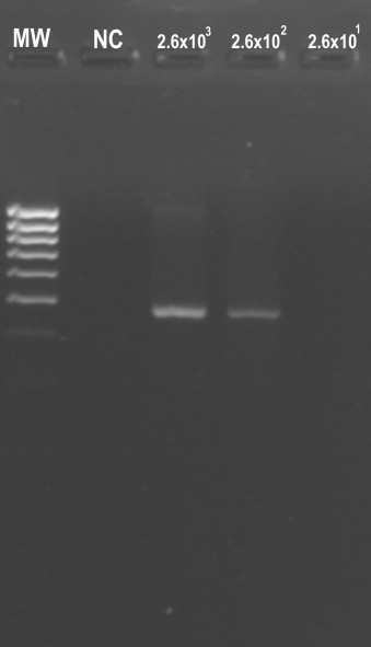
Sensitivity of the conventional RT-PCR using primers described by Ward et al. (1990). (MW) MassRuler™ low range DNA ladder. (NC) Negative control. Virus dilutions are expressed in FFU/mL.
Furthermore, influence of the serum before the nucleic acids extraction step, was evaluated in the assay sensitivity. No significant difference was observed between the qRT-PCR DUGV assay carried out with water or serum (data not shown). In our hands it took us ∼5 h to carry out the conventional RT-PCR, including gel migration, versus ∼2 h for the qRT-PCR.
3.3. Field study
Among the 498 captured ticks, one Dugbe virus RNA was detected in one tick using the qRT-PCR assay (0.2%). The Ct value was 40.6. The sequence of this amplicon confirmed the presence of DUGV genome. This DUGV genome was retrieved from a Hyalomma impeltatum female adult tick (Z-491) collected on a 3 year old male zebu.
4. Discussion
DUGV, a member of the Bunyaviridae family is detected frequently in tick-borne virus surveys in Africa (Guilherme et al., 1996), however little data are available in the literature that provides a comprehensive view of virus circulation. Thus, the detection of DUGV in field samples of arthropods and animal sera is needed to provide greater understanding of the virus epidemiology. Also, as described for other arboviruses, a rapid, specific and sensitive test is necessary for effective surveillance of new DUGV circulating strains. Currently DUGV identification is based on virus isolation. However, a conventional RT-PCR (Ward et al., 1990) and ELISAs (Burt et al., 1996, Ward et al., 1992) have also been described for specific detection of DUGV in samples. To date, RNA DUGV quantification is not available; therefore a qRT-PCR assay would be useful. The specificity of the assay was also important to discriminate the closely related DUGV, Hazara virus and CCHFV. Nevertheless, since Kupe virus was not sought, a genomic amplification of Kupe virus cannot be excluded. The 16 out of 62 nucleotide differences displayed when the primers and probe were aligned with the Kupe virus sequence suggested that they are not specific for Kupe virus. Of note the nucleotide difference was 18 out of 62 for both Hazara and CCHFV.
Furthermore, by comparison with the conventional RT-PCR (Ward et al., 1990), the DUGV real-time detection system described above was more sensitive (increasing the sensitivity of 3 log10 FFU/mL), rapid (approximately 2 h vs. 5 h) and minimized the potential for cross-contamination by using a one tube detection system. Our field study highlights the value of increased sensitivity because the single positive sample (Z-491) was detected at a Ct of 40.6. Such a low Ct would not have been detected using the conventional RT-PCR. Even if the value was out of the RNA in vitro transcript range, the amplicon amount was sufficient for sequencing. The prevalence of DUGV positive tick in our study (0.2%) was comparable to previous observations of tick pools obtained from Africa (Sang et al., 2006).
The detection of DUGV genome in Hyalomma impeltatum tick confirms the presence of DUGV in the Chad sub-region. Hyalomma ticks are known vectors of both DUGV (Booth et al., 1991) and CCHFV (Bridgen et al., 2002) suggesting the potential of tick-borne infection including CCHFV in slaughter houses in Africa.
In conclusion, a sensitive and specific qRT-PCR assay was developed to detect and quantify DUGV RNAs in infected cell supernatants, extracts from ticks and potentially sera. In this study the presence of DUGV genome in ticks captured in this region is described for the first time. The method described in this study might be useful for DUGV epidemiological survey in animals and humans.
Acknowledgements
This work was partly funded by IRBA and Fondation Mérieux. We thank M. Beassem, K. Matsumoto and J.-L. Marié for their helpful collaboration in this study. We also thank Jon Davis for reviewing the manuscript.
References
- Begum F., Wisseman C.L., Casals J. Tick-borne viruses of West Pakistan. II. Hazara virus, a new agent isolated from Ixodes redikorzevi ticks from the Kaghan Valley, W. Pakistan. Am. J. Epidemiol. 1970;92:192–194. doi: 10.1093/oxfordjournals.aje.a121197. [DOI] [PubMed] [Google Scholar]
- Booth T.F., Gould E.A., Nuttall P.A. Structure and morphogenesis of Dugbe virus (Bunyaviridae Nairovirus) studied by immunogold electron microscopy of ultrathin cryosection. Virus Res. 1991;21:1999–2212. doi: 10.1016/0168-1702(91)90033-r. [DOI] [PubMed] [Google Scholar]
- Boyd A., Fazakerley J.K., Bridgen A. Pathogenesis of Dugbe virus infection in wild-type and interferon-deficient mice. J. Gen. Virol. 2006;87:2005–2009. doi: 10.1099/vir.0.81767-0. [DOI] [PubMed] [Google Scholar]
- Bridgen A., Dalrymple D.A., Elliott R.M. Dugbe nairovirus S segment: correction of published sequence and comparison of five isolates. Virology. 2002;294:364–371. doi: 10.1006/viro.2001.1324. [DOI] [PubMed] [Google Scholar]
- Burt F.J., Spencer D.C., Leman P.A., Patterson B., Swanepoel R. Investigation of tick-borne viruses as pathogens of humans in South Africa and evidence of Dugbe virus infection in a patient with prolonged thrombocytopenia. Epidemiol. Infect. 1996;116:353–361. doi: 10.1017/s0950268800052687. [DOI] [PMC free article] [PubMed] [Google Scholar]
- Bustin S.A. Absolute quantification of mRNA using real-time reverse transcription polymerase chain reaction assays. J. Mol. Endocrinol. 2000;25:169–193. doi: 10.1677/jme.0.0250169. [DOI] [PubMed] [Google Scholar]
- Causey O.R. Supplement to the catalogue of Arthropod-borne viruses. Am. J. Trop. Med. Hyg. 1970;19:1123–1124. (No. 226) [Google Scholar]
- Crabtree M.B., Sang R., Miller B.R. Kupe virus, a new virus in the family bunyaviridae, genus nairovirus, Kenya. Emerg. Infect. Dis. 2009;15(2):147–154. doi: 10.3201/eid1502.080851. [DOI] [PMC free article] [PubMed] [Google Scholar]
- David-West T.S., Porterfield J.S. Dugbe virus: a tick-borne arbovirus from Nigeria. J. Gen. Virol. 1974;23:297–307. doi: 10.1099/0022-1317-23-3-297. [DOI] [PubMed] [Google Scholar]
- Davies F.G. Nairobi sheep disease. Parasitologia. 1997;39:95–98. [PubMed] [Google Scholar]
- Elliott R.M., Bouloy M., Calisher C.H., Goldbach R., Moyer J.T., Nichol S.T., Pettersson R., Plyuisnin A., Schmaljohn C.S. Bunyaviridae. In: van Regenmortel M.H.V., Fauquet C.M., Bishop D.H.L., Carstens E.B., Estes M.K., Lemon S.M., Maniloff J., Mayo M.A., McGeoch D.J., Pringle C.R., Wickner R.B., editors. Virus Taxonomy. Seventh Report of the International Commitee on Taxonomy of Viruses. Academic Press; San Diego: 2000. pp. 599–621. [Google Scholar]
- Guilherme J.M., Gonella-Legall C., Legall F., Nakoume E., Vincent J. Seroprevalence of five arboviruses in Zebu cattle in the Central African Republic. Trans. R. Soc. Trop. Med. Hyg. 1996;90:31–33. doi: 10.1016/s0035-9203(96)90468-x. [DOI] [PubMed] [Google Scholar]
- Marriott A.C., el-Ghorr A.A., Nuttall P.A. Dugbe nairovirus M RNA:nucleotide sequence and coding strategy. Virology. 1992;190:606–615. doi: 10.1016/0042-6822(92)90898-y. [DOI] [PubMed] [Google Scholar]
- Marriott A.C., Nuttall P.A. Large RNA segment of Dugbe nairovirus encodes the putative RNA polymerase. J. Gen. Virol. 1996;77:1775–1780. doi: 10.1099/0022-1317-77-8-1775. [DOI] [PubMed] [Google Scholar]
- Peyrefitte C.N., Perret M., Garcia S., Rodrigues R., Bagnaud A., Lacote S., Crance J.-M., Vernet G., Garin D., Bouloy M., Paranhos-Baccalà G. Differential activation profiles of Crimean-Congo hemorrhagic fever virus versus Dugbe virus infected antigen presenting cells. J. Gen. Virol. 2010;91:189–198. doi: 10.1099/vir.0.015701-0. [DOI] [PubMed] [Google Scholar]
- Sang R., Onyango C., Gachoya J., Mabinda E., Konongoi S., Ofula V., Dunster L., Okoth F., Coldren R., Tesh R., Travassos da Rosa A., Finkbeiner S., Wang D., Crabtree M., Miller B. Tickborne arbovirus surveillance in marquet livestock, Nairobi, Kenya. Emerg. Infect. Dis. 2006;12:1074–1080. doi: 10.3201/eid1207.060253. [DOI] [PMC free article] [PubMed] [Google Scholar]
- Swanepoel R., Shepherd A.J., Leman P.A., Shepherd S.P., McGillivray G.M., Erasmus M.J., Searle L.A., Gill D.E. Epidemiologic and clinical features of Crimean-Congo hemorrhagic fever in southern Africa. Am. J. Trop. Med. Hyg. 1987;36:120–132. doi: 10.4269/ajtmh.1987.36.120. [DOI] [PubMed] [Google Scholar]
- Thompson J.D., Higgings D.G., Gibson T.J. CLUSTALW: improving the sensitivity of progressive multiple sequence alignments through sequence weighting, position specific gap penalties and weight matrix choice. Nucleic Acids Res. 1994;22:4673–4680. doi: 10.1093/nar/22.22.4673. [DOI] [PMC free article] [PubMed] [Google Scholar]
- Walker A.R., Bouattour A., Camicas J.L., Estrada-Peña A., Horak I.G., Latif A.A., Pegram R.G., Preston P.M. The University of Edinburgh; Scotland, UK: 2003. Ticks of Domestic Animals in Africa: A Guide to Identification of Species. (Bioscience Reports) [Google Scholar]
- Ward V.K., Marriott A.C., Booth T.F., el-Ghorr A.A., Nuttall P.A. Detection of an arbovirus in an invertebrate and a vertebrate host using the polymerase chain reaction. J. Virol. Methods. 1990;30:291–300. doi: 10.1016/0166-0934(90)90071-m. [DOI] [PubMed] [Google Scholar]
- Ward V.K., Marriott A.C., Polyzoni T., el-Ghorr A.A., Antoniadis A., Nuttall P.A. Expression of the nucleocapsid protein of Dugbe virus and antigenic cross-reactions with other nairoviruses. Virus Res. 1992;24:223–229. doi: 10.1016/0168-1702(92)90009-x. [DOI] [PubMed] [Google Scholar]