

Abstract
Background Several types of fixation materials may be used for the radial styloid fractures such as Kirschner wire fixation, screw fixation, volar plate fixation, and fragment-specific radial buttress plate fixation. However, each of these fixation techniques has certain complications usually related to either the surgical dissection or the application of fixation and symptomatic permanent hardware. Implant removal secondary to irritation of prominent screw heads or bulky plates is not uncommon after radial styloid fracture fixation.
Case Description Herein, two patients with an isolated radial styloid fracture who were treated with bioabsorbable magnesium (alloy: MgYREZr) screws are presented. In both patients, the fracture union was achieved without any complication and need for implant removal.
Literature Review This is the first report on the use of magnesium screws for this indication.
Clinical Relevance Magnesium bioabsorbable compression screw fixation may be an alternative solution that eliminates removal operations due to symptomatic hardware in radial styloid fractures.
Keywords: magnesium, bioabsorbable, screw, biomaterial, distal radius fracture, radial styloid fracture
Distal radius fractures are the most common type of fracture that accounts for one-sixth of all emergency department visits. These fractures usually result from a fall onto an outstretched hand, and multiple fracture patterns may occur in respect to comminution, articular extension, associated soft tissue injuries, and radioulnar involvement. 1 Radial styloid fractures, also called “ chauffeur's fractures ,” are intra-articular fractures extending to the metaphysis, producing a triangular shape fragment. Although these fractures are quite innocent-looking fractures, they may be associated with severe ligamentous ruptures that result in carpal instability. 2
Nondisplaced fractures can be treated with plaster cast immobilization. However, because these fractures are intra-articular, surgical treatment is recommended in case of displacement, or articular incongruity greater than 2 mm. Several types of fixation materials may be used for the radial styloid fractures. Acceptable treatment modalities for isolated radial styloid fractures include Kirschner wire fixation, screw fixation, volar plate fixation, and fragment-specific radial buttress plate fixation. However, each of these techniques has some disadvantages and specific complications. Kirschner wire fixation is usually associated with pin tract infection, and they may need to remain in place until there is sufficient evidence of fracture healing. This condition may also result in joint stiffness because it prevents early rehabilitation. Plate fixation necessitates deep surgical dissection, and plate removal late after the fracture union is not uncommon. Therefore, screw fixation, preferably with a percutaneous or limited open technique, is generally advocated. But prominent screw heads may also cause tendon irritation, and/or tenosynovitis that eventually necessitate implant removal. 3
Bioabsorbable fixation materials (pins and screws) can be a solution to overcome all these disadvantages mentioned above and complications as they eliminate subsequent implant removal. Previously, polydioxanone (PDS) pins have been used for the fixation of distal radius fractures. Hoffmann et al reported the long-term results of 40 distal radius fracture cases treated with PDS pins. The authors concluded that these implants should not be preferred for this indication due to severe local foreign body reaction that negatively affects the final clinical outcome. 4 Today, the development of bioabsorbable materials used in orthopedic surgery continues with the introduction of new generation biomaterials such as magnesium (Mg) screws. Mg screw fixation has already begun to be used by the foot surgeons, especially in the fixation of corrective osteotomies for hallux valgus. In addition, there is a limited number of publications about their use in acute fractures. 5 In the current literature, there has been no report about the use of Mg screws in distal radius fractures. Herein, the clinical and radiological results of two patients with distal radius styloid fracture treated with bioabsorbable Mg screws were reported.
Case Report #1
A 29-year-old male motorcycle driver admitted to the emergency department with left wrist pain after a traffic accident. He was conscious, and there was no cranial, thoracic, and abdominal injury. On physical examination, there was swelling and tenderness over his left wrist, particularly over the radial styloid. Direct radiographic examination demonstrated a distal radial styloid fracture (AO classification type 23B1.1). Under brachial axillary block anesthesia, a gentle closed reduction was performed, and the fracture was fixed with two (2.7 mm Ø) Mg bioabsorbable headless compression screws (MAGNEZIX CS, Syntellix AG, Hannover, Germany) percutaneously through small incisions on the skin. Short-arm plaster splint was applied for 4 weeks after which the patient received 2 weeks of physiotherapy including passive and active wrist range of motion exercises. At the 2nd month follow-up, the patient was free of pain with normal wrist movements. There was a radiolucent zone around the screws at the 2nd month; however, this zone gradually disappeared and could not be seen at the 26th-month radiographic control. At the final follow-up 48 months after the initial injury, direct radiographs showed the complete union of the fracture and disintegration of the bioabsorbable screws ( Fig. 1 ). Computerized tomography (CT) examination showed no remaining radiolucency around the screws; furthermore, the density of the screws was equal to the cortical bone ( Fig. 2 ). The patient gained full range of wrist motion and strength without pain and turned back his previous level of activity and work.
Fig. 1.
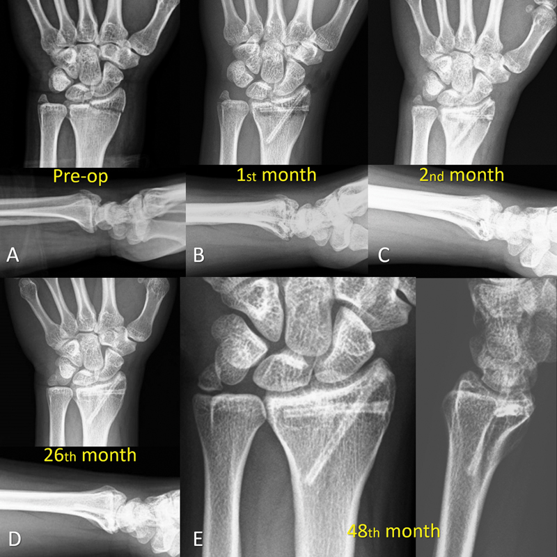
( A ) Anteroposterior and lateral wrist radiographs at initial admission showing the radial styloid fracture. ( B ) Fixation was achieved with two crossed magnesium screw. Radiographs at 2nd month ( C ) and 26th month ( D ) showed the union of the fracture with the gradual disappearance of gas around the screws. ( E ) Final follow-up at the 48th month.
Fig. 2.

( A ) Axial, ( B ) coronal, and ( C ) sagittal computed tomography examination at 48th month demonstrated union of the fracture and absence of gas around the screws. ( D ) The density of the screw (ROI 1) and cortical bone (ROI 2) was similar (1795 ± 606 HU vs. 1794 ± 249 HU). HU, Hounsfield unit; ROI, region of interest.
Case Report #2
A 21-year-old construction worker fell from height (1.5 m) on his left hand and brought to the emergency department with pain, swelling, and deformity in his left wrist. On admission, the wrist was remarkably swollen and deformed. But the neurovascular examination was intact. Direct radiographs of the wrist showed a displaced radial and ulnar styloid fracture with wrist subluxation. CT was taken to visualize the carpal bones in detail and search for other carpal injuries. There was no other accompanying injury other than radial and ulnar styloid fracture (AO classification type 23B1.1) ( Fig. 3 ). Under general anesthesia, the fracture reduced with simple traction and was fixed with two Mg bioabsorbable headless compression screws (MAGNEZIX CS) percutaneously. The wrist was immobilized with a short-arm plaster cast for 5 weeks and removed afterward. Home-based physiotherapy consisted of wrist movements, and strengthening exercises were prescribed for a further 3 weeks. Sequential direct radiographic examination showed radiolucent zone around the screws with decreasing amount with time. At the 6th month follow-up, the fracture was united completely ( Fig. 4 ). But the CT examination at the 6th month showed a radiolucent zone around the screws. In density measurement, this radiolucent zone was consistent with gas ( Fig. 5 ). Despite these radiographic findings, the patient was free of complaints, had normal wrist movements, and returned to his previous job ( Fig. 6 ).
Fig. 3.

Anteroposterior ( A ) and lateral ( B ) radiographs showing the displaced radial styloid fracture. ( C ) Coronal computerized tomography view after the closed reduction. The white arrows show the fracture line.
Fig. 4.

Serial radiographic examination from the 1st day to the 6th month ( A – D ). Note the gradual disappearance of radiolucent zones around the screws without loss of reduction.
Fig. 5.

( A ) Axial, ( B ) coronal, and ( C ) sagittal computed tomography examination at 6th month demonstrated union of the fracture and radiolucent zones around the screws. ( D ) The density of the radiolucent zone (ROI 2) and air (ROI 1) was very close to each other (−995 ± 7.7 HU vs. −798 ± 118 HU). HU, Hounsfield unit; ROI: region of interest.
Fig. 6.

Clinical appearance of the patient: ( A ) wrist extension and ( B ) flexion at the final follow-up.
Discussion
Several muscles, tendons, and neurovascular structures pass through the wrist, and these structures are in close contact with the surface of the underlying bones. Furthermore, soft tissue support is weak particularly on the dorsal aspect of the wrist. Consequently, skin irritation, tenosynovitis, and even tendon ruptures are frequently encountered due to permanent metallic implants used in wrist fracture treatment. Some authors recommend routine implant removal procedure late after healing of the wrist fractures due to high rate of symptomatic hardware. 6 Therefore, the use of bioabsorbable implants is a rational approach as it will prevent these late complications and subsequent implant removal operations in distal radius fractures.
Previously, bioabsorbable plates, screws, and pins were used in fixation of wrist fractures. But almost all of these studies used implants made of polymer-based implants. In contrast, bioabsorbable Mg screws were used in the patients presented in this report. Mg screws are a relatively new biomaterial that has been used in fracture and osteotomy fixation in a variety of indications over the past decade. In fact, the clinical use of bioabsorbable implants made of pure Mg was tried in fracture fixation in the early 1900s but was abandoned due to rapid degradation and extensive gas formation within both soft tissues and bone. Mg turns into Mg (OH) 2 and H 2 when contacted with tissue fluids, particularly with water (H 2 O). After that, it loses its primary strength and ability to keep the bone fragments in the desired position. Although the resulting corrosion products were not toxic, this initial version of pure Mg plates and screws unfortunately failed. 7
However, with the new generation of Mg alloys and implant surface treatments, the corrosion time has been extended to an acceptable level, and it is now available for clinical use on the market since 2013. Previously, successful results were obtained with this new biomaterial in foot and ankle surgery. 8 However, there is no article on the use of these screws for the treatment of wrist fractures.
In both of our cases, the fractures were completely united. As mentioned in earlier publications on Mg screws, a radiolucent area is formed around the screw during the corrosion process, but afterward, this area disappeared completely. 5 In our second case, CT examination at the 6th month showed that radiolucent area seen on the direct radiographs was consistent with gas. The density of the air and the radiolucent zone was almost similar. Despite the presence of the gas around the screws, there was no displacement of the fracture reduction which was achieved initially. Furthermore, the patient was free of symptoms with excellent wrist functions. CT examination of the first case clearly demonstrated the complete absorbance of the gas around the screws in the long term. Density measurement of the screw and the cortical bone was identical. These findings suggest that Mg screws were degraded by gas forming process in the early period and eventually turned into native cortical bone. Also, degradation products did not interfere with fracture union nor cause any clinical problems. Usually, total degradation takes place within 1 to 2 years after implantation. Slow degradation time is an advantage for bioabsorbable materials because the implant should sufficiently maintain its mechanical strength until the fracture union is achieved.
Mg screws are not suitable for fixation of all type of fracture patterns. Because these screws are small (3.2 mm in diameter and maximum 40 mm in length), they cannot provide enough stability for large fragments. Furthermore, use in the spinal column, transepiphyseal fixation in children, infection in the operating area, and history of any known allergy to one of the ingredients are absolute contraindications. Although there is no clinically available data, the presence of osteoporosis and epilepsy is listed as relative contraindications advocated by the manufacturer.
In conclusion, Mg screws are an alternative bioabsorbable material that can be used in distal radius fractures when screw fixation is indicated such as a radial styloid fracture. These screws provide compression, can be applied percutaneously, and implant removal operations are abandoned.
Footnotes
Conflict of Interest None declared.
References
- 1.MacIntyre N J, Dewan N. Epidemiology of distal radius fractures and factors predicting risk and prognosis. J Hand Ther. 2016;29(02):136–145. doi: 10.1016/j.jht.2016.03.003. [DOI] [PubMed] [Google Scholar]
- 2.Helm R H, Tonkin M A. The chauffeur's fracture: simple or complex? J Hand Surg [Br] 1992;17(02):156–159. doi: 10.1016/0266-7681(92)90078-g. [DOI] [PubMed] [Google Scholar]
- 3.Reichel L M, Bell B R, Michnick S M, Reitman C A. Radial styloid fractures. J Hand Surg Am. 2012;37(08):1726–1741. doi: 10.1016/j.jhsa.2012.06.002. [DOI] [PubMed] [Google Scholar]
- 4.Hoffmann R, Krettek C, Hetkämper A, Haas N, Tscherne H. [Osteosynthesis of distal radius fractures with biodegradable fracture rods. Results of two years follow-up] Unfallchirurg. 1992;95(02):99–105. [PubMed] [Google Scholar]
- 5.Kose O, Turan A, Unal M, Acar B, Guler F. Fixation of medial malleolar fractures with magnesium bioabsorbable headless compression screws: short-term clinical and radiological outcomes in eleven patients. Arch Orthop Trauma Surg. 2018;138(08):1069–1075. doi: 10.1007/s00402-018-2941-x. [DOI] [PubMed] [Google Scholar]
- 6.Gyuricza C, Carlson M G, Weiland A J, Wolfe S W, Hotchkiss R N, Daluiski A. Removal of locked volar plates after distal radius fractures. J Hand Surg Am. 2011;36(06):982–985. doi: 10.1016/j.jhsa.2011.03.032. [DOI] [PubMed] [Google Scholar]
- 7.Waizy H, Seitz J M, Reifenrath J et al. Biodegradable magnesium implants for orthopedic applications. J Mater Sci. 2013;48:39–50. [Google Scholar]
- 8.Plaass C, von Falck C, Ettinger S et al. Bioabsorbable magnesium versus standard titanium compression screws for fixation of distal metatarsal osteotomies - 3 year results of a randomized clinical trial. J Orthop Sci. 2018;23(02):321–327. doi: 10.1016/j.jos.2017.11.005. [DOI] [PubMed] [Google Scholar]