

Abstract
Background Ulnar carpometacarpal (CMC) joint dislocations and fracture–dislocations are uncommon injuries that are often overlooked. Most authors advocate surgical stabilization in order to prevent a secondary dislocation assuming that these injuries are inherently unstable.
Case Description This is a series of eight ulnar CMC joint dislocations and fracture–dislocations treated by closed reduction and splint immobilization after assessing the joint stability. Mean follow-up was 30.2 months, and minimum follow-up was 12 months. Satisfactory results were obtained in range of motion, grip strength, pain, DASH (Disabilities of the Arm, Shoulder and Hand) questionnaire, and time to return to working activities. In the same period, the closed reduction and cast failed two (20%) cases that were referred for surgery.
Literature Review There is little published literature on the nonoperative treatment of these injuries. Most of them are isolated case reports, whereas the largest series reports four cases. All of them have reported satisfactory results.
Clinical Relevance Based on our results, we believe that if the diagnosis of an ulnar CMC joint dislocation or fracture–dislocation is early accomplished and a concentric and stable reduction is initially achieved, the nonoperative treatment may be a successful option to take into account but requiring a close follow-up for the first week.
Keywords: carpal, dislocation, fracture, metacarpal, ulnar
Carpometacarpal (CMC) joint dislocations and fracture–dislocations are uncommon injuries that are often overlooked because of the diffuse hand swelling obscuring the deformity and the misinterpretation of subtle radiographic features as normal. 1
Two different scenarios could be defined related to these injuries: (1) patients suffering from a high-energy trauma including sudden violent impacts, motor vehicle accidents, or falls from tall heights who are transferred to the trauma or emergency area with diffuse swelling of the hand and (2) patients who came to the emergency area complaining of localized swelling and tenderness over the involved CMC joints after fighting or punching a blunt object. The second situation usually increases the index of suspicion for these injuries, but both situations require a meticulous physical examination and adequate radiological views. 1 2 3 4
Most authors recommend surgical stabilization in order to prevent a secondary dislocation assuming that these injuries are inherently unstable and that closed reduction and cast immobilization will be risky. 2
Case Report
This is a series of eight ulnar CMC joint dislocations and fracture–dislocations in a 6-month period. They were seven males and one female. The average age at the time of injury was 23 years (range: 19–28 years). All patients came to the emergency department after striking a clenched fist against a solid object. In all cases, the diagnosis was confirmed by posteroanterior (PA), oblique, and lateral radiographs.
This series include three isolated CMC joint dislocations of the fourth and fifth metacarpal and five fracture–dislocations: fracture of the base of the fourth metacarpal with dorsal dislocation of fifth metacarpal in three cases, and fracture of the base of the fourth and fifth metacarpal with an associated dislocation of the fourth and fifth metacarpals in two cases ( Fig. 1 ). There were no major fractures. The affected hand was the dominant one in all cases.
Fig. 1.
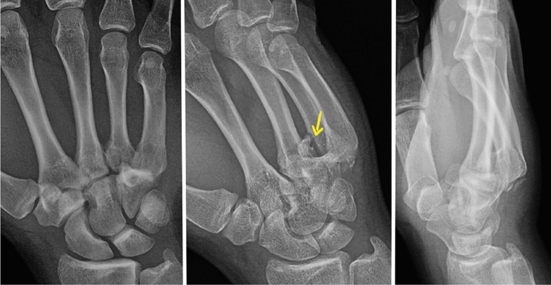
Posteroanterior, oblique, and true lateral radiographic views of case 2. Fracture of the volar aspect of the fourth metacarpal base (best identified in the oblique view, arrow), and articular fracture of the fifth metacarpal with an associated dislocation of the fourth and fifth metacarpals.
An anesthetic ulnar nerve block was performed at the wrist level, and a closed reduction was carried out by traction and pushing the base of the dislocated metacarpal into its place. Joint stability was assessed by active motion of the wrist and metacarpophalangeal joints by the patient. In all cases, postreduction PA, oblique, and lateral radiographs confirmed a concentric reduction.
After confirming the joint stability, a dorsal splint was placed in five cases ( Fig. 2 ) and a Bennett-type splint was used in three. The metacarpophalangeal joint was left free in all cases, and active motion was encouraged from the beginning.
Fig. 2.
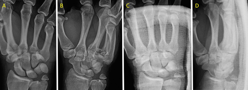
(A,B) Posteroanterior and oblique radiographic views of case 5. Comminuted fracture of the volar and radial aspect of the fourth metacarpal's base with an associated dislocation of the fifth metacarpal. (C,D) Posteroanterior and oblique radiographic views at the third week confirming a concentric reduction.
Weekly radiographic controls were performed during the first 3 weeks to ensure adequate alignment of the CMC joints. Minimum follow-up was 12 months, and average follow-up was 30.2 months.
The splint was maintained for 4 weeks, and a buddy taping was recommended for daily activities for another 3 weeks. No physical therapy was required in any case.
At the final follow-up, the range of motion of the ulnar digits assessed by the pulp-to-palm distance was 0 mm in all cases. Visual analog scale for pain was 0 at rest, 0 in daily life activities, and 1.6 on average in physical-demanding activities (range: 0–3).
DASH (Disabilities of the Arm, Shoulder and Hand) questionnaire was fulfilled by all patients at the final follow-up, with a reported score of 2.27 (range: 0–4.5). The average grip strength assessed using a Jamar dynamometer was 40 kg (range: 36–52) in the affected dominant hand and 37 kg (range: 28–50) in the nondominant and noninjured hand. As we had no previous data on the injury, we are not able to assess if the grip strength decreased. The average time to return to their working activities was 10 weeks (range: 7–13). There were no major complications at the final follow-up.
In our series, taking into account a total of 10 consecutive cases, the incidence of failure of reduction and cast was 2/10 (20%). In one case, a recurrence of the dislocation was confirmed during the clinical assessment of stability, and in the second one, a secondary dislocation was diagnosed at consultation 1 week after the injury. We did not find any predictive factor for the loss of reduction in our series, although it was too small to find significant differences if they were.
Discussion
Stability of the CMC joints is provided by the specific anatomical configuration of the joints and several volar and dorsal ligamentous attachments. 5 The CMC joints of the ring and small fingers are at a higher risk of dislocation probably because of the greater mobility due to their saddle joint configuration and looser ligamentous attachments. 1 CMC joint dislocations are uncommon injuries, with a reported incidence of 1 to 2% of carpal injuries, 6 although its real incidence is probably not well established as these injuries are often overlooked. Most of them are fracture–dislocations due to avulsion of the ligaments, and the size of the fracture fragments depends on the position, direction, and transmission of the trauma forces through the metacarpals. 5
Ulnar CMC joint dislocations and fracture–dislocations can be easily missed because of the diffuse swelling of the hand and the misinterpretation of the initial radiographs. Thus, posteroanterior, true lateral, and oblique radiographic views should be requested as routine views in any wrist trauma. Besides this, additional oblique views or a computed tomography (CT) scan may be helpful if there is any doubt or to better understand the fracture pattern. 1 3
A dislocated CMC joint can lead to chronic pain and reduced the grip strength; therefore, immediate reduction is recommended to achieve proper function of the hand. There is no consensus regarding optimal treatment of these injuries, but most authors advocate surgical treatment either by open reduction and internal fixation or by closed reduction and percutaneous pinning to prevent secondary dislocation. 2 7
In our series, satisfactory results were obtained by closed reduction and cast immobilization in 8 of 10 cases. We believe that an advantage of the conservative treatment is that in case of failure as in two of our patients, any surgical treatment remains possible under good conditions.
There are few cases of ulnar CMC joint dislocations and fracture–dislocations treated nonoperatively by closed reduction and splint immobilization, but all of them have reported satisfactory results. 1 7 8 9 10 Storken et al 1 reported four cases of acute fourth and fifth CMC joint dislocations treated conservatively by closed reduction and splinting, showing that conservative treatment through immediate reduction and splint immobilization may be enough to achieve good results as in our series.
Our series has limitations. It is a retrospective study, and the sample size was small. All clinical examinations were carried out by the authors and not by independent researchers, and this could produce an evaluation bias. We assessed patient self-perceived disability using a closed questionnaire, but this is not a specific and validated system for hand traumatic injuries and could also produce an evaluation bias. Finally, the follow-up is not long enough to assess the long-term clinical and radiological results (posttraumatic osteoarthritis).
We believe that if the diagnosis of an ulnar CMC joint dislocation or fracture–dislocation is accomplished early, a concentric and stable closed reduction can be initially achieved, and there are no major fractures, the nonoperative treatment may be a successful option to take into account with good functional results and a quick recovery but requiring a close follow-up for the first weeks.
Funding Statement
Funding None.
Conflict of Interest None declared.
Note
The authors confirm that they have not published the same or a very similar study with the same or very similar results and major conclusions in any other journal.
References
- 1.Storken G, Bogie R, Jansen E JP. Acute ulnar carpometacarpal dislocations. Can it be treated conservatively? A review of four cases. Hand (N Y) 2011;6(04):420–423. doi: 10.1007/s11552-011-9347-3. [DOI] [PMC free article] [PubMed] [Google Scholar]
- 2.Day C S, Stern P J. Philadelphia, PA: Elsevier Churchill Livingstone; 2011. Fractures of the metacarpals and phalanges; pp. 239–290. [Google Scholar]
- 3.Fisher M R, Rogers L F, Hendrix R W. Systematic approach to identifying fourth and fifth carpometacarpal joint dislocations. AJR Am J Roentgenol. 1983;140(02):319–324. doi: 10.2214/ajr.140.2.319. [DOI] [PubMed] [Google Scholar]
- 4.Garcia-Elias M, Bishop A T, Dobyns J H, Cooney W P, Linscheid R L. Transcarpal carpometacarpal dislocations, excluding the thumb. J Hand Surg Am. 1990;15(04):531–540. doi: 10.1016/s0363-5023(09)90011-9. [DOI] [PubMed] [Google Scholar]
- 5.Yoshida R, Shah M A, Patterson R M, Buford W L, Jr, Knighten J, Viegas S F. Anatomy and pathomechanics of ring and small finger carpometacarpal joint injuries. J Hand Surg Am. 2003;28(06):1035–1043. doi: 10.1016/s0363-5023(03)00373-3. [DOI] [PubMed] [Google Scholar]
- 6.Gangloff D, Mansat P, Gaston A, Apredoaei C, Rongières M.Carpometacarpal dislocation of the fifth finger: descriptive study of 31 cases [in French] Chir Main 200726(4-5):206–213. [DOI] [PubMed] [Google Scholar]
- 7.Frick L, Mezzadri G, Yzem I, Plotard F, Herzberg G.Acute carpometacarpal joint dislocation of the long fingers: study of 100 cases[in French] Chir Main 20113005333–339. [DOI] [PubMed] [Google Scholar]
- 8.Jumeau H, Lechien P, Dupriez F. Conservative treatment of carpometacarpal dislocation of the three last fingers. Case Rep Emerg Med. 2016;2016:4.962021E6. doi: 10.1155/2016/4962021. [DOI] [PMC free article] [PubMed] [Google Scholar]
- 9.Anjum R, Roy A, Farooque K, Sharma V. An isolated pure dislocation of fifth carpometacarpal joint: case report and review of literature. J Orthop Case Rep. 2017;7(02):14–16. doi: 10.13107/jocr.2250-0685.728. [DOI] [PMC free article] [PubMed] [Google Scholar]
- 10.Beekhuizen S, de Witte P B, Rutgers M, Ohanis D. Isolated ulnopalmar dislocation of the fifth carpometacarpal joint. BMJ Case Rep. 2018:pii: bcr-2018–225363. doi: 10.1136/bcr-2018-225363. [DOI] [PMC free article] [PubMed] [Google Scholar]