Abstract
Purpose
Infusion of highly heterogeneous populations of autologous tumor-infiltrating lymphocytes (TILs) can result in tumor regression of exceptional duration. Initial tumor regression has been associated with persistence of tumor-specific TILs one month after infusion, but mechanisms leading to long-lived memory responses are currently unknown. Here we studied the dynamics of bulk tumor-reactive CD8+ T cell populations in patients with metastatic melanoma following treatment with TILs.
Experimental Design
We analyzed the function and phenotype of tumor-reactive CD8+ T cells contained in serial blood samples of sixteen patients treated with TILs
Results
Polyfunctional tumor-reactive CD8+ T cells accumulated over time in the peripheral lymphocyte pool. Combinatorial analysis of multiple surface markers (CD57, CD27, CD45RO, PD-1 and LAG-3) showed a unique differentiation pattern of polyfunctional tumor-reactive CD8+ T cells, with highly specific PD-1 upregulation early after infusion. The differentiation and functional status appeared largely stable for up to 1 year post-infusion. Despite some degree of clonal diversification occurring in vivo within the bulk tumor-reactive CD8+ T cells, further analyses showed that CD8+ T cells specific for defined tumor-antigens had similar differentiation status.
Conclusions
We demonstrated that tumor-reactive CD8+ T cell subsets which persist after TIL therapy are mostly polyfunctional, display a stable partially differentiated phenotype and express high levels of PD-1. These partially differentiated PD-1+ polyfunctional TILs have a high capacity for persistence and may be susceptible to PD-L1/PD-L2-mediated inhibition.
Introduction
Ideal cancer treatment should induce durable tumor regression to give a meaningful life extension. In melanoma, work performed in the context of previous clinical trials based on transfer of autologous tumor-infiltrating lymphocytes (TILs) have identified a number of factors influencing the likelihood of tumor regression (1–5). However, despite the high degree of efficacy, over 50% of patients with significant tumor regression achieve only a temporary, partial remission (1). Sustained tumor responses may be dependent on the long-term fate of the infused tumor-specific T cells. However, the attributes of these persisting T cell populations are largely unknown. Current TIL treatment regimens are based on a single infusion of expanded T cells derived from an autologous tumor metastasis. These T cells are not selected for any specific class or type of antigen, so this treatment takes advantage of a multitarget T cell attack directed to multiple and mostly unknown antigenic specificities, including mutant neo-antigens (6). Current methods reveal the existence of neo-antigen specific T cells in TILs from most patients with melanoma; however, whole tumor-cell recognition assays indicate that the frequency of naturally occurring tumor-specific T cells may largely exceed those observed for the neo-antigens evaluated (7). Libraries of known shared antigens have been constructed and validated (8), but greatly underestimate the frequency of tumor-specific cells (9). Here, we evaluated the immunological dynamics (immunodynamics) of the total repertoire of tumor-reactive CD8+ T cells identified with whole-tumor cell recognition assays, regardless of the type or class of melanoma antigens recognized. This comprehensive analysis allowed the identification of several common features of CD8+ T cell responses that persisted for up to 1 year after infusion.
Methods
Patients and clinical trial
All protocols were approved by the Scientific Ethics Committee of the Capital Region of Denmark. Written informed consent was obtained from patients before any procedure, according to the Declaration of Helsinki. Patients were treated with TIL immunotherapy in the clinical trial NCT00937625. This clinical trial consisted of two consecutive sub-studies: a pilot study (n=6), with a low dose of IL-2 administered after T-cell infusion (2 MIU/day subcutaneously for 14 days) (10); an amended study (n=25), with an intensified regimen of IL-2 (decrescendo i.v. regimen or intermediate dose) (5). Blood samples were collected at serial time points after infusion of TILs: at discharge (about 1-2 weeks after infusion) and at approximately 1 month, 2 months, 4 months, 6 months, 9 months (this sample was analyzed only in functional evaluation, see below) and 12 months, unless the patient was excluded from the protocol at a previous time point. Peripheral blood mononuclear cells (PBMCs) were isolated with standard methods, and cryopreserved at -140 °C until use. We included sixteen selected patients where previous tumor recognition assays had identified sufficient numbers of tumor reactive T cells in the peripheral blood after infusion (described in (5)). Phenotypic assessment of T cells with defined antigen specificity and clonotypic analysis of tumor-reactive TILs were conducted each on three selected patients. All other analyses included at least thirteen patients. We were unable to include the three remaining patients (total cohort of 16) in all analyses due to insufficient sample material.
Polyfunctionality and Phenotype of tumor-reactive CD8+ T cells
Evaluation of T cell responses was performed as previously described (5,11). Briefly, TILs or PBMCs were co-cultured with autologous short-term cultured melanoma cell lines (available for 10/16 patients), freshly cryopreserved autologous tumor single cell suspensions (1/16 patients) or partially HLA-matched allogeneic melanoma cell lines (5/16 patients, in this case TILs and PBMCs were tested only for recognition of the cell line where TILs had previously shown the highest level of recognition, (see (5) for details). For phenotypic assessment of polyfunctional tumor-reactive T cells, we combined assessment of tumor cell recognition with staining of surface phenotype markers. We also compared the phenotype of non-tumor reactive CD8+ T cells in addition to polyfunctional tumor-reactive T cells.
For phenotypic assessment of T cells with defined antigen specificity, we combined staining with fluorochrome conjugated peptide-MHC (p-MHC) multimers and staining of surface phenotype markers. Details on reagents and additional description of the methodologies are available as Supplemental data.
Flow Cytometry data processing and analysis
The relative fractions of cells were analyzed with BD FACS Diva 6 (only for enumeration of CD107a+ cells within the TNF+/IFNγ+ gate) or FlowJo 9.7.1 (FlowJo LLC, Ashland, OR) sub-categorizing flow cytometry events with parallel Boolean gating (gates drawn solely on the parameters of interest). For enumeration of CD107a+ cells within the TNF+/IFNγ+ gate, data were plotted in excel and displayed on exponential scale. Logarithmic r2 values are calculated separately for each individual patient.
For functional characterization, the population of cells negative for all three functional markers was removed from the analysis. Thus boolean combination gates of the three functional markers resulted in 7 individual gates (eight minus one, which is the only group negative for all functional markers), each showing the percentage of CD8+ T cells expressing a unique combination of the three markers. Data were exported into Pestle 1.7 (courtesy of Dr. Mario Roederer, ImmunoTechnology Section, VRC/NIAID/NIH, Bethesda, MD, USA) and formatted, according to the manufacturer’s instructions. Analysis and presentation of distributions was performed using Simplified Presentation of Incredibly Complex Evaluations (SPICE) version 5.3, downloaded from http://exon.niaid.nih.gov (12). Background subtraction was performed with Pestle 1.7, according to the manufacturer’s instructions. In SPICE, thresholds were set at 0.1. Comparison of distributions was performed using a Student’s T test and a partial permutation test as described (12). To compare bar charts, a Wilcoxon signed rank-test assuming unequal variances was carried out, with p<0.05 considered significant. Other statistical analyses were performed with GraphPad Prism 5 (GraphPad Software, La Jolla, CA, USA). For the analysis of single markers phenotype, TP cells were gated and data were exported in SPICE via FlowJo and Pestle. Bar charts were compared as described above.
For combinatorial analysis of phenotype, a “differentiation” scoring system was established according to the model proposed by Gattinoni et al. (13), which identifies “early” effector and “late” effector cells. Despite CCR7 is not included in the analysis, expression of CD45RO was used as marker of earlier differentiation because we and others have previously demonstrated that the vast majority of TILs consist of T cells with an effector memory (CD45RO+ CD45RA- CCR7-) phenotype (14), and loss of CD45RO in this population is likely to represent a transition to TEMRA cells (15). The model of Speiser et al. (16) which identifies PD-1+ cells as terminally differentiated cells was also incorporated into these analyses. Since LAG-3 was largely detected only in TP cells contained in TILs infusion products, this marker was removed from further analyses. The following groups were established: Group 0 (early effector cell) contained the CD27+, CD45RO+, CD57- and PD-1- while group 4 (late effector cell) contained the CD27-, CD45RO-, CD57+ and PD-1+ fraction. Group 1 contained one of the markers from group 4, group 2 two of the markers from group 4 and group 3 three of the markers from group 4. In brief, a score from 0 to 4 was assigned to TP (triple positive) cells, with 0 representing the less differentiated cells to 4 the most differentiated. This is shown in Supplementary Figure S6A, where intra-sample analyses (i.e. the frequency of TP cells contained in each group, for each sample) are demonstrated. Furthermore, one single combined “Differentiation Score” was given to each sample (each patient, at each time point), by multiplying the arcsine transformed value of the frequency of cell subpopulations contained in the groups described above, with a pre-defined score for each group which was 0 for early effector cells (group 0) and 4 for late effector cells (group 4) as described above (intermediate values 1-3 were given to cells with 1, 2 or 3 phenotypic markers). The differentiation score of each sample is demonstrated in Figure 3B. Supplementary Figure S1 shows a graphical representation of the scoring system and the unique T cell subpopulations analyzed are reported in Table S1. In addition, boolean combination gates of the four phenotype markers resulting in 16 individual gates were analyzed and processed as described above in functional characterization. These data are shown in Supplementary Figure S7
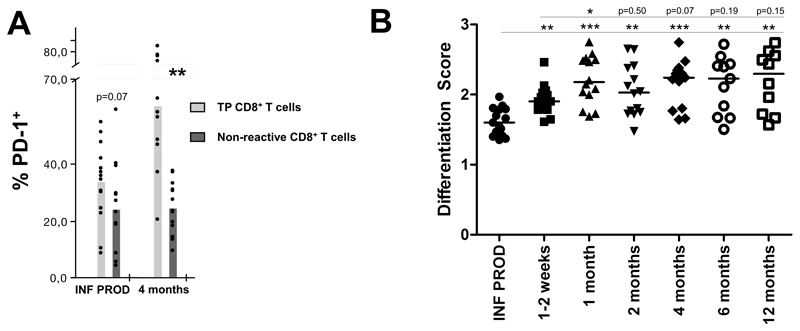
Figure 3. PD-1 expression and differentiation score of tumor-reactive CD8+ T cells phenotype after cell transfer.
(A) The percentage of cells expressing PD-1 among the TP tumor reactive and non tumor-reactive CD8+ T cells is shown. Infusion products n=15, peripheral blood 4 months after infusion n=13 (B) The scatter plot shows the differentiation score given to each sample at each time point. * p<0.05; ** p<0.01; *** p<0.001
Clonotypic analysis of tumor-reactive CD8+ T cells
Autologous tumor cells were plated one day before sorting in RPMI medium supplemented with 100 U/mL Penicillin, 100 μg/mL Streptomycin, 2 mM L-Glutamine and 5% heat-inactivated fetal calf serum (all from Life Technologies, Paisley, UK). TIL infusion products or PBMCs were stimulated with autologous tumor one day after defrosting, in order to avoid TCR repertoire bias due to culturing and expansion in vitro. TIL samples or PBMCs were co-incubated with autologous tumor at a 1:1 ratio in the presence of 15 µL of anti-TNF PE-Cy7 (BD Biosciences, Oxford, UK), 15 µL CD107a PE (BD Biosciences, Oxford, UK) and 30 µM of TAPI-0 (Calbiochem) for 5h at 37 °C, 5% CO2. Following incubation, cells were stained with LIVE/DEAD® Fixable Violet Dead Cell Stain Kit (Life Technologies), anti-CD3 PerCP (Miltenyi Biotec), and anti-CD8 APC-Vio 770 (Miltenyi Biotec). Cells from each experimental condition were washed and the live CD3+TNF-+CD107a+ fraction was sorted directly into 1.5 mL microcentrifuge tubes containing 350 µL of lysis buffer (Qiagen, Hilden, Germany). RNA extraction was carried out using the RNEasy Micro kit (Qiagen, Hilden, Germany). cDNA was synthesized using the 5’/3’ SMARTer kit (Clontech, Paris, France) according to the manufacturer’s instructions. The SMARTer approach used a Murine Moloney Leukaemia Virus (MMLV) reverse transcriptase, a 3’ oligo-dT primer and a 5’ oligonucleotide to generate cDNA templates which were flanked by a known, universal anchor sequence. PCR was then set up using a single primer pair. A TCR-β constant region-specific reverse primer (Cβ-R1, 5’-GAGACCCTCAGGCGGCTGCTC-3’, Eurofins Genomics, Ebersberg, Germany) and an anchor-specific forward primer (Clontech, Paris, France) were used in the following PCR reaction: 2.5 µL template cDNA, 0.25 µL High Fidelity Phusion Taq polymerase, 10 µL 5X Phusion buffer, 0.5 µL DMSO (all from Thermo Fisher Scientific, UK), 1 µL dNTP (50 mM each, Life Technologies, Paisley, UK), 1 µL of each primer (10 µM), and nuclease-free water for a final reaction volume of 50 µL. Subsequently, 2.5 µL of the first PCR products were taken out to set up a nested PCR as above, using a nested primer pair (Cβ-R2, 5’-TGTGTGGCCAGGCACACCAGTGTG-3’, Eurofins Genomics, Ebersberg, Germany and anchor-specific primer from Clontech, Paris, France). For both PCR reactions, cycling conditions were as follows: 5 min at 94°C, 30 cycles of 30 s at 94 °C, 30 s at 63 °C, 90 s at 72 °C, and a final 10 min at 72 °C. The final PCR products were loaded on a 1% agarose gel and purified with the QIAEX II gel extraction kit (Qiagen, Hilden, Germany). Purified products were barcoded, pooled and sequenced on an Illumina MiSeq instrument.
Results
Patient characteristics
Patient characteristics are summarized in Table 1. Out of sixteen TIL-treated patients selected on the basis of the availability of samples and measurable antitumor responses in the peripheral blood, 10 had response evaluation criteria in solid tumors (RECIST) version 1.0 confirmed responses and three additional patients had minor tumor regressions including unconfirmed partial responses. Two patients had stable disease and only one patient, M27, was classified as pure progressor after infusion of TILs. The predominance of responding patients in this cohort was largely expected, as we have previously shown that the detection of tumor-reactive CD8+ T cells in the peripheral blood lymphocyte (PBL) pool after TIL infusion is strongly associated with tumor regression (5).
Table 1. Patient Characteristics.
| Patient | Progression free survival (months) | Tumor Response (target lesions) | RECIST | Tumor recognition assays | |
|---|---|---|---|---|---|
| M | 11 | 13.1 | -100 | CR | Autologous Cell Line |
| M | 15 | +47 | -100 | CR | Autologous Cell Line |
| M | 18 | 3,9 | -23 | SD | Allogeneic Cell Line |
| M | 20 | 12 | -54.9 | PR | Allogeneic Cell Line |
| M | 24 | +36 | -100 | PR | Autologous Cell Line |
| M | 26 | +32 | -100 | CR | Allogeneic Cell Line |
| M | 27 | 2 | 24.7 | PD | Autologous Cell Line |
| M | 22 | +28 | -100 | PR | Autologous Cell Line |
| M | 31 | 11.3 | -61.3 | PR | Allogeneic Cell Line |
| M | 34 | 2.8 | -5.8 | SD | Autologous Cell Line |
| M | 36 | 8.2 | -53.8 | PR | Allogeneic Cell Line |
| M | 37 | 3 | 2,6 | SD | Autologous Cell Line |
| M | 42 | +22 | -100 | CR | Autologous Fresh Tumor Suspension |
| M | 43 | 1.9 | -76.6 | PD* | Autologous Cell Line |
| M | 45 | 11.3 | -35.7 | PR | Autologous Cell Line |
| M | 47 | 3.3 | -21.4 | SD | Autologous Cell Line |
this patient is classified as PD because of the appearance of one new lesion after treatment
Polyfunctionality of tumor-reactive CD8+ T cells
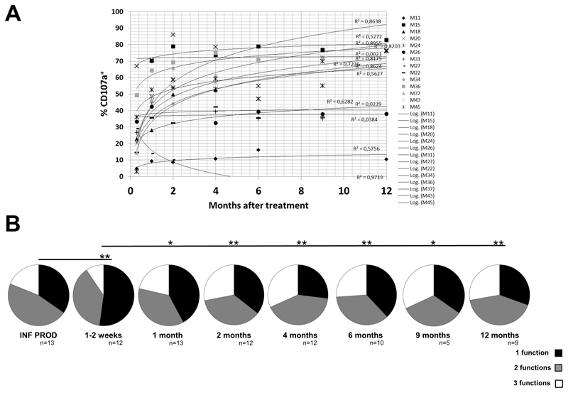
It is believed that T cells expressing multiple functions may mediate superior immune responses (17) and polyfunctionality is known to correlate with antigen-sensitivity and TCR binding affinity (18). We have previously shown that over 95% of CD8+ T cells expressing at least one out of seven antitumor functions express either TNF, IFNγ or CD107a (11). Thus, we limited our analyses to these three functional markers. A retrospective analysis of our previously published dataset on TIL recognition of autologous melanoma cells (11) indicates that the fraction of CD8+ T cells expressing CD107a was lower than the fraction producing cytokines (Supplementary Figure S2A, S2B and S2C), and the majority of CD107a+ cells (approximately 65%) also produced at least one cytokine (Supplementary Figure S2D). This suggests that CD107a mobilization was mainly associated with cytokine production, while cytokine production could occur independently of CD107a upregulation. Thus, we initially focused on CD8+ T cells expressing cytokines, and to increase the sensitivity of the analysis, primary on those expressing simultaneously TNF and IFNγ (double positive or DP cells). Notably, among DP cells, the percentage of cells expressing CD107a increased over time in all (n=13; median r2 0.63) but one patient (Figure 1A). CD107a is a surrogate marker of lytic granule release and, therefore, the capacity of T cells to directly destroy tumor cells (19). In this context, it is interesting that the only patient where the CD107a+ fraction decreased was the one patient who, despite detection of tumor-reactive CD8+ T cells in post-infusion blood samples, had a significantly progressive disease at first evaluation after treatment, and was quickly excluded from the study (patient M27, Table 1). In light of this finding, we analyzed whether the frequency of CD8+ T cells expressing all three functions (polyfunctional T cells, triple positive cells or TP cells) within the whole pool of tumor-reactive CD8+ T cells (expressing at least one function after recognition of whole tumor cells) showed a similar increasing trend. An initial drop in the frequency of TP cells was observed from TIL infusion products to the first blood sample, obtained 1-2 weeks after infusion (Figure 1B, Supplementary Figure S3A and S3B). However, TIL infusion products are cultured in vitro with high doses of IL-2, which may potentially explain this phenomenon of hyper-functionality of TIL infusion products. The fraction of TP cells in patient blood samples tended to gradually increase until ~4 months evaluation and then stabilized thereafter for up to 1 year (Figure 1B, Supplementary Figure S3A and S3B). These data indicate that polyfunctional T cells accumulate within the peripheral pool of tumor-reactive CD8+ T cells and may establish long-lived immune responses.
Figure 1. Accumulation of polyfunctional tumor-reactive CD8+ T cells after cell transfer.
(A) Frequency of CD107a+ cells among TNF+ / IFN-γ+ (double positive, DP) CD8+ T cells after cell transfer. Trendlines (logarithmic) are shown for each patient, and individual r2 values are indicated. (B) Pie charts showing the relative frequency of tumor-reactive CD8+ T cells expressing one (black), two (grey) or three (blue) simultaneous functions.
Phenotype of polyfunctional CD8+ T cells – single markers
Next, we investigated whether any change in phenotype of polyfunctional tumor-reactive CD8+ T cells occured in vivo after infusion. For these analyses we focused on known markers of T cell differentiation and immune checkpoints thought to constrain T cell responses. The phenotypes of polyfunctional CD8+ T cells and of non-tumor reactive CD8+ T cells were analyzed in parallel.
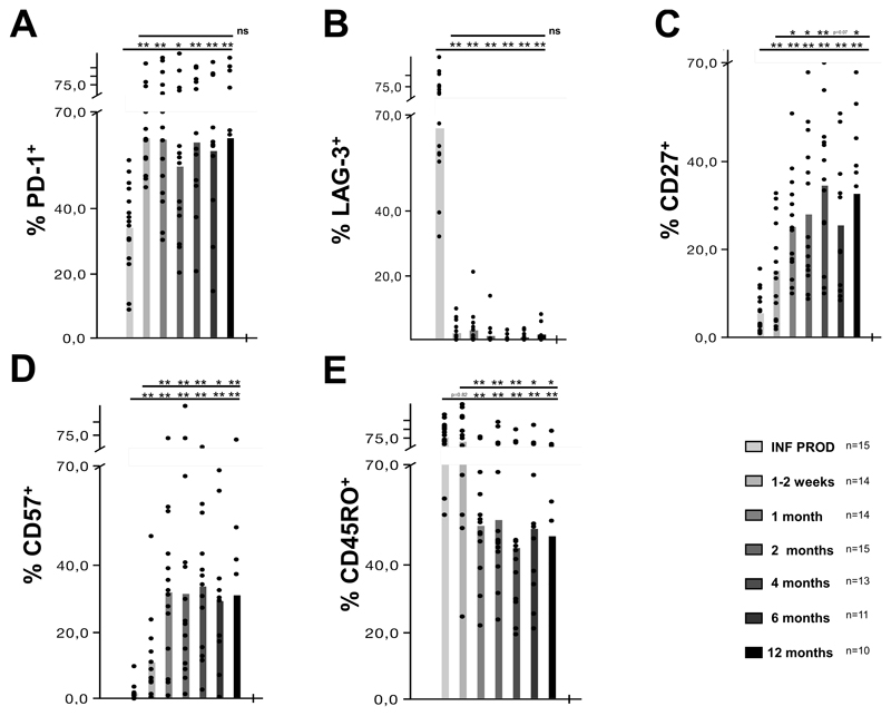
Programmed death-1 (PD-1) is a cell surface immune regulatory molecule (or immune checkpoint) which negatively regulate T cell responses (20). PD-1 was expressed on nearly 35% of the TP cells in the infusion product, which increased to about 65% shortly after infusion and stabilized thereafter (Figure 2A). No increase was observed in non-tumor reactive CD8+ T cells (Figure 3A and Supplementary Figure S4A), indicating that PD-1 accumulation is a highly specific feature of tumor-reactive cells.
Figure 2. Phenotype of polyfunctional tumor-reactive CD8+ T cells after cell transfer.
Frequency (%) of cells positive for surface markers among polyfunctional (triple positive, TP) tumor-reactive CD8+ T cells (A), PD-1, (B) LAG-3, (C) CD27, (D) CD57 and E (CD45RO). Dots show individual patients and bars show mean values. * p<0.05; ** p<0.01
LAG-3 is another cell surface molecule functioning as an immune checkpoint (21), which may work synergistically with PD-1 to downregulate T cell responses (22). Interestingly, while around 65% of the TP in the infusion product expressed LAG-3 very few cells expressed this marker in vivo even a week post infusion (Figure 2B). This surprisingly dramatic change is consistent with previous data reported in a clinical trial with infusion of gene-engineered T cells (23). It is likely that some phenotypic changes, such as loss of LAG-3 expression post infusion, could simply be the result of having removed the high doses of IL-2 used to culture cells in vivo. Indeed, a similar pattern was observed in non-tumor reactive CD8+ T cells (Supplementary Figure S4B). Alternatively, LAG-3+ cells might have reduced capacity to survive in vivo or might exhibit differential migration patterns so as not to be present in peripheral blood samples. In any case, it does not appear that LAG- is expressed at high levels on circulating tumor-reactive CD8+ T cells, but LAG-3 may be upregulated several hours after T cell activation upon recognition of tumor cells (Borch TH, personal communication). The majority of CD8+ T cells in heavily expanded TILs consist of antigen-experienced effector memory T cells (14). Thus, three additional surface markers were analyzed in order to characterize the differentiation status of TP cells (CD27 and CD45RO representing less differentiated cells – CD57 representing more differentiated cells). In vivo accumulation of CD27+ cells after T cell infusion has been reported in other studies (24,25) and the size of the CD27+CD8+ T cell pool in bulk TILs was highly associated with their ability to mediate tumor regression (26). Thus, CD27 may be a key molecule for maintaining immunological memory. Our data show that the mean frequency of TP cells expressing CD27 increased significantly from around 5% in the TIL infusion product, with a progressive trend up to 30-35% 4, 6 and 12 months after infusion (Figure 2C). Very low expression of CD27 in infused TILs may be a consequence of the high doses of IL-2 used to drive massive TIL expansion (26), and indeed a similar increase in CD27 was observed in non-tumor reactive CD8+ T cells (Supplementary Figure S4C and S5A). Therefore, it cannot be excluded that the observed phenomenon might be explained, at least in part, by IL-2 withdrawal, as previously reported in vitro (26). Similarly, CD57 expression exhibited a progressive trend from around 2-5% in the infusion product to almost 37% 12 months after treatment (Figure 2D). In contrast, around 75% of TP cells in the infusion product expressed CD45RO, which was mirrored 1 week later in the PBMCs before dropping to about 50% at over 1 month post infusion (Figure 2E). Parallel increase in CD57+ and decrease in CD45RO+ were detected in non-tumor reactive CD8+ T cells (Supplementary Figure S4D, S4E, S5B and S5C). Comparison of phenotype markers in TP and non-tumor reactive CD8+ T cells revealed difference only for CD57 in the infusion products (Supplementary Figure S5B), and major differences in the peripheral pools for PD-1 (Figure 3A) and CD57 (Supplemenraty Figure S5B), reflecting quantitatively different changes in the two sub-populations. However, in this context it should be highlighted that changes in non-tumor reactive CD8+ T cell populations are likely to represent a replacement of the infused cells with de novo generated populations, including naïve CD8+ T cells (including CD45RO- T cells, which for non-tumor reactive CD8+ T cells may represent naïve CD45RO- CCR7+ cells), during bone marrow recovery after lymhodepleting chemotherapy.
In summary, our data showed a characteristic change in the phenotype of bulk tumor-reactive TP CD8+ T cells, with rapid and highly specific accumulation of PD-1. Other phenotypic markers such as LAG-3, CD27, CD57 and CD45RO displayed similar early changes post-infusion in both TP and non-tumor reactive CD8+ T cells. This altered phenotype remained largely stable for up to 1 year after infusion. Thus, we next looked to dissect the complex picture emerging from analyses of individual phenotype markers using combinatorial analyses.
Phenotype of polyfunctional CD8+ T cells – combinatorial analysis
Given the complex picture obtained from analysis of single markers, a differentiation scoring system was established. TP CD8+ T cells were categorized into five groups with a score from 0 to 4, with 0 representing early effector cells and 4 representing the most differentiated effector cells (described in Methods/Flow Cytometry data processing and analysis, see also Supplementary Figure S1 and Supplementary Table S1). LAG-3 was excluded from these analyses, because it could not be detected in all post-infusion samples. The frequency of cells in the five groups changed early post-infusion, with an increase in more differentiated cells up to 1 month after infusion that remained largely stable for up to 1 year (Supplementary Figure S6). The differentiation score, which was given on a per sample-basis (one single combined score for each sample), confirmed only early changes with progressive differentiation to up to at least 1 month after infusion and stabilization of the phenotype thereafter without further differentiation (Figure 3B). Full combinatorial analyses of the unique combination of surface markers is shown in Supplementary Figure S7. In summary, while the frequency of less differentiated TP CD8+ T cells seemed to drop progressively during the period prior to 1 month after infusion, a similar phenotypic pattern of TP CD8+ T cells from 1 month to 12 months after infusion.
Clonotype analysis of tumor-reactive CD8+ T cells
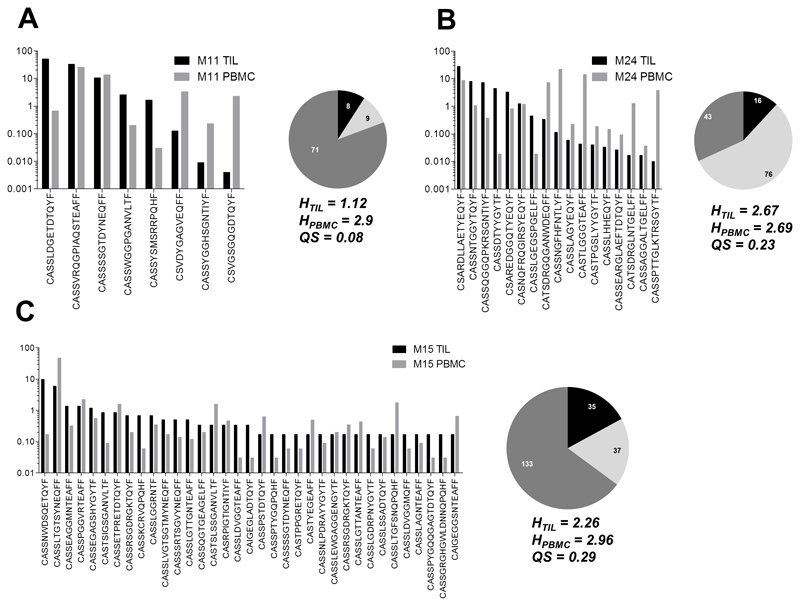
Having established that several phenotypic markers on tumor-reactive CD8+ T cells were preserved up to 12 months post-infusion, we next sought to determine whether TCR-β chain clonotypes from the TIL infusion product would persist in the periphery over time. We reasoned that the persistence of TCR-β chains found in the infusion product, in the periphery, would suggest the establishment of long-term memory, alternatively continuous antigen-specific stimulation. Three patients with prolonged tumor regression (M11, M15 and M24) were selected for clonotypic analyses of the tumor-reactive (CD107a+TNF+) T cell populations in the infusion product and peripheral blood 6 months after infusion. The degree to which the TIL and peripheral blood repertoires overlapped varied across patients. Despite individual differences in overall repertoire diversity and composition, several tumor-reactive clonotypes found in the infusion product were detectable in peripheral blood six months post-treatment (Figure 4). While the relative frequency of certain TCR-β chain clonotypes was lower in the blood samples, compared to TIL infusion product, other clonotypes expanded over time. Interestingly, each patient had several tumor-reactive CD8+ T cell clones in the 6-month sample that were not detectable in the infusion product. In all three patients, TCR diversity appeared to rise post-treatment as indicated by an increase in Shannon entropy. Thus, whereas many tumor-reactive T cell clones can persist for at least 6 months after infusion, other T cell populations arise in the periphery over time from de novo recruitment or from expansion of rare clones present in the infusion product at frequencies below our limits of detection. The presence of tumor-reactive clonotypes in the blood that were not present in the infusion product might suggest epitope spreading.
Figure 4. Tumor-reactive CD8+ T cell clonotypes from infusion products persist in the blood up to 6 months post-treatment.
The frequency of TCR-β chain sequences found in both TIL (solid bar) and peripheral blood samples (white bar) is shown for A) patient M11, B) patient M15, and C) patient M24. The absolute number of shared (black) and unique (light grey for TIL; dark grey for PBMC) TCR-β chain sequences for each patient is shown as pie charts. Out-of-frame chains and ambiguous sequences were discarded from analysis. Shannon entropy (H) was calculated for TIL and PBMC samples, and compositional similarity measured by the Sørensen coefficient (QS). H values increase with TCR diversity; QS increases with repertoire overlap.
Phenotype of CD8+ T cells with defined tumor-antigen specificity
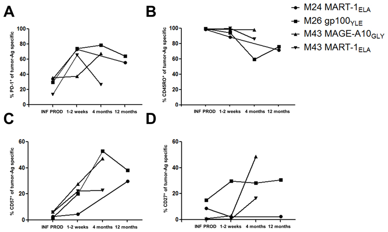
In order to gain insights on whether the observed phenotype changes occur in the repertoires of infused CD8+ T cells targeting a defined antigen, and whether similar phenotype changes could be detected in resting cells (phenotype of TP cells might be influenced by T cell activation upon recognition of naturally presented tumor-antigens), we carried out similar phenotype analyses focusing on CD8+ T cells recognizing known tumor-associated antigens as detected by p-MHC multimer staining. Samples from three patients with high-frequencies of CD8+ T cells with defined tumor-antigen specificity were selected for these analyses (Supplementary Figure S8A). We observed a similar trend as seen in TP CD8+ T cells, with patterns of strong LAG-3 downregulation (data not shown), increase of PD-1 (Figure 5A), decrease of CD45RO (Figure 5B) and increase of CD57 (Figure 5C). Similar to TP CD8+ T cells, the increase of PD-1 appeared larger in tumor-antigen specific CD8+ T cells (Supplementary Figure S8B). CD27 did not increase as much as observed in whole pool of tumor-reactive CD8+ T cells (Figure 5D). However current data does not allow to establish whether the more pronounced changes observed in tumor-reactivity experiments were due to the activated state of TP CD8+ T cells or whether this discrepancy was simply due to the small sample size of p-MHC experiments. Combinatorial analysis of surface markers of these populations was similar to that seen on TP CD8+ T cells (compare pie charts in Supplementary Figure S7 with Supplementary Figure S8C).
Figure 5. Phenotype of CD8+ T cells with defined tumor-antigen specificity.
The figure shows the frequency of the (A) PD-1+, (B) CD45RO+, (C) CD57+ and (D) CD27+ subpopulations among CD8+ p-MHC multimer+ T cells.
Discussion
Optimization of cellular immunotherapies requires a detailed understanding of the dynamic processes leading to establishment of long-term immunological memory. We conducted a detailed, sequential, multidimensional analysis of bulk tumor-reactive T cell populations derived from melanoma patients treated with TILs, dissecting dynamic functional and phenotypic changes of antitumor CD8+ T cells. Polyfunctionality is a highly desirable feature of potent CD8+ T cells, well known in infections (17), but only recently described in cancer immunity (11,27). This feature is known to correlate with antigen-sensitivity and TCR affinity for cognate antigen (18,28). We showed that polyfunctional tumor-reactive CD8+ T cells accumulate in the peripheral blood of patients treated with TILs and that individual clonotypes within the TILs persist in patient peripheral blood 6 months after treatment. The increase of polyfunctional (TP) cells over time may indicate that these populations have a very high capacity to survive in vivo and/or that mono-functional CD8+ T cells acquire further functions over time. Interestingly, the relative increase of the polyfunctional T cell subset occurred mainly in the first few months after infusion, setting the premises for long-term memory. This expanding population of tumor-reactive T cells exhibited a partially differentiated cellular phenotype, which remained stable for up to 1 year post-infusion and included the highly specific feature of early PD-1 upregulation.
After withdrawal of exogenous IL-2, which is known to overcome PD-L1-mediated inhibition (29), this increase in PD-1 expression in tumor-reactive cells may render them sensitive to PD-L1. Thus, impaired T cell activity via the PD-1 pathway may be responsible for relapses observed in some patients who initially responded to TIL-treatment (1). The other markers we studied displayed similar early changes post-infusion on both TP and non-tumor reactive CD8+ T cells. For example, CD27 is an unstable marker that can be markedly affected by cytokines and exposure to (or even self-expression of) its ligand CD70 (26). Consequently, expression of CD27 can be sporadic and fluctuating during the life of clonal T cells. Thus, accumulation of CD27+ cells in vivo might represent a slow increase in CD27 expression following withdrawal of exogenously supplied IL-2. The kinetic of increase in CD27 is seen within the first month after withdrawal of IL-2 and thus consistent with in vitro studies from Huang and colleagues (26), and in parallel to similar increase in CD27 expression on non-tumor reactive CD8+ T cells. High levels of CD27 were associated with clinical responses following ACT (1,26) thus, overall, increasing or maintaining a sufficient level CD27 expression highlights the its role as a key marker of immunological memory. Upregulation of CD57 is normally interpreted as continued progression to a terminally differentiated phenotype (30). The population of CD27+CD57+ cells that we described here is unusual but has been described previously in patients with melanoma (31).
Importantly, the pattern of polyfunctionality and phenotypic markers that is established within the first month after TIL therapy is then maintained for at least a year. In this context, we proposed a differentiation scoring system based on combinatorial expression of CD27, CD45RO, CD57 and PD-1. This system is imperfect due to the known instability of some markers such as CD27, and the absence of other potentially important markers such as CD62L and CCR7. Nevertheless, this system proved useful for visualizing the global phenotypic dynamics up to 1 year following infusion of TILs (Figure 3B).
We also investigated whether individual T cells within the infusion product could persist in patient blood after treatment by examining the clonotypic architecture of the tumor-specific response in the infusion product and patient blood 6 months after treatment. These results confirmed that some tumor-reactive T cell clones from the TIL infusion product persisted and dominated the anti-tumor response in vivo. In addition new tumor-reactive clonotypes were detected suggesting that these might have been expanded post infusion as the result of epitope spreading as the chosen patients successfully rejected their tumors. Ongoing work in our laboratories is aimed at determining the individual antigen-specificities of some key clones in order to confirm whether they recognize new epitopes that were not recognized by the original TIL infusion product. Additional analyses indicated that the phenotypic profile observed in the bulk tumor-reactive CD8+ T cell repertoire was similarly reflected within the responses to known tumor-antigens, It therefore seems reasonable to conclude that all non de novo T cell subpopulations, that contributed to the bulk CD8+ T cell response against tumor persisting over time, had a similar phenotypic profile.
Our discovery of biomarkers correlating the fate of tumor-specific CD8+ T cells after adoptive transfer is of high interest, because these findings could have direct implications on the selection of CD8+ T cells for future adoptive immunotherapy studies or combination therapies. By using highly homogeneous T cell clone populations specific for melanoma differentiation antigens, Chandran et al. have recently shown that tumor-specific CD8+ T cells expressing IL-7 receptor and c-myc have superior capacity for persisting in vivo (32). Our present data does not allow determination of whether polyfunctionality and the observed partial differentiation phenotype can be used to predict the fate of pre-infusion TILs, or whether this is simply a natural differentiation pathway of long-lived memory CD8+ T cells. Additional detailed studies will be required to definitively link cell phenotype to cell fate in vivo. Nonetheless, the specific and strong accumulation over time of PD-1 in polyfunctional T cells suggest that combined TIL/checkpoint inhibitor blockade or the use of PD-1 gene-edited TILs (33) should be explored in future clinical trials, especially to prevent disease relapses. With current adoptive cell therapy protocols, disease relapses occurred in up to 63% of patients with an initial objective response (analysis of clinical data presented in (1)).
In summary, we have begun to dissect the immunological dynamics of T cell persistence and differentiation in vivo following adoptive transfer of TILs. The changes we observed mainly took place shortly after infusion, with early PD-1 upregulation as a highly specific feature of polyfunctional tumor-reactive CD8+ T cells, and remained stable for up to a year. These PD-1+ polyfunctional T cells may have a high capacity for persistence and generation of immunological memory in patients with metastatic melanoma treated with T cell therapy, and be crucial in keeping patients tumor free. Our results have important implications for the next generation adoptive immunotherapy protocols as well as immunological monitoring of patients treated with T cell based immunotherapy.
Supplementary Material
Statement of Translational Relevance.
Infusion of autologous tumor-infiltrating lymphocytes can induce dramatic and durable clinical responses in patients with widely metastatic cancers. Persistence of tumor-reactive T cells in the circulation is strongly associated with tumor regression, thus the identification of T cell subsets with greater persistence may pave the way to more effective cellular immunotherapies.
In this study, we identified a CD8+ T cell subset with high persistence. This tumor-reactive CD8+ T cell subset generates polyfunctional immune responses and exhibits a stable but partially differentiated phenotype. Sustained upregulation of PD-1 in polyfunctional tumor-reactive CD8+ T cells make them susceptible to PD-L1/PD-L2 mediated inhibition, and interfering with this signaling pathway may prolong clinical responses. Our results suggest that these partially differentiated, PD-1+, polyfunctional TILs may hold the key to continuous immune surveillance. Next-generation immunotherapy protocols to induce polyfunctional TILs with reduced sensitivity to PD-1 signaling may result in improved clinical outcomes.
Acknowledgements
The Danish Cancer Society, The Capital Region of Denmark Research Foundation and Danielsen Foundation are acknowledged for financial support. We thank all the staff of the Department of Oncology, Herlev Hospital, in particular Dr. Eva Ellebaek, for inclusion and treatment of patients in the clinical trial. The technicians of the CCIT lab are acknowledged for blood sample processing and storage. We thank the creators of SPICE for providing powerful data analysis tools. AKS is a Wellcome Trust Senior Investigator.
Footnotes
Conflict of interest disclosures
None of the authors declares competing conflict of interest
References
- 1.Rosenberg S, Yang JC, Sherry RM, Kammula US, Hughes MS, Phan GQ, et al. Durable complete responses in heavily pretreated patients with metastatic melanoma using T-cell transfer immunotherapy. [cited 2012 Nov 5];Clin cancer Res. 2011 17:4550–7. doi: 10.1158/1078-0432.CCR-11-0116. [Internet] [DOI] [PMC free article] [PubMed] [Google Scholar]
- 2.Besser MJ, Shapira-Frommer R, Itzhaki O, Treves AJ, Zippel D, Levy D, et al. Adoptive Transfer of Tumor Infiltrating Lymphocytes in Metastatic Melanoma Patients: Intent-to-Treat Analysis and Efficacy after Failure to Prior Immunotherapies. [cited 2013 May 24];Clin cancer Res. 2013 doi: 10.1158/1078-0432.CCR-13-0380. [Internet] [DOI] [PubMed] [Google Scholar]
- 3.Dudley ME, Gross Ca, Somerville RPT, Hong Y, Schaub NP, Rosati SF, et al. Randomized Selection Design Trial Evaluating CD8+-Enriched Versus Unselected Tumor-Infiltrating Lymphocytes for Adoptive Cell Therapy for Patients With Melanoma. [cited 2013 May 13];J Clin Oncol. 2013 doi: 10.1200/JCO.2012.46.6441. [Internet] [DOI] [PMC free article] [PubMed] [Google Scholar]
- 4.Radvanyi LG, Bernatchez C, Zhang M, Fox P, Miller P, Chacon J, et al. Specific lymphocyte subsets predict response to adoptive cell therapy using expanded autologous tumor-infiltrating lymphocytes in metastatic melanoma patients. [cited 2012 Nov 7];Clin cancer Res. 2012 doi: 10.1158/1078-0432.CCR-12-1177. [Internet] [DOI] [PMC free article] [PubMed] [Google Scholar]
- 5.Andersen R, Donia M, Ellebæk E, Borch TH, Kongsted P, Iversen TZ, et al. Long-lasting complete responses in patients with metastatic melanoma after adoptive cell therapy with tumor-infiltrating lymphocytes and an attenuated IL-2 regimen. [cited 2016 Mar 28];Clin Cancer Res. 2016 doi: 10.1158/1078-0432.CCR-15-1879. [Internet] [DOI] [PubMed] [Google Scholar]
- 6.Robbins PF, Lu Y-C, El-Gamil M, Li YF, Gross C, Gartner J, et al. Mining exomic sequencing data to identify mutated antigens recognized by adoptively transferred tumor-reactive T cells. [cited 2013 May 22];Nat Med. 2013 2013 doi: 10.1038/nm.3161. [Internet] [DOI] [PMC free article] [PubMed] [Google Scholar]
- 7.Gros A, Parkhurst MR, Tran E, Pasetto A, Robbins PF, Ilyas S, et al. Nat Med. Nature Publishing Group; 2016. Prospective identification of neoantigen-specific lymphocytes in the peripheral blood of melanoma patients. [DOI] [PMC free article] [PubMed] [Google Scholar]
- 8.Andersen RS, Thrue CA, Junker N, Lyngaa R, Donia M, Ellebæk E, et al. Dissection of T-cell antigen specificity in human melanoma. [cited 2013 Jan 29];Cancer Res. 2012 72:1642–50. doi: 10.1158/0008-5472.CAN-11-2614. [Internet] [DOI] [PubMed] [Google Scholar]
- 9.Lyngaa R, Donia M, Andersen R, Bjoern J, Andersen RS, Ellebaek E, et al. Type, frequency and breadth of tumor associated antigen reactivity in tumor infiltrating lymphocyte from metastatic melanoma patients. CIMT Annu Meet; 2016. page Abstract 150. [Google Scholar]
- 10.Ellebaek E, Iversen TZ, Junker N, Donia M, Engell-Noerregaard L, Met O, et al. Adoptive cell therapy with autologous tumor infiltrating lymphocytes and low-dose Interleukin-2 in metastatic melanoma patients. [cited 2012 Nov 8];J Transl Med. 2012 10:169. doi: 10.1186/1479-5876-10-169. [Internet] [DOI] [PMC free article] [PubMed] [Google Scholar]
- 11.Donia M, Andersen R, Kjeldsen JW, Fagone P, Munir S, Nicoletti F, et al. Aberrant expression of MHC Class II in melanoma attracts inflammatory tumor specific CD4+ T cells which dampen CD8+ T cell antitumor reactivity. Cancer Res. 2015 doi: 10.1158/0008-5472.CAN-14-2956. [DOI] [PubMed] [Google Scholar]
- 12.Roederer M, Nozzi JL, Nason MC. SPICE: exploration and analysis of post-cytometric complex multivariate datasets. [cited 2013 Sep 25];Cytom Part A. 2011 79:167–74. doi: 10.1002/cyto.a.21015. [Internet] [DOI] [PMC free article] [PubMed] [Google Scholar]
- 13.Gattinoni L, Powell DJ, Rosenberg Sa, Restifo NP. Adoptive immunotherapy for cancer: building on success. [cited 2012 Nov 2];Nat Rev Immunol. 2006 6:383–93. doi: 10.1038/nri1842. [Internet] [DOI] [PMC free article] [PubMed] [Google Scholar]
- 14.Donia M, Junker N, Ellebaek E, Andersen MH, Straten PT, Svane IM. Characterization and comparison of “Standard” and “Young” tumor infiltrating lymphocytes for adoptive cell therapy at a Danish Translational Research Institution. [cited 2012 Dec 14];Scand J Immunol. 2011 :157–67. doi: 10.1111/j.1365-3083.2011.02640.x. [Internet] [DOI] [PubMed] [Google Scholar]
- 15.Sallusto F, Geginat J, Lanzavecchia A. Central memory and effector memory T cell subsets: function, generation, and maintenance. Annu Rev Immunol. 2004;22:745–63. doi: 10.1146/annurev.immunol.22.012703.104702. [DOI] [PubMed] [Google Scholar]
- 16.Speiser DE, Utzschneider DT, Oberle SG, Münz C. T cell differentiation in chronic infection and cancer : functional adaptation or exhaustion? Nat Rev Immunol. 2014;14:768–74. doi: 10.1038/nri3740. [DOI] [PubMed] [Google Scholar]
- 17.Seder Ra, Darrah Pa, Roederer M. T-cell quality in memory and protection: implications for vaccine design. [cited 2014 Nov 14];Nat Rev Immunol. 2008 8:247–58. doi: 10.1038/nri2274. [Internet] [DOI] [PubMed] [Google Scholar]
- 18.Tan MP, Gerry AB, Brewer JE, Melchiori L, Bridgeman JS, Bennett AD, et al. T cell receptor binding affinity governs the functional profile of cancer-specific CD8+ T cells. [cited 2016 May 3];Clin Exp Immunol. 2015 180:255–70. doi: 10.1111/cei.12570. [Internet] [DOI] [PMC free article] [PubMed] [Google Scholar]
- 19.Betts MR, Brenchley JM, Price DA, De Rosa SC, Douek DC, Roederer M, et al. Sensitive and viable identification of antigen-specific CD8+ T cells by a flow cytometric assay for degranulation. [cited 2016 May 3];J Immunol Methods. 2003 281:65–78. doi: 10.1016/s0022-1759(03)00265-5. [Internet] [DOI] [PubMed] [Google Scholar]
- 20.Chikuma S. Int J Clin Oncol. Vol. 21. Springer Japan; 2016. Basics of PD-1 in self-tolerance, infection, and cancer immunity; pp. 448–55. [Internet] Available from: [DOI] [PubMed] [Google Scholar]
- 21.Matsuzaki J, Gnjatic S, Mhawech-Fauceglia P, Beck A, Miller A, Tsuji T, et al. Tumor-infiltrating NY-ESO-1-specific CD8+ T cells are negatively regulated by LAG-3 and PD-1 in human ovarian cancer. [cited 2013 Feb 25];Proc Natl Acad Sci U S A. 2010 107:7875–80. doi: 10.1073/pnas.1003345107. [Internet] [DOI] [PMC free article] [PubMed] [Google Scholar]
- 22.Woo S-R, Turnis ME, Goldberg MV, Bankoti J, Selby M, Nirschl CJ, et al. Immune inhibitory molecules LAG-3 and PD-1 synergistically regulate T-cell function to promote tumoral immune escape. [cited 2013 Mar 5];Cancer Res. 2012 72:917–27. doi: 10.1158/0008-5472.CAN-11-1620. [Internet] [DOI] [PMC free article] [PubMed] [Google Scholar]
- 23.Abate-Daga D, Hanada K, Davis JL, Yang JC, Rosenberg Sa, Morgan Ra. Expression profiling of TCR-engineered T cells demonstrates overexpression of multiple inhibitory receptors in persisting lymphocytes. [cited 2014 Nov 1];Blood. 2013 122:1399–410. doi: 10.1182/blood-2013-04-495531. [Internet] [DOI] [PMC free article] [PubMed] [Google Scholar]
- 24.Powell DJJ, Dudley ME, Robbins PF, Rosenberg Sa. Transition of late-stage effector T cells to CD27+ CD28+ tumor-reactive effector memory T cells in humans after adoptive cell transfer therapy. [cited 2013 Apr 4];Blood. 2005 105:241–50. doi: 10.1182/blood-2004-06-2482. [Internet] [DOI] [PMC free article] [PubMed] [Google Scholar]
- 25.Chapuis A, Thompson J, Margolin K, Rodmyre R, Lai I, Dowdy K, et al. Transferred melanoma-specific CD8 + T cells persist, mediate tumor regression, and acquire central memory phenotype. [cited 2013 Sep 8];Proc Natl Acad Sci. 2012 109:4592–7. doi: 10.1073/pnas.1113748109. [Internet] Available from: http://www.pnas.org/content/109/12/4592.short. [DOI] [PMC free article] [PubMed] [Google Scholar]
- 26.Huang J, Kerstann KW, Ahmadzadeh M, Li YF, El-Gamil M, Rosenberg SA, et al. Modulation by IL-2 of CD70 and CD27 expression on CD8+ T cells: importance for the therapeutic effectiveness of cell transfer immunotherapy. [cited 2012 Dec 14];J Immunol. 2006 176:7726–35. doi: 10.4049/jimmunol.176.12.7726. [Internet] [DOI] [PMC free article] [PubMed] [Google Scholar]
- 27.Berinstein NL, Karkada M, Oza AM, Odunsi K, Villella JA, Nemunaitis JJ, et al. Survivin-targeted immunotherapy drives robust polyfunctional T cell generation and differentiation in advanced ovarian cancer patients. [cited 2016 Mar 28];Oncoimmunology. 2015 4:e1026529. doi: 10.1080/2162402X.2015.1026529. [Internet] [DOI] [PMC free article] [PubMed] [Google Scholar]
- 28.Tan MP, Dolton GM, Gerry AB, Brewer JE, Bennett AD, Pumphrey NJ, et al. Human leucocyte antigen class I-redirected anti-tumour CD4 + T cells require a higher T cell receptor binding affinity for optimal activity than CD8 + T cells. [cited 2016 Nov 21];Clin Exp Immunol. 2016 doi: 10.1111/cei.12828. [Internet] Available from: [DOI] [PMC free article] [PubMed] [Google Scholar]
- 29.Carter L, Fouser LA, Jussif J, Fitz L, Deng B, Wood CR, et al. PD-1:PD-L inhibitory pathway affects both CD4(+) and CD8(+) T cells and is overcome by IL-2. [cited 2013 Jun 21];Eur J Immunol. 2002 32:634–43. doi: 10.1002/1521-4141(200203)32:3<634::AID-IMMU634>3.0.CO;2-9. [Internet] [DOI] [PubMed] [Google Scholar]
- 30.Strioga M, Pasukoniene V, Characiejus D. CD8+ CD28- and CD8+ CD57+ T cells and their role in health and disease. [cited 2016 Jun 5];Immunology. 2011 134:17–32. doi: 10.1111/j.1365-2567.2011.03470.x. [Internet] [DOI] [PMC free article] [PubMed] [Google Scholar]
- 31.Wu RC, Liu S, Chacon Ja, Wu S, Li Y, Sukhumalchandra P, et al. Detection and characterization of a novel subset of CD8+CD57+ T cells in metastatic melanoma with an incompletely differentiated phenotype. [cited 2013 Aug 15];Clin cancer Res. 2012 18:2465–77. doi: 10.1158/1078-0432.CCR-11-2034. [Internet] [DOI] [PMC free article] [PubMed] [Google Scholar]
- 32.Chandran SS, Paria BC, Srivastava AK, Rotherme LD, Stephens DJ, Kammula US. Tumor-specific effector CD8+ T cells that can establish immunological memory in humans after adoptive transfer are marked by expression of IL7 receptor and c-myc. Cancer Res. 2015;75:3216–26. doi: 10.1158/0008-5472.CAN-15-0584. [DOI] [PMC free article] [PubMed] [Google Scholar]
- 33.Beane JD, Lee G, Zheng Z, Mendel M, Abate-Daga D, Bharathan M, et al. Clinical scale zinc finger nuclease mediated gene editing of PD-1 in tumor infiltrating lymphocytes for the treatment of metastatic melanoma. Mol Ther. 2015;23:1380–90. doi: 10.1038/mt.2015.71. [Internet] Available from: [DOI] [PMC free article] [PubMed] [Google Scholar]
Associated Data
This section collects any data citations, data availability statements, or supplementary materials included in this article.


