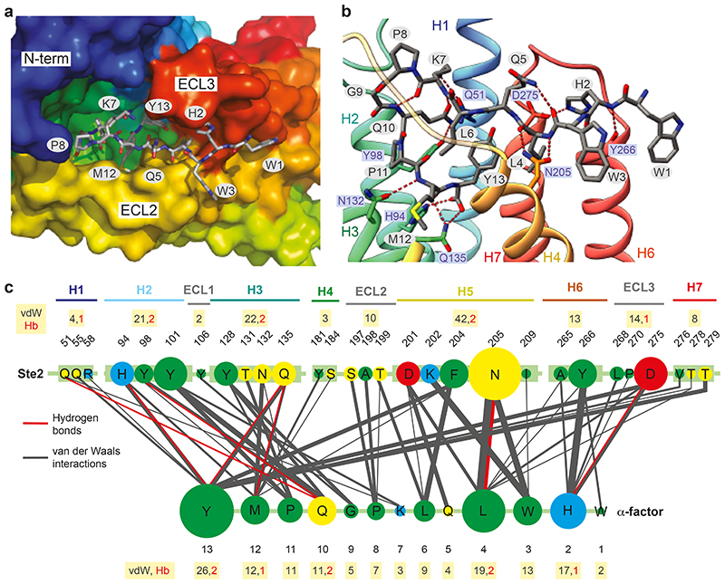
Fig. 3. α-Factor binding site.
a,b view of the orthosteric binding pocket; Ste2A (rainbow colouration); α-factor (sticks; carbon, grey; oxygen, red; nitrogen, blue). a, view from the extracellular surface. b, view in the membrane plane. c, Interactions between α-factor and Ste2. The circle size and line thickness are proportional to the number of interactions. Panels a-b were prepared using PyMOL (Schrödinger Inc).

