Highlights
-
•
BAG3 down-modulation by siRNA technology restored p53.
-
•
Reduced BAG3 expression sensitized HeLa but not C33A (HPV-negative) cells to PEITC.
-
•
Reduced BAG3 expression was associated with a decrease of E6 viral protein levels.
Keywords: BAG3, HeLa cells, HPV18, E6, p53, PEITC
Abstract
BAG3 is a multi-functional component of tumor cell pro-survival machinery, and its biological functions have been largely associated to proteasome system. Here, we show that BAG3 down-modulation resulted in reduced cell viability and enhanced PEITC-induced apoptosis largely more extensively in HeLa (HPV18+) rather than in C33A (HPV−) cervical carcinoma cell lines. Moreover, we demonstrate that BAG3 suppression led to a decrease of viral E6 oncoprotein and a concomitant recovery of p53 tumor suppressor, the best recognized target of E6 for proteasome degradation. E6 and p53 expression were modulated at protein level, since their respective mRNAs were unaffected. Taken together our findings reveal a novel role for BAG3 as host protein contributing to HPV18 E6-activated pro-survival strategies, and suggest a possible relevance of its expression levels in drug/radiotherapy-resistance of HPV18-bearing cervical carcinomas.
Introduction
High-risk human papillomavirus (HPV) infection is the major risk factor of a large number of human cancers, among which the most prevalent is cervical cancer. HPV subtypes 16 and 18 are the most carcinogenic and are responsible for the majority of cervical cancer cases [1]. HPV-encoded oncoproteins E5, E6 and E7 are the primary viral factors responsible for initiation and progression of cervical cancer. They act largely by overcoming negative growth regulation by host cell proteins and by inducing genomic instability ([2] and references therein). E5 has lower transforming activity than E6 and E7, but it is able to potentiate the activity of the other two viral oncoproteins during the early steps of viral infection/transformation. E6 and E7 act in combination to promote degradation by proteasome of proteins involved in cell cycle arrest and DNA damage sensing/repair and of pro-apoptotic proteins as well. The first and best characterized proteins targeted by E7 for proteasome degradation are tumor suppressor proteins of the pRB family, which in turn leads to the increase of p53 and apoptosis [3]. E6 counteracts p53 elevation by targeting it for proteasome degradation mainly through association with cellular ubiquitin ligase E6-associated protein (E6AP) [4]. Moreover, HPV oncoproteins have been shown to up-regulate several proteins important for cancer development [5]. Such complex network of interactions between high-risk HPV viral proteins and host proteins ultimately results in a host cell survival advantage necessary for virus replication and cancer development ([2], [6], [7], [8] and references therein).
The anti-apoptotic cellular machinery includes several proteins, among which is the BAG family molecular chaperone regulator 3 (BAG3). BAG3 protein is a member of the human BAG family of molecular co-chaperone proteins. All BAG proteins contain an evolutionarily conserved domain (BAG domain), through which they bind to heat shock protein cognate 70/heat-shock protein 70 (Hsc70/Hsp70) ATPase domain and to other partners (steroid hormone receptors, RAF-1, and others) ([9] and references therein). In addition, BAG3 contains a WW domain, a proline-rich region (PXXP), and two conserved IPV (Ile-Pro-Val) motifs, that can mediate binding to other proteins. Due to the presence of several protein–protein interaction domains in its structure, BAG3 is implicated in several cellular processes, including cell survival, proliferation, migration, and apoptosis [10], [11]. bag3 gene expression, which is constitutive only in a few normal cell types (e.g. skeletal muscle and heart), can be induced by stressors, such as oxidants, high temperature, and serum deprivation in other normal cell types. The pro-survival role of BAG3 is signified by its over-expression in several human tumors (e.g. pancreatic cancer, melanoma, and leukemia), where it appears to exert an anti-apoptotic role [12]. Recent studies demonstrated that BAG3 is required for efficient growth of different viruses, including varicella-zoster virus [13], HIV-1 [14], Epstein–Barr virus [15], herpes simplex virus [16], polyomavirus JC [17], SARS-CoV [18], and adenovirus [19]. Moreover, we recently demonstrated a positive correlation between BAG3 expression and the presence of Bovine Papilloma Virus in equine sarcomas [20].
To the best of our knowledge, there are only two studies reporting changes of BAG3 expression in high-risk HPV-harboring cells and results were contradictory, possibly depending on the experimental model employed. BAG3 has been proposed as candidate biomarker for early detection of cervical neoplasia by Ranamukhaarachchi et al. [21] on the basis of its up-regulation during dysplastic differentiation of keratinocytes derived from a clinical biopsy of HPV16+ cervical epithelium. Conversely, lowered BAG3 expression has been observed in SiHa cells, harboring HPV16, compared to a normal keratinocyte cell line [22].
With this study we aimed to investigate whether BAG3 is involved in survival and resistance to pro-apoptotic stimuli of high-risk HPV18-infected cells. Here, we demonstrated that down-modulation of BAG3 protein sensitized HPV18+ HeLa, but not HPV– C33A cells to phenethyl isothiocyanate (PEITC)-induced apoptosis. The impact of BAG3 suppression on E6-dependent p53 inactivation machinery in HeLa cells was also explored.
Materials and methods
Reagents and antibodies
Fetal Bovine Serum (FBS) was from GIBCO (Life Technologies, Grand Island, NY, USA). Protein A/G-Sepharose was from Santa Cruz Biotechnology (Santa Cruz, CA, USA). Trizol Reagent, RNAse H, SuperScript® II Reverse Transcriptase, random primers, and dNTP mix were purchased from Invitrogen (Life Technologies). SYBR Green I Master Mix and DNase I were from Roche Applied Science (Mannheim, Germany). Primers (custom synthesized) and all the other reagents were from Sigma-Aldrich (St. Louis, MO, USA).
The polyclonal (TOS-2) antibody against human BAG3 protein was provided by Biouniversa, Italy. Anti-GAPDH (mouse monoclonal, sc-32233), anti-α-tubulin (mouse monoclonal, sc-32293), anti-E6-AP (rabbit polyclonal, sc-25509), and immune control IgG were from Santa Cruz Biotechnology; anti-p53, clone E26 (rabbit monoclonal) were from Millipore (Billerica, MA, USA), anti-HPV16 E6/18 E6 (C1P5) (mouse monoclonal) was from Abcam (Cambridge, CB4 0FL, UK). Peroxidase-conjugated secondary antibodies were from Jackson ImmunoResearch (West Grove, PA).
Cells and BAG3 siRNA transfection
Cervical cancer cell lines HeLa and C33A were obtained from American Type Culture Collection (ATCC) (Manassas, VA, USA). All cells were maintained in EMEM and DMEM (BioWhittaker, Lonza, NJ, USA) media, respectively supplemented with 10% (v/v) FBS, 2 mM L-glutamine and antibiotics at 37 °C in humidified atmosphere at 5% CO2. To ensure logarithmic growth, cells were sub-cultured every 3 days.
A specific small interfering RNA (siRNA) (5′-AAGGUUCAGACCAUCUUGGAA-3′) targeting BAG3 mRNA and a control, scramble (scr) RNA (5′-CAGUCGCGUUUGCGACUGG-3′) were obtained from Dharmacon (Thermo Fisher Scientific, Lafayette, CO, USA). HeLa and C33A cells, at a cell density of 1 × 10⁵/ml, were transfected with siRNA and scrRNA at a final concentration of 100 nM using Lipofectamine™ RNAiMAX reagent (Invitrogen, Life Technologies). Cells were harvested at indicated time points and BAG3 silencing was monitored in all the experiments by Western blotting.
Western blotting and immunoprecipitation
Cell whole lysates for immunoblot analysis were prepared according to the standard protocol. Protein concentration was determined by DC Protein Assay (Bio-Rad, Berkeley, CA, USA), using bovine serum albumin (BSA) as a standard. Proteins were fractionated on SDS-PAGE, transferred into nitrocellulose membranes, and immunoblotted with appropriate primary antibodies. Signals were visualized with appropriate horseradish peroxidase-conjugated secondary antibodies and enhanced chemiluminescence (Amersham Biosciences-GE Healthcare, NY, USA). Densitometry of bands was performed with ImageJ software (http://rsbweb.nih.gov/ij/download.html).
E6 detection was achieved by immunoprecipitation of a large amount of cell lysate (4 mg of proteins) substantially according to Hsu et al. [23]. Briefly, lysates were incubated with 2 µg of anti-HPV16 E6/18 E6 antibody or immune control IgG at 4 °C overnight on a tube rotator. Thirty-five microliters of protein A/G-Sepharose was then added and the reaction mixtures were incubated further for 2 h at 4 °C. The immune complexes were pelleted and washed five times with 0.1% Tween/PBS solution. After final centrifugation, pellets were suspended in 25 µl of Laemmli buffer.
Cell viability, cell cycle and apoptosis
The number of viable cells was quantified by MTT ([3-(4,5-dimethylthiazol-2-yl)-2,5-diphenyl tetrazolium bromide]) assay. Absorption at 550 nm was assessed using a microplate reader (LabSystems, Vienna, VA, USA). In some experiments cell viability was also checked by Trypan Blue exclusion assay using a Bürker counting chamber.
Cellular DNA content was evaluated by propidium iodide (PI) staining of permeabilized cells according to the available protocol and flow cytometry (BD FACSCalibur flow cytometer, Becton Dickinson, San Jose, CA, USA). Data from 5000 events per sample were collected. The percentages of the elements in the hypodiploid region were calculated using the CellQuest software and those in G0/G1, S and G2/M phases of the cell cycle were determined using the MODFIT software.
Apoptosis was determined by Human Annexin V/FITC kit (Bender MedSystem, Wien, Austria) according to the manufacturer's instructions. Green (Annexin V-FITC) and red (PI) fluorescence of individual cells were measured by flow cytometry. Electronic compensation was required to exclude overlapping of the two emission spectra.
Phase contrast microscopic analysis were performed by using a Zeiss Axiovert 200 microscope (Zeiss, Oberkochen, Germany) equipped with a 40× objective (λexc, 351 nm; λem, 380 nm), and images were acquired from randomly selected fields.
Adhesion assay
ScrRNA and BAG3siRNA-loaded cells were harvested at 72 h post-transfection. After centrifugation to remove dead cells, HeLa and C33A cells (at a cell density of 1 × 10⁵/ml and 2 × 10⁵/ml, respectively) were added to wells, previously coated with 10 µg/ml fibronectin (FN), and allowed to adhere for 90 min. The wells were washed three times with PBS, to remove non adherent cells. The number of cells was then determined by evaluating endogenous acid phosphatase with p-nitrophenyl phosphate as substrate [24]. In each experiment reagents were checked using known amounts of cells.
RNA isolation and quantitative real-time RT-PCR (qRT-PCR)
Total RNA was isolated using Trizol Reagent according to the manufacturer's instructions and spectrophotometrically quantified. RNA integrity was assessed by agarose gel electrophoresis. Three micrograms of RNA were reverse transcribed, and qRT-PCR was performed with Light-Cycler® 480 (Roche Diagnostics GmbH, Mannheim, Germany) using SYBR Green detection in a total volume of 20 µl with 1 µl of forward and reverse primers (10 mM) and 10 µl of SYBR Green I Master Mix. Reactions included an initial cycle at 95 °C for 10 min, followed by 40 cycles of denaturation at 95 °C for 10 s, annealing at 56 °C for 5 s, extension at 72 °C for 15 s. The 18S RNA was used as an internal standard. The following primer sets were used for qRT-PCR to assay specific mRNAs: i) forward E6 HPV18: 5′-ACC CTA CAA GCT ACC TGA TC-3′, reverse E6 HPV18: 5′-GTG TCT CCA TAC ACA CAG AGT C-3′; forward 18S: 5′-CGA TGC TCT TAG CTG AGT GT-3′, reverse 18S: 5′-GGT CCA AGA ATT TCA CCT CT-3′; ii) forward p53: 5′-CCA CTT CAC CGT ACT AAC CA-3′, reverse p53: 5′-GTC AAG TTC TAG ACC CCA TG-3′. Fold changes of E6 and p53 mRNA levels were determined by calculating ratios between scrRNA and BAG3siRNA and control normalized signals.
Statistical analysis
Data reported in each figure are the mean values ± SD from at least three experiments, performed in duplicate, showing similar results. Differences between treatment groups were analyzed by Student's t-test. Differences were considered significant when p < 0.05.
Results
BAG3 pro-survival role in HeLa cells
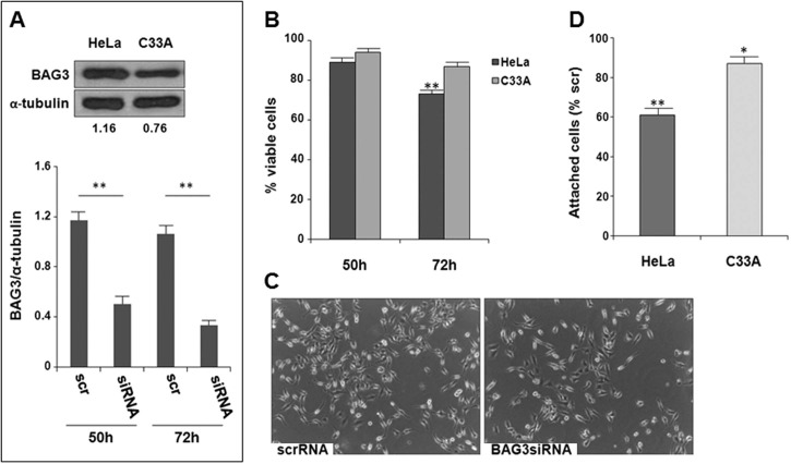
Firstly we examined BAG3 basal expression in HeLa (HPV18+) and C33A (HPV–) cervical carcinoma cell lines [25]. The blot in Fig. 1A shows that BAG3 is expressed in both cell line, even though to a higher extent in HeLa cells. BAG3 was efficiently down-modulated in both HeLa and C33A cells using a BAG3-specific siRNA (BAG3siRNA). In Fig. 1A (lower panels) we reported only the time-dependent reduction of BAG3 expression in HeLa cells as representative of results obtained in C33A cells. At 50 h and 72 h post-transfection (p.t.), BAG3 levels in BAG3siRNA-loaded cells were reduced by about 40% and more than 70%, respectively, compared to scramble RNA (scrRNA)-transfected cells. The slight increase of BAG3 protein in scrRNA-loaded cells with respect to control (untransfected) cells was possibly due to transfection procedure-induced stress. Notably, we found that, under our experimental conditions, BAG3 down-modulation caused a reduction of cell number specifically in HeLa cells (Fig. 1B). Phase-contrast microscopy analysis of BAG3-silenced HeLa cell revealed the presence of cells loosing the flat/spindle-shaped morphology and an increase in the number of cells floating in the medium (Fig. 1C). Figure 1D shows that BAG3 silencing reduced the adhesion efficiency to FN-coated plates more markedly in HeLa than in C33A cells, thus confirming that BAG3 contributes in promoting cell-matrix contacts specifically in HeLa.
Fig. 1.
BAG3 down-modulation affects HeLa cell viability. (A) BAG3 expression in HeLa and C33A cells (upper panel): BAG3/α-tubulin densitometry ratios are indicated; the blot is representative of at least two separate experiments with similar results. BAG3 down-modulation in HeLa cells in function of the time post-transfection (p.t) (lower panel): BAG3 levels were monitored by Western blotting and densitometry data of samples are expressed as fractions of BAG3/α-tubulin ratio in control cells normalized to 1. (B) Effect of BAG3 down-modulation on HeLa and C33A viability; on the Y axis the percentages of viable cells in BAG3-silenced cell samples versus scrRNA-transfected cells (% cell number of scrRNA versus control: 90 ± 3 at 50 h, 94 ± 3.3 at 72 h). (C) Representative phase contrast microscopy images (40× magnification) of scrRNA- and BAG3siRNA-transfected HeLa at 72 h p.t. (D) Effect of BAG3 down-modulation on HeLa and C33A adhesion to FN-coated plates. All results in A, B and D are the mean values ± SD from at least three experiments performed in duplicate (*p < 0.05, **p < 0.001).
PEITC effects on HeLa and C33A cell proliferation and viability
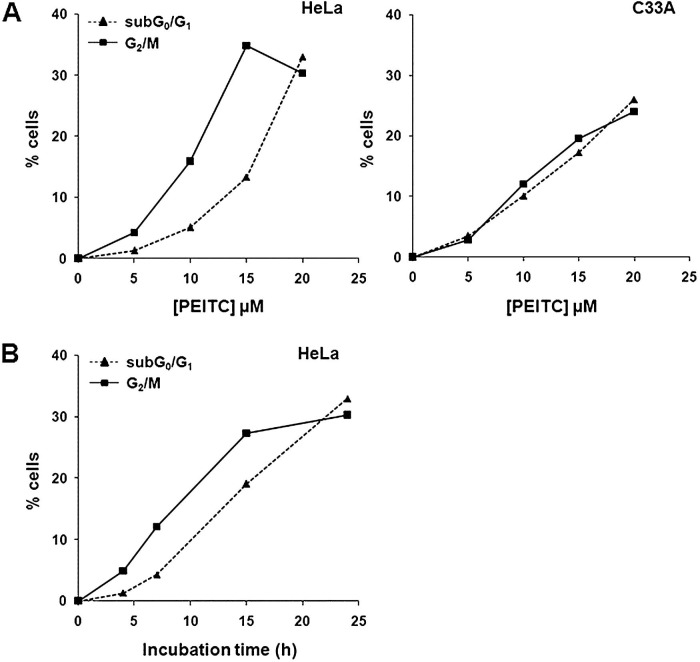
In preliminary experiments we confirmed that PEITC efficiently inhibited HeLa and C33A cell growth [26]. Under our experimental conditions, PEITC Ic50 (half-maximal inhibitory concentration) value at 24 h was about 12 µM in both the two cell lines (dose–response curves not shown). To gain more insight into the mechanisms underlying PEITC cell growth inhibition potential in HeLa and C33A cells, we titrated the effects of such drug on cell cycle progression and apoptotic death (Fig. 2A ). In C33A cells, apoptosis and cell cycle arrest were induced almost simultaneously, while in HeLa cells the block in G2/M seemed to precede the onset of apoptosis. The time-course of apoptosis and cell cycle arrest induction by 20 µM PEITC (Fig. 2B) suggested that apoptotic death occurred subsequently to G2/M arrest. As the percentages of cells accumulated in G2/M increased, the percentage of subG0/G1 cells started to grow. The reduction of G2/M cell population at 24 h treatment with this high PEITC dose (panels A and B in Fig. 2), concomitant to an increase of cells in S and G0/G1 (data not shown), was due to the apoptotic fate of arrested cells, rather than to a recovery of cell cycle progression. Apoptosis-induced DNA fragmentation in G2/M cells generates, indeed, subG2/M populations artifactually integrated in FACS analysis as S or G0/G1 peaks.
Fig. 2.
PEITC-induced G2/M and apoptosis in cervical cancer cell lines. (A) HeLa and C33A cells were exposed to increasing amounts of PEITC for 24 h; the percentages of hypodiploid and G2/M arrested cells were evaluated by flow cytometry analysis of PI-stained nuclei. (B) HeLa cells were exposed to a fixed dose of PEITC (20 µM) for the times indicated. Data in A and B have been subtracted for the corresponding values in control cells (exposed to vehicle only): HeLa, subG0/G1 ≤ 2%; G2/M 14.74 ± 1.1%; C33A, subG0/G1 ≤ 2.2%; G2/M 13.04 ± 1.5%. Data are the mean values from three experiments: SD values never exceeded 12% of the means.
The more pronounced G2/M arrest in HeLa compared to C33A cells was found to correlate with a more extensive PEITC-induced tubulin disruption (Supplementary Fig. S1).
BAG3 down-modulation enhances PEITC-induced apoptosis in HeLa cells
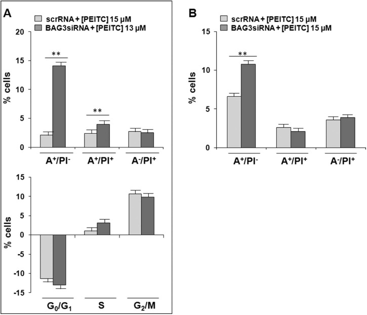
The effect of BAG3 down-modulation on PEITC-induced apoptosis in HeLa and C33A cells was then evaluated. To avoid the possibility of false positive results due to BAG3 silencing-induced reduction of HeLa cell number (see Fig. 1B) we adopted the following experimental conditions: i) BAG3siRNA- and scrRNA-transfected HeLa and C33A cells received vehicle or PEITC as early as at 62 h p.t. and were incubated with the drug further for 10 h; ii) only for HeLa cells: the amount of PEITC added to BAG3siRNA-transfected cells was about 15% lower than that used for scrRNA-transfected cells. PEITC-induced apoptosis in the different samples was monitored by the Annexin V/PI test to better discriminate between apoptotic and necrotic death. Results summarized in Fig. 3A show that BAG3 down-modulation allowed overcoming the low/delayed basal HeLa cell response to PEITC-triggered pro-death signals, this resulting in a marked and earlier increase of cells undergoing apoptosis. Notably, the percentages of apoptotic cells (phosphatidyl serine exposing cells, A+/PI– and AP+/PI+) was increased by about sevenfold in HeLa cells, while by less than twofold in C33A cells (Fig. 3B). BAG3 down-modulation did not, instead, affect the extent of PEITC-induced G2/M block either in HeLa (Fig. 3A, lower panel) or C33A cells (not shown).
Fig. 3.
Effects of BAG3 down-modulation on HeLa and C33A cell susceptibility to PEITC-induced apoptosis. (A) ScrRNA- and BAG3siRNA-transfected HeLa and C33A cells received PEITC or vehicle at 62 h post-transfection (PEITC doses are indicated) and incubated for further 10 h. Left panel: apoptosis was assessed by Annexin V/PI assay (A+/PI–, early apoptotic cells; A+/PI+, late apoptotic/necrotic cells; A–/PI+, necrotic cells) and flow cytometry. On the Y axis: the percentage in each quadrant of PEITC-treated scrRNA- and BAG3siRNA-transfected cells subtracted for the percentage in the respective quadrants of control cells. Right panel: cell cycle progression was evaluated by flow cytometry analysis of PI stained nuclei. On the Y axis: the percentage of PEITC-treated scrRNA- and BAG3siRNA-transfected cells with a specific DNA content subtracted for the respective percentage in control cells. (B) Effect of BAG3 silencing on PEITC-induced apoptosis in C33A. Treatment conditions and procedures are same as in A. All results in A and B are the mean values ± SD from at least three experiments performed in duplicate (**p < 0.001).
The involvement of BAG3 in PEITC-induced response in HeLa cells was also signified by the bi-phasic effect of the drug on BAG3 levels (Fig. 4 ). In agreement with the stress-inducible nature of BAG3, its expression resulted in an increase at PEITC doses lower than 15 µM. The appearance of the additional immunoreactive BAG3 band with a slightly higher molecular weight in the doublet has been already reported in response to others stress-inducing chemicals [12]. Notably, the increase of BAG3 expression was followed by a marked suppression of BAG3 signal at 20 µM PEITC. Such a reduction was not simply due to gross toxicity since in C33A the same highly cytotoxic dose of PEITC had low, if any, effect on BAG3 levels.
Fig. 4.
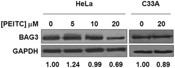
PEITC affects BAG3 expression in HeLa cells. BAG3 levels in HeLa and C33A cells exposed for 8 h to increasing concentrations of PEITC. Densitometry data of PEITC-treated cells are expressed as fractions of BAG3/GAPDH ratio in control cells (vehicle only) normalized to 1. Blots shown are from one experiment representative of at least two with similar results.
BAG3 silencing and PEITC restore p53 levels in HeLa cells
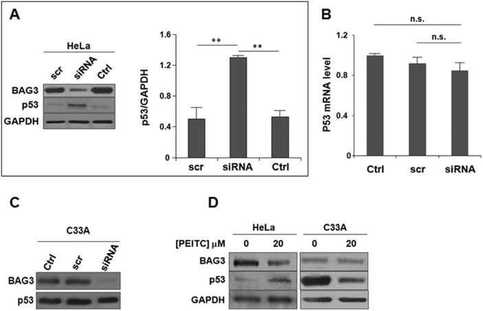
The levels of p53 in HeLa cells are kept low due to E6/E6AP-enhaced proteasome degradation [4]. Therefore to detect p53 signal up to 100 µg of HeLa cell lysate proteins were loaded onto the gel. Figure 5A clearly shows that the p53 faint signal detected in control and scrRNA-transfected HeLa cell lysates became more intense in BAG3siRNA-transfected cells. qRT-PCR analysis revealed no changes of p53 mRNA levels, thus indicating that p53 was increased at protein level (Fig. 5B). In C33A cells, the intensity of p53 band was, instead, unaffected by BAG3 down-modulation (Fig. 5C).
Fig. 5.
BAG3 down-modulation and PEITC restore p53 levels in HeLa cells. (A) p53 levels in control (Ctrl), scrRNA (scr)- and BAG3siRNA (siRNA)-transfected HeLa cells harvested at 72 h post-transfection. Eighty micrograms of proteins were loaded in each lane. Data are expressed as p53/GAPDH densitometry ratios and represented as the mean values ± SD from at least three experiments (**p < 0.001). BAG3 silencing was checked. (B) p53 mRNA levels by qRT-PCR. p53 mRNA levels (Y axis) in scrRNA (scr) and BAG3siRNA (siRNA) are expressed relative to p53 mRNA level in control cells. Data are the mean values ± SD from two independent experiments performed in duplicate. (C) Effect of BAG3 silencing on p53 levels in C33A cells. (D) BAG3 and p53 levels in C33A and HeLa cells exposed to 20 µM PEITC for 10 h. The blots in C and D are from one experiment representative of at least two with similar results.
An increase of the p53 signal was also observed in HeLa cells exposed to 20 µM PEITC (Fig. 5D, left panel), a concentration of the drug causing a marked and early apoptotic response and effective in reducing per se BAG3 protein levels (see Fig. 4). The elevation of p53 was specific for HeLa cells, since the treatment of C33A cells with the same dose of PEITC led, instead, to a reduction of p53 protein (Fig. 5D, right panel).
Effect of BAG3 silencing on E6 and E6AP levels
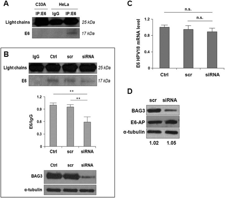
Next, we explored the effect of reduced BAG3 expression on the levels of both viral E6 oncoprotein and cellular E6AP protein. Due to the low expression of E6 protein in HeLa cells, the samples were enriched in E6 protein by immunoprecipitating a large amount of cell lysates (details in Materials and methods). Using this experimental trick, we successfully demonstrated the presence of an E6 immunoreactive band (apparent MW of 17 kDa) in HeLa cells. The signal was specific for E6 because it was absent in C33A cells (Fig. 6A ). BAG3 down-modulation led to a slight, but highly reproducible, decrease of E6 immunoreactive signal compared to control (non transfected) and scrRNA-transfected HeLa cells (Fig. 6B). E6 was reduced at protein level, since BAG3 silencing did not significantly affect E6 mRNA levels (Fig. 6C). Unlike E6, E6AP was unaffected by BAG3 down-modulation (Fig. 6D).
Fig. 6.
Effect of BAG3 down-modulation on E6 viral protein and cellular E6AP levels. (A) HeLa and C33A lysates were immunoprecipitated with an anti-E6 antibody and blotted for E6. (B) E6 levels in vehicle (Ctrl), scrRNA (scr)- and BAG3siRNA (siRNA)-transfected HeLa cells harvested at 72 h post-transfection. Densitometry data of samples are expressed as fractions of E6/IgG (light chains) ratio in control cells normalized to 1 and represented as the mean values ± SD of four separate experiments. An aliquot of each lysate was used to check BAG3 silencing (lower blots). (C) E6 mRNA levels by qRT-PCR. E6 mRNA levels (Y axis) in scrRNA (scr) and BAG3siRNA (siRNA) are expressed relative to E6 mRNA level in control cells. Data are the mean values ± SD from three independent experiments. (D) E6AP levels in the indicated samples (15 µg protein/lane). E6AP to α-tubulin densitometry ratios are indicated. The blot, probed also for BAG3 to confirm the down-regulation of the protein, is representative of at least two experiments with similar results.
Discussion
BAG3 is a stress-inducible anti-apoptotic protein over-expressed in cancers and tumor cell lines [9]. BAG3 influences cell survival by interacting with different molecular partners involved in cell development, apoptosis, autophagy, cell migration, and tumor metastasis. In several cancer cell models, the reduction of BAG3 expression leads to the sensitization to many apoptosis inducers, while its overexpression reduces sensitivity to apoptosis. In view of the demonstrated association of BAG3 biological functions with the proteasome–ubiquitination system [27], [28] and the known ability of the high risk oncoprotein E6 to target numerous host cell proteins for proteasome degradation [29], [30], we explored the role of BAG3 in HPV18-infected HeLa cells survival and resistance to drug-induced apoptosis.
BAG3 was found to be expressed at higher levels in HPV18+ HeLa than in HPV− C33A cervical carcinoma cell lines and its down-modulation by a specific siRNA had a more negative impact on HeLa cell growth. These findings were taken as preliminary evidence of possible major pro-survival functions of BAG3 in the HPV18+ cell line. To support this hypothesis, we next examined whether BAG3-down modulation might also differently affect HeLa and C33A susceptibility to chemical-induced apoptosis.
PEITC is a natural compound from cruciferous vegetables, widely investigated for its low systemic toxicity, but relevant anti-tumor potential ([31] and references therein). According to previous data [26], we found that PEITC efficiently inhibited HeLa and C33A cell growth by promoting apoptotic death and cell cycle arrest in G2/M phases. However, the two carcinoma cell lines displayed a different susceptibility to PEITC cytostatic and cytotoxic effects. The comparison of PEITC dose–response curves indicated that the reduction of cell number in HeLa cells was due mainly to cell cycle arrest, while in C33A cells to both apoptosis and G2/M block. Moreover, the kinetics of G2/M arrest and apoptosis induction by a fixed PEITC dose showed that in HeLa cells the onset of apoptosis was delayed and occurred mainly in G2/M arrested cells. Notably, we found that BAG3 down-modulation sensitized HeLa cells to PEITC-triggered pro-apoptotic signals, this resulting in a marked and early increase of cells undergoing apoptosis. The enhanced apoptotic response to PEITC appeared to be cell type-specific, since BAG3 suppression increased only moderately the response in C33A cells.
The tumor suppressor p53 controls the expression of several proteins involved in apoptosis or in the control of cell cycle, and the inhibition of p53 transactivation functions promotes cell death and/or cell cycle arrest [32]. The presence of mutate p53 gene or perturbation of its functions are, indeed, common features of several human cancers [33]. Cervical carcinoma C33A (HPV–) cells express mutate p53, while HeLa (HPV18+) cells express wild-type p53 [34]. However, the viral E6 oncoprotein strongly affects the p53 status in HPV-infected cells mainly by promoting p53 proteasome degradation [35] and inhibiting its transactivation [36]. The restoration of p53 in high risk HPV+ cells has been shown to positively correlate with increased susceptibility to undergo apoptosis in response to chemical and physical agents [37], [38]. Regarding PEITC, literature data suggest that the involvement of p53 pathways in PEITC cell growth inhibition is cell type-specific [39], [40], [41], [42], [43]. Here, we demonstrated that BAG3 suppression, in addition to enhancing the apoptotic response of HeLa cells to PEITC, caused a significant increase of p53. This result could be taken as evidence of a role of restored p53 pathways in PEITC-induced apoptosis in HeLa cells. A possible relationship between BAG3 suppression and p53 recovery for the commitment of HeLa cells toward a death fate was also supported, even if not conclusively proven, by the observation that the treatment of HeLa cells with a highly cytotoxic PEITC concentration resulted in a decrease of BAG3 concomitant to an increase of p53.
Conversely, the restoration of p53 in HeLa cells silenced for BAG3 did not affect the extent of G2/M arrest induced by PEITC. The lack of any role of p53 in PEITC cytostatic effects was not completely surprising in view of the well established ability of PEITC to target tubulin and, consequently, to promote its disruption [44]. Accordingly, the more marked and earlier tubulin decrease in HeLa than in C33A cells well correlated with the more pronounced G2/M arrest (Supplementary Fig. S1).
Unlike in HeLa cells, in C33A cells the levels of p53 were unaffected by BAG3 down-regulation and the treatment with highly cytotoxic PEITC doses reduced mutate p53 protein. This result was in agreement with the previous study of Wang et al. [45], who also demonstrated that the selective depletion of mutate p53 proteins resulted in a higher basal susceptibility to PEITC-induced apoptosis of cells expressing mutate p53 compared to cells with wild-type p53. In contrast, we found that BAG3 suppression and the resulting recovery of p53 made HeLa more susceptible than C33A cells to PEITC pro-apoptotic signals. Although the results of Wang et al. [45] could not be generalized to all cell types, we cannot exclude that other mechanisms, in addition to the recovery of wild-type p53, contributed to BAG3 silencing-induced HeLa cells sensitization to PEITC cytotoxic effects.
The recovery of p53 in HeLa cells, induced by suppressing BAG3 expression, was mediated by post-transcriptional mechanisms, since the levels of its mRNA were unaffected. E6-mediated p53 proteasome degradation is the main, if not exclusive, post-transcriptional mechanism responsible of the low p53 levels in HPV-infected cells [35], [46]. It has been shown that E6 promotes p53 proteasome degradation not only by the well established ubiquitination-dependent pathway [4], but also by ubiquitination-independent pathways [47]. Moreover, E6 can inactivate p53 also by inhibiting post-translational modifications, i.e. phosphorylation [48] and acetylation [36], required not only for p53 transactivation, but also for its stability [49], [50]. Appropriate experimental conditions allowed us to detect E6, expressed at very low levels in HeLa cells, and to demonstrate that the suppression of BAG3 caused the decrease of E6. This was in support of a BAG3 silencing-induced impairment of p53 degradation machinery as one of the mechanisms responsible for the elevation of the transcriptional factor in HeLa cells. The cellular E3 ubiquitin ligase, E6AP, which cooperates with E6 for p53 ubiquitination and subsequent proteasome degradation [4], was, instead, unaffected by BAG3 down-modulation. The intracellular levels of E6 are regulated by the balance between expression and proteasome degradation [51], [52]. We showed that BAG3 silencing affected E6 at protein level. On the basis of this result and by considering the well known association of BAG3 functions with the proteasome system [27], [28], we could hypothesize that BAG3 suppression allows a more efficient proteasome degradation of viral E6 oncoprotein [51], [52]. However, BAG3 has not been recognized as one of E6 partners [8]. Thus, it might contribute to E6 stability by assisting/sustaining the binding of the viral oncoprotein to those partners, namely E6AP [53], 14-3-3ζ [54], and PDZ domain-containing proteins [55], shown to be relevant for its own stabilization. In the present study, due to the low basal levels of viral E6 oncoprotein and, consequently, of p53 tumor suppressor in HeLa cell model, we were unable to investigate in depth the possible mechanisms by which BAG3 silencing promoted p53 elevation and E6 reduction in these cells. Further studies in cells over-expressing E6 or p53 are needed to address these questions.
In conclusion, the present study reveals a role of BAG3 in sustaining HPV18 E6-mediated p53 disruption and suggests this as one of the mechanisms involved in BAG3-silenced HeLa cells sensitization to PEITC. The higher BAG3 expression in HPV18+ HeLa compared to HPV– C33A might, in turn, result from an E6-activated positive feedback loop to sustain infected cell survival. Moreover, the demonstrated ability of PEITC to promote per se p53 recovery, possibly by depleting BAG3, might provide a rationale for the previously shown PEITC-induced sensitization of HeLa cells to apoptosis induced by cisplatin, a chemotherapeutic method routinely used in cervical cancer treatment [26]. Since cisplatin has also been reported to prevent p53 degradation [56], the combined treatment of cells with the two drugs could actually result in a fast and marked p53 restoration in HeLa cells and, consequently, apoptotic death.
Authors' contributions
The work presented here was carried out in collaboration between all authors. RC and ER carried out the laboratory experiments, analyzed the data, and interpreted the results. AB and DG performed statistical analysis, participated in literature searches, and data presentation. MAB conceived the biochemical study design, coordinated the experiments, and drafted the manuscript. All authors approved the final manuscript to be submitted.
Conflicts of interest
BIOUNIVERSA s.r.l., which produces anti-BAG3 antibodies, provided them free of charges for this study. AB is shareholder of the company BIOUNIVERSA that provided some of the used antibodies. The remaining authors declare no conflict of interest.
Acknowledgments
We thank Prof. M. C. Turco for helpful discussion. This work was supported by PRIN grant n. 2008LTY389 from Ministero dell'Università e della Ricerca Scientifica, Italy, and by an Internal Grant of the University of Salerno, Italy. The study sponsors had no involvement in the writing of the manuscript; and in the decision to submit the manuscript for publication.
Footnotes
Supplementary data to this article can be found online at doi:10.1016/j.canlet.2014.08.022.
Appendix. Supplementary material
The following is the supplementary data to this article:
PEITC-induced tubulin degradation in HeLa and C33A cells. The decreasing of α-tubulin was evaluated in HeLa and C33A cells incubated with15 µM PEITC for the indicated times. GAPDH was used as loading control.
References
- 1.Smith J.S., Lindsay L., Hoots B., Keys J., Franceschi S., Winer R. Human papillomavirus type distribution in invasive cervical cancer and high-grade cervical lesions: a meta-analysis update. Int. J. Cancer. 2007;121:621–632. doi: 10.1002/ijc.22527. [DOI] [PubMed] [Google Scholar]
- 2.Moody C.A., Laimins L.A. Human papillomavirus oncoproteins: pathways to transformation. Nat. Rev. Cancer. 2010;10:550–560. doi: 10.1038/nrc2886. [DOI] [PubMed] [Google Scholar]
- 3.Demers G.W., Espling E., Harry J.B., Etscheid B.G., Galloway D.A. Elevated wild-type p53 protein levels in human epithelial cell lines immortalized by the human papillomavirus type 16 E7 gene. Virology. 1994;198:169–174. doi: 10.1006/viro.1994.1019. [DOI] [PubMed] [Google Scholar]
- 4.Talis A.L., Huibregtse J.M., Howley P.M. The role of E6AP in the regulation of p53 protein levels in human papillomavirus (HPV)-positive and HPV-negative cells. J. Biol. Chem. 1998;273:6439–6445. doi: 10.1074/jbc.273.11.6439. [DOI] [PubMed] [Google Scholar]
- 5.Fragoso-Ontiveros V., Alvarez-García R.M., Contreras-Paredes A., Vaca-Paniagua F., Alonso Herrera L., López-Camarillo C. Gene expression profiles induced by E6 from non-European HPV18 variants reveals a differential activation on cellular processes driving to carcinogenesis. Virology. 2012;432:81–90. doi: 10.1016/j.virol.2012.05.029. [DOI] [PubMed] [Google Scholar]
- 6.Tungteakkhun S.S., Duerksen-Hughes P.J. Cellular binding partners of the human papillomavirus E6 protein. Arch. Virol. 2008;153:397–408. doi: 10.1007/s00705-007-0022-5. [DOI] [PMC free article] [PubMed] [Google Scholar]
- 7.Pim D., Bergant M., Boon S.S., Ganti K., Kranjec C., Massimi P. Human papillomaviruses and the specificity of PDZ domain targeting. FEBS J. 2012;279:3530–3537. doi: 10.1111/j.1742-4658.2012.08709.x. [DOI] [PubMed] [Google Scholar]
- 8.White E.A., Kramer R.E., Tan M.J., Hayes S.D., Harper J.W., Howley P.M. Comprehensive analysis of host cellular interactions with human papillomavirus E6 proteins identifies new E6 binding partners and reflects viral diversity. J. Virol. 2012;86:13174–13186. doi: 10.1128/JVI.02172-12. [DOI] [PMC free article] [PubMed] [Google Scholar]
- 9.Rosati A., Graziano V., De Laurenzi V., Pascale M., Turco M.C. BAG3: a multifaceted protein that regulates major cell pathways. Cell Death Dis. 2011;2:141–144. doi: 10.1038/cddis.2011.24. [DOI] [PMC free article] [PubMed] [Google Scholar]
- 10.McCollum A.K., Casagrande G., Kohn E.C. Caught in the middle: the role of Bag3 in disease. Biochem. J. 2009;425:1–3. doi: 10.1042/BJ20091739. [DOI] [PMC free article] [PubMed] [Google Scholar]
- 11.Zhu H., Liu P., Li J. BAG3: a new therapeutic target of human cancers? Histol. Histopathol. 2012;27:257–261. doi: 10.14670/HH-27.257. [DOI] [PubMed] [Google Scholar]
- 12.Rosati A., Ammirante M., Gentilella A., Basile A., Festa M., Pascale M. Apoptosis inhibition in cancer cells: a novel molecular pathway that involves BAG3 protein. Int. J. Biochem. Cell Biol. 2007;39:1337–1342. doi: 10.1016/j.biocel.2007.03.007. [DOI] [PubMed] [Google Scholar]
- 13.Kyratsous C.A., Silverstein S.J. BAG3, a host cochaperone, facilitates varicella-zoster virus replication. J. Virol. 2007;81:7491–7503. doi: 10.1128/JVI.00442-07. [DOI] [PMC free article] [PubMed] [Google Scholar]
- 14.Rosati A., Khalili K., Deshmane S.L., Radhakrishnan S., Pascale M., Turco M.C. BAG3 protein regulates caspase-3 activation in HIV-1-infected human primary microglia cells. J. Cell. Physiol. 2009;218:264–267. doi: 10.1002/jcp.21604. [DOI] [PMC free article] [PubMed] [Google Scholar]
- 15.Young P., Anderton E., Paschos K., White R., Allday M.J. Epstein-Barr virus nuclear antigen (EBNA) 3A induces the expression of and interacts with a subset of chaperones and co-chaperones. J. Gen. Virol. 2008;89:866–877. doi: 10.1099/vir.0.83414-0. [DOI] [PMC free article] [PubMed] [Google Scholar]
- 16.Kyratsous C.A., Silverstein S.J. The co-chaperone BAG3 regulates herpes simplex virus replication. Proc. Natl. Acad. Sci. U.S.A. 2008;105:20912–20917. doi: 10.1073/pnas.0810656105. [DOI] [PMC free article] [PubMed] [Google Scholar]
- 17.Basile A., Darbinian N., Kaminski R., White M.K., Gentilella A., Turco M. Evidence for modulation of BAG3 by polyoma-virus JC early protein. J. Gen. Virol. 2009;90:1629–1640. doi: 10.1099/vir.0.008722-0. [DOI] [PMC free article] [PubMed] [Google Scholar]
- 18.Zhang L., Zhang Z.P., Zhang X.E., Lin F.S., Ge F. Quantitative proteomics analysis reveals BAG3 as a potential target to suppress severe acute respiratory syndrome coronavirus replication. J. Virol. 2010;84:6050–6059. doi: 10.1128/JVI.00213-10. [DOI] [PMC free article] [PubMed] [Google Scholar]
- 19.Gout E., Gutkowska M., Takayama S., Reed J.C., Chroboczek J. Co-chaperone BAG3 and adenovirus penton base protein partnership. J. Cell. Biochem. 2010;111:699–708. doi: 10.1002/jcb.22756. [DOI] [PMC free article] [PubMed] [Google Scholar]
- 20.Cotugno R., Gallotta D., d'Avenia M., Corteggio A., Altamura G., Roperto F. BAG3 protects bovine papillomavirus type 1-transformed equine fibroblasts against pro-death signals. Vet. Res. 2013;44:61–72. doi: 10.1186/1297-9716-44-61. [DOI] [PMC free article] [PubMed] [Google Scholar]
- 21.Ranamukhaarachchi D.G., Unger E.R., Vernon S.D., Lee D., Rajeevan M.S. Gene expression profiling of dysplastic differentiation in cervical epithelial cells harboring human papillomavirus 16. Genomics. 2005;85:727–738. doi: 10.1016/j.ygeno.2005.02.008. [DOI] [PubMed] [Google Scholar]
- 22.Kim Y.W., Suh M.J., Bae J.S., Bae S.M., Yoon J.H., Hur S.Y. New approaches to functional process discovery in HPV 16-associated cervical cancer cells by gene ontology. Cancer Res. Treat. 2003;35:304–313. doi: 10.4143/crt.2003.35.4.304. [DOI] [PubMed] [Google Scholar]
- 23.Hsu C.H., Peng K.L., Jhang H.C., Lin C.H., Wu S.Y., Chiang C.M. The HPV E6 oncoprotein targets histone methyltransferases for modulating specific gene transcription. Oncogene. 2012;31:2335–2349. doi: 10.1038/onc.2011.415. [DOI] [PMC free article] [PubMed] [Google Scholar]
- 24.Belisario M.A., Tafuri S., Di Domenico C., Squillacioti C., Della Morte R., Lucisano A. H2O2 activity on platelet adhesion to fibrinogen and protein tyrosine phosphorylation. Biochim. Biophys. Acta. 2000;1495:183–193. doi: 10.1016/s0167-4889(99)00160-3. [DOI] [PubMed] [Google Scholar]
- 25.Yee C. Presence and expression of human papillomavirus sequences in human cervical carcinoma cell lines. Am. J. Pathol. 1985;119:361–366. [PMC free article] [PubMed] [Google Scholar]
- 26.Wang X., Govind S., Sajankila S.P., Mi L., Roy R., Chung F.L. Phenethyl isothiocyanate sensitizes human cervical cancer cells to apoptosis induced by cisplatin. Mol. Nutr. Food Res. 2011;55:1572–1581. doi: 10.1002/mnfr.201000560. [DOI] [PMC free article] [PubMed] [Google Scholar]
- 27.Doong H., Rizzo K., Fang S., Kulpa V., Weissman A.M., Kohn E.C. CAIR-1/BAG-3 abrogates Heat Shock Protein-70 chaperone complex-mediated protein degradation. J. Biol. Chem. 2003;278:28490–28500. doi: 10.1074/jbc.M209682200. [DOI] [PubMed] [Google Scholar]
- 28.Chen Y., Yang L.N., Cheng L., Tu S., Guo S.J., Le H.Y. Bcl2-associated athanogene 3 interactome analysis reveals a new role in modulating proteasome activity. Mol. Cell. Proteomics. 2013;12:2804–2819. doi: 10.1074/mcp.M112.025882. [DOI] [PMC free article] [PubMed] [Google Scholar]
- 29.Di Domenico F., De Marco F., Perluigi M. Proteomics strategies to analyze HPV-transformed cells: relevance to cervical cancer. Expert Rev. Proteomics. 2013;10:461–472. doi: 10.1586/14789450.2013.842469. [DOI] [PubMed] [Google Scholar]
- 30.de Freitas A.C., Coimbra E.C., Leitão M.C.G. Molecular targets of HPV oncoproteins: potential biomarkers for cervical carcinogenesis. Biochim. Biophys. Acta. 1845;2014:91–103. doi: 10.1016/j.bbcan.2013.12.004. [DOI] [PubMed] [Google Scholar]
- 31.Singh S.V., Singh K. Cancer chemoprevention with dietary isothiocyanates mature for clinical translational research. Carcinogenesis. 2012;33:1833–1842. doi: 10.1093/carcin/bgs216. [DOI] [PMC free article] [PubMed] [Google Scholar]
- 32.Levine A.J., Oren M. The first 30 years of p53: growing ever more complex. Nat. Rev. Cancer. 2009;9:749–758. doi: 10.1038/nrc2723. [DOI] [PMC free article] [PubMed] [Google Scholar]
- 33.Muller P.A.J., Vousden K.H. p53 mutations in cancer. Nat. Cell Biol. 2013;15:2–8. doi: 10.1038/ncb2641. [DOI] [PubMed] [Google Scholar]
- 34.Scheffner M., Münger K., Byrne J.C., Howley P.M. The state of the p53 and retinoblastoma genes in human cervical carcinoma cell lines. Proc. Natl. Acad. Sci. U.S.A. 1991;88:5523–5527. doi: 10.1073/pnas.88.13.5523. [DOI] [PMC free article] [PubMed] [Google Scholar]
- 35.Scheffner M., Werness B.A., Huibregtse J.M., Levine A.L., Howley P.M. The E6 oncoprotein encoded by human papillomavirus types 16 and 18 promotes the degradation of p53. Cell. 1990;63:1129–1136. doi: 10.1016/0092-8674(90)90409-8. [DOI] [PubMed] [Google Scholar]
- 36.Thomas M.C., Chiang C.M. E6 oncoprotein represses p53-dependent gene activation via inhibition of protein acetylation independently of inducing p53 degradation. Mol. Cell. 2005;17:251–264. doi: 10.1016/j.molcel.2004.12.016. [DOI] [PubMed] [Google Scholar]
- 37.Abdulkarim B., Sabri S., Deutsch E., Chagraoui H., Maggiorella L., Thierry J. Antiviral agent Cidofovir restores p53 function and enhances the radiosensitivity in HPV-associated cancers. Oncogene. 2002;21:2334–2346. doi: 10.1038/sj.onc.1205006. [DOI] [PubMed] [Google Scholar]
- 38.Li W., Anderson R.A. Star-PAP controls HPV E6 regulation of p53 and sensitizes cells to VP-16. Oncogene. 2014;33:928–932. doi: 10.1038/onc.2013.14. [DOI] [PMC free article] [PubMed] [Google Scholar]
- 39.Huang C., Ma W.Y., Li J., Hecht S.S., Dong Z. Essential role of p53 in phenethyl isothiocyanate-induced apoptosis. Cancer Res. 1998;581:4102–4106. [PubMed] [Google Scholar]
- 40.Xiao D., Singh S.V. Phenethylisothiocyanate-induced apoptosis in p53-deficient PC-3 human prostate cancer cell line is mediated by extracellular signal-regulated kinases. Cancer Res. 2002;62:3615–3619. [PubMed] [Google Scholar]
- 41.Moon Y.J., Brazeau D.A., Morris M.E. Dietary phenethylisothiocyanate alters gene expression in human breast cancer cells. Evid. Based Complement. Alternat. Med. 2011:2011. doi: 10.1155/2011/462525. [DOI] [PMC free article] [PubMed] [Google Scholar]
- 42.Antony M.L., Kim S.H., Singh S.V. Critical role of p53 upregulated modulator of apoptosis in benzyl isothiocyanate-induced apoptotic cell death. PLoS ONE. 2012;7:e32267. doi: 10.1371/journal.pone.0032267. [DOI] [PMC free article] [PubMed] [Google Scholar]
- 43.Yeh Y.T., Yeh H., Su S.H., Lin J.S., Lee K.J., Shyu H.W. Phenethylisothiocyanate induces DNA damage-associated G2/M arrest and subsequent apoptosis in oral cancer cells with varying p53 mutations. Free Radic. Biol. Med. 2014;74:1–13. doi: 10.1016/j.freeradbiomed.2014.06.008. [DOI] [PubMed] [Google Scholar]
- 44.Mi L., Gan N., Cheema A., Dakshanamurthy S., Wang X., Yang D.C. Cancer preventive isothiocyanates induce selective degradation of cellular alpha- and beta-tubulins by proteasomes. J. Biol. Chem. 2009;284:17039–17051. doi: 10.1074/jbc.M901789200. [DOI] [PMC free article] [PubMed] [Google Scholar]
- 45.Wang X., Di Pasqua A.J., Govind S., McCracken E., Hong C., Mi L. Selective depletion of mutant p53 by cancer chemopreventive isothiocyanates and their structure-activity relationships. J. Med. Chem. 2011;54:809–816. doi: 10.1021/jm101199t. [DOI] [PMC free article] [PubMed] [Google Scholar]
- 46.Hengstermann A., Linares L.K., Ciechanover A., Whitaker N.J., Scheffner M. Complete switch from Mdm2 to human papillomavirus E6-mediated degradation of p53 in cervical cancer cells. Proc. Natl. Acad. Sci. U.S.A. 2001;98:1218–1223P. doi: 10.1073/pnas.031470698. [DOI] [PMC free article] [PubMed] [Google Scholar]
- 47.Camus S., Menéndez S., Cheok C.F., Stevenson L.F., Laín S., Lane D.P. Ubiquitin-independent degradation of p53 mediated by high-risk human papillomavirus protein E6. Oncogene. 2007;26:4059–4070. doi: 10.1038/sj.onc.1210188. [DOI] [PMC free article] [PubMed] [Google Scholar]
- 48.Ajay A.K., Meena A.S., Bhat M.K. Human papillomavirus 18 E6 inhibits phosphorylation of p53 expressed in HeLa cells. Cell. Biosci. 2012;2:2. doi: 10.1186/2045-3701-2-2. [DOI] [PMC free article] [PubMed] [Google Scholar]
- 49.Ashcroft M., Vousden K.H. Regulation of p53 stability. Oncogene. 1999;18:7637–7643. doi: 10.1038/sj.onc.1203012. [DOI] [PubMed] [Google Scholar]
- 50.Chao C., Wu Z., Mazur S.J., Borges H., Rossi M., Lin T. Acetylation of mouse p53 at lysine 317 negatively regulates p53 apoptotic activities after DNA damage. Mol. Cell. Biol. 2006;26:6859–6869. doi: 10.1128/MCB.00062-06. [DOI] [PMC free article] [PubMed] [Google Scholar]
- 51.Kehmeier E., Ruhl H., Voland B., Stoppler M.C., Androphy E., Stoppler H. Cellular steady-state levels of ‘‘high-risk’’ but not ‘‘low-risk’’ human papillomavirus (HPV) E6 proteins are increased by inhibition of proteasome-dependent degradation independent of their p53- and E6AP-binding capabilities. Virology. 2002;292:72–87. doi: 10.1006/viro.2002.1502. [DOI] [PubMed] [Google Scholar]
- 52.Stewart D., Kazemi S., Li S., Massimi P., Banks L., Koromilas A.E. Ubiquitination and proteasome degradation of the E6 proteins of human papillomavirus types 11 and 18. J. Gen. Virol. 2004;85:1419–1426. doi: 10.1099/vir.0.19679-0. [DOI] [PubMed] [Google Scholar]
- 53.Tomaić V., Pim D., Banks L. The stability of the human papillomavirus E6 oncoprotein is E6AP dependent. Virology. 2009;393:7–10. doi: 10.1016/j.virol.2009.07.029. [DOI] [PubMed] [Google Scholar]
- 54.Boon S.S., Banks L. High-risk human papillomavirus E6 oncoproteins interact with 14-3-3ζ in a PDZ binding motif-dependent manner. J. Virol. 2013;87:1586–1595. [Google Scholar]
- 55.Nicolaides L., Davy C., Raj K., Kranjec C., Banks L., Doorbar J. Stabilization of HPV16 E6 protein by PDZ proteins, and potential implications for genome maintenance. Virology. 2011;414:137–145. doi: 10.1016/j.virol.2011.03.017. [DOI] [PubMed] [Google Scholar]
- 56.Wesierska-Gadek J., Schloffer D., Kotala V., Horky M. Escape of p53 protein from E6 mediated degradation in HeLa cells after cisplatin therapy. Int. J. Cancer. 2002;101:128–136. doi: 10.1002/ijc.10580. [DOI] [PubMed] [Google Scholar]
Associated Data
This section collects any data citations, data availability statements, or supplementary materials included in this article.
Supplementary Materials
PEITC-induced tubulin degradation in HeLa and C33A cells. The decreasing of α-tubulin was evaluated in HeLa and C33A cells incubated with15 µM PEITC for the indicated times. GAPDH was used as loading control.


