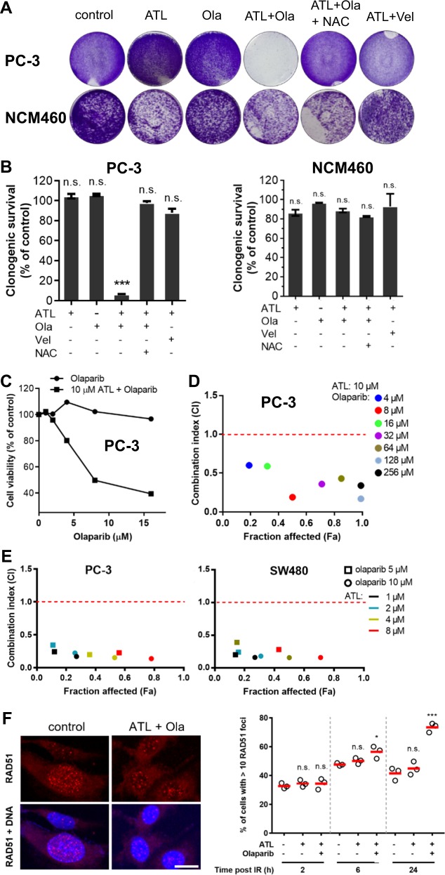
Fig. 2. Olaparib and ATL synergize to result in synthetic lethality in HR-proficient cancer cells.
a Colony formation assay. PC-3 and NCM460 cells were treated by 10 μM ATL, 10 μM olaparib (Ola), or combination of 10 μM ATL and 10 μM Ola or 10 μM ATL and 10 μM veliparib (Vel) for 7 days. The combination of 10 μM ATL and 10 μM Ola, but not 10 μM ATL and 10 μM Vel, completely inhibited the clonogenic growth of PC-3 but not the NCM460 cells and 10 mM NAC blocked the inhibition. b Quantification of colony formation assay. Cells stained by crystal violet were dissolved in 70% ethanol and absorbance at 595 nm was measured using a microplate reader. Data were presented as mean ± SD of three independent experiments. c MTT proliferation assay. PC-3 cells were treated by 1, 2, 4, 8, or 16 μM olaparib alone or combined with 10 μM ATL for 72 h. Olaparib in combination with 10 μM ATL dose-dependently inhibited the growth of PC-3 cells, while olaparib alone had no impact. d Determination of combination index (CI) values. PC-3 cells were treated by 10 μM ATL and the indicated concentrations of olaparib for 72 h. The CI values were determined by the Chou-Talalay method using the CompuSyn software. e CI values between lower concentrations of ATL and olaparib. PC-3 or SW480 cells were treated by 5 or 10 μM olaparib and the indicated concentrations of ATL for 72 h. f RAD51 foci formation after ionizing radiation (IR) exposure. PC-3 cells were irradiated with 3 Gy X-rays, treated with vehicle control, 10 μM ATL alone or in combination with 10 μM olaparib (Ola), and immunostained at the indicated time points. Left: representative micrographs of PC-3 cells stained with anti-RAD51 and counterstained with DAPI 2 h post IR (scale bar: 10 μm). Right: quantification of the percentage of cells with more than 10 RAD51 foci at the indicated time points post IR. n.s. not significant, *p < 0.05, ***p < 0.001 vs. vehicle control.

