Abstract
Influenza A (H1N1) is an acute respiratory infectious disease caused by mutant influenza A virus subtype H1N1. The pathogen is a new virus emerging after virus gene recombination of swine influenza, avian influenza, and human influenza. Influenza A (H1N1) is transmitted commonly via respiratory droplets and direct or indirect contact. Clinically, influenza A (H1N1) is characterized by influenza-like symptoms such as fever, cough, and rhinorrhea. But in rare serious cases, the condition may progress rapidly, with occurrence of viral pneumonia, respiratory failure, and multiple organ failure. Even death occurs in serious cases, with a mortality rate of 23–25 %. On April 30, 2009, influenza A (H1N1) was officially listed into Class B infectious diseases in China. Since then, it has been managed according to the Infectious Disease Prevention and Control Act in China. Meanwhile, it has been introduced into the category of quarantinable infectious disease for management based on the Frontier Health and Quarantine Law of China.
Keywords: Swine Influenza, Severe Acute Respiratory Syndrome, Swine Influenza Virus, Viral Pneumonia, Severe Influenza
Influenza A (H1N1) is an acute respiratory infectious disease caused by mutant influenza A virus subtype H1N1. The pathogen is a new virus emerging after virus gene recombination of swine influenza, avian influenza, and human influenza. Influenza A (H1N1) is transmitted commonly via respiratory droplets and direct or indirect contact. Clinically, influenza A (H1N1) is characterized by influenza-like symptoms such as fever, cough, and rhinorrhea. But in rare serious cases, the condition may progress rapidly, with occurrence of viral pneumonia, respiratory failure, and multiple organ failure. Even death occurs in serious cases, with a mortality rate of 23–25 %. On April 30, 2009, influenza A (H1N1) was officially listed into Class B infectious diseases in China. Since then, it has been managed according to the Infectious Disease Prevention and Control Act in China. Meanwhile, it has been introduced into the category of quarantinable infectious disease for management based on the Frontier Health and Quarantine Law of China.
Etiology
Influenza A (H1N1) virus belongs to the family of Orthomyxoviridae, the species of influenza A (H1N1) virus. The typical virus particle is an enveloped sphere with a diameter of 80–120 nm. On the envelope, there are many glycoprotein protrusions arrayed radially, including column-like hemagglutinin (HA), mushroom-shaped neuraminidase (NA), and matrix protein M2. A symmetrically spiral nucleocapsid is inside the virus envelope, with a diameter of 10 nm and ringlike structures in both ends. As a new mutant, influenza A (H1N1) virus is a single-negative-stranded RNA with a genome weighing about 13.6 kb. The virus strain of influenza A (H1N1) is the result of recombined eight gene fragments of influenza virus in different sizes, including HI, NP, and NS from classical swine influenza virus, PB2 and PA from avian influenza virus in North America, PB1 from human influenza A (H3N2) virus, and N1 and M from swine influenza virus in Europe and Asia. A scholarly study on ferret models was reported to have findings that influenza A (H1N1) virus is more pathogenic than seasonal influenza virus because it survives in the small intestine while seasonal influenza virus fails to. Influenza A (H1N1) virus is sensitive to common disinfectants such as ethanol, iodophor, and tincture of iodine and can be inactivated by oxidant, olefin acid, halogen compounds, and calcium oxychloride. Influenza A (H1N1) virus is heat intolerable, which can be inactivated at a temperature of 56 °C for 30 min. Its infectivity can be rapidly destructed by direct sunlight for 40–48 h or ultraviolet radiation.
Epidemiology
General Introduction of Its Epidemiology
Global Prevalence
In March 2009, an outbreak of human-infected swine influenza occurred in Mexico, with following rapid spread worldwide. The WHO initially nominated the disease as human-infected swine influenza, which is renominated as influenza A (H1N1) later. By mid-June 2009, the cases of influenza A (H1N1) have been reported in 74 countries and regions across the world, with a total of 28,744 reported cases including 144 cases of deaths. At that time, the WHO upgraded the warning level of influenza to 6 again to declare a pandemic of influenza in the world. By August 6, 2010, the cases of influenza A (H1N1) have been reported in 214 countries, with a total of 18,449 reported cases of death.
Prevalence of Influenza A (H1N1) in China
In mainland China, the first imported case of influenza A (H1N1) was reported on May 11, 2009. During May to August in 2009, the reported cases of influenza A (H1N1) are more commonly imported, with a low level of prevalence. However, since the end of August 2009, the prevalence of influenza A (H1N1) in mainland China progresses rapidly, with a widespread that is predominantly outbreaks in schools. In early December 2009, its prevalence reached its peak, with following decline of reported new cases. In early January 2010, the prevalence of influenza A (H1N1) is close to its prevalence level during the same period of the previous years. By March 31, 2010, a total of above 127,000 cases of definitively diagnosed influenza A (H1N1) had been reported in 31 provinces of China, including 126,000 cases of domestic infection and 1,228 imported cases. A total of 122,000 cases had been cured, with 4,859 cases receiving therapies in hospitals, 46 cases being treated at home, and 800 cases of death.
Source of Infection
The main source of infection is patients with influenza A (H1N1), and asymptomatic patients are also infectious. No evidence has been found to prove infection of humans from animals. After the infection, an incubation period usually lasts for 2–7 days. Most patients discharge viruses during the period from 1 day prior to the onset of the disease to 5–7 days after its onset or to the day prior to the absence of symptoms. Patients of young children and individuals with compromised immunity or serious symptoms might have a longer period of infectivity.
Routes of Transmission
Influenza A (H1N1) is mainly transmitted along with droplets via the respiratory tract. Sometimes, it also spreads via direct or indirect contact of the mucosa at the mouth, nose, and eyes. Contacts to respiratory secretions and body fluids from patients with influenza A (H1N1) or to contaminated utensils might also cause infection. The route of transmission along with aerosol via the respiratory tract needs further evidence to be proved.
Susceptible Population
Populations generally susceptible to influenza A (H1N1) are those who lack antibodies to fight against this new virus mutant. Most patients are at the age of 25–45 years, predominantly young adults and occasionally elderly people and children.
Population at High Risk of Developing Severe Influenza A (H1N1)
In the reported severe cases earlier, patients aged 19–49 years account for 35 %, indicating that severe influenza A (H1N1) commonly occurs in young adults. Most experts believe that the high-risk populations of severe influenza A (H1N1) include pregnancies, especially those infected in the first 3 months during pregnancy, children under the age of 2 years, and patients with chronic pulmonary diseases.
According to the guideline of influenza A (H1N1) (2010 edition) issued by the Ministry of Health in China, the following populations tend to develop severe influenza A (H1N1) after the occurrence of influenza-like symptoms:
Pregnancies
Patients with the following diseases or conditions: chronic respiratory diseases, cardiovascular diseases (with hypertension excluded), renal diseases, hepatic diseases, blood disorders, neurological diseases and neuromyopathies, metabolic disturbance and endocrinopathies, immunosuppression (including individuals using an immune suppressor or with immunodeficiency due to HIV infection), long-term use of aspirin, and under the age of 19 years
Obese persons with a body weight index exceeding 30
Children under the age of 5 years, especially children under the age of 2 years
The elderly people aged above 65 years
For these five populations with influenza-like symptoms, focused attention should be paid clinically. They should receive immediate detection for nucleic acids of influenza A (H1N1) virus and other necessary examinations.
Epidemiologic Features
Demographic Features
According to a report by the WHO, the cases with definitive diagnosis of influenza A (H1N1) are commonly young adults all over the world, with its incidence rates in males and females being close to 1:1.
Regional Distribution
The prevalence of influenza A (H1N1) occurs globally in 214 countries and regions, with serious prevalence in both southern and northern hemispheres. Initial studies have demonstrated that during the early period of global prevalence, the severity of prevalence is related to airline transportations in Mexico. During the disseminating period of the prevalence, the early regional distribution of influenza A (H1N1) is related to the communication frequency with the epidemic foci.
Chronological Distribution
In May 2009, influenza A (H1N1) rapidly spreads worldwide, with a peak prevalence in July in the southern hemisphere and a peak prevalence in October and November in the northern hemisphere.
Pathogenesis and Pathological Changes
Pathogenesis
Influenza A (H1N1) virus contains α-2,6-sialic acid glycosides and α-2,3-sialic acid glycosides. After its infection to the human body, it can replicate in epithelial cells of the whole respiratory tract to invade bronchi, bronchioles, and alveolar epithelial cells in large quantities. The hemagglutinin binds to the sialic acid on the surface of cells to invade the cells, with following replication of genetic materials in a large quantity to produce viral proteins. The cell membrane envelopes the genetic materials of the virus and viral protein, giving rise to another sphere from the cell-like budding. The new virus adheres to the cell via the bond between hemagglutinin and sialic acid. After that, neuraminidase hydrolyzes the sialic acid to release the new virus from its host cell and the released new virus can invade another cell. By autopsy of the death cases from influenza A (H1N1), the pathological examination of the pulmonary tissues demonstrated extensive epithelial degeneration and necrosis of bronchi and bronchioles as well as exudates containing neutrophils and monocytes filling the bronchi, bronchioles, and alveoli. The critical factors determining the onset and development of influenza A (H1N1) include the loading dose of influenza A (H1N1) virus, the intrapulmonary infiltration of the inflammatory cells, and the release of inflammatory mediators. The early animal studies about influenza A (H1N1) virus collected in 1918 have demonstrated infiltration of large quantities of neutrophils and macrophages in rat pulmonary tissues, waterfall-like releases of enzymes and cytokines (including IFN-α, IFN-γ, TNF-α, MIP-1α, and MIP-2) by inflammatory cells, and mutually enhanced pathological effects by cytokines and inflammatory cells. Consequently, pulmonary lesions, pulmonary dysfunction, decreased body weight, and general fatigue occur. The studies on animals inoculated with HA DNA influenza virus vaccine indicated that the increase of released IFN-γ is more than the released IL-4 and the replication of influenza A (H5N1) virus declines, both of which have quantity-effect relationship with the amount of vaccines. These findings indicated that the immune reactions against influenza virus infection have bias on Th cellular immune reactions and the production of cytokine Th2 is related to large quantity replications of influenza virus. By clinical trials, it has been found that strong cytokine IRF-3 reaction can reduce IFN-β, IFN-λ1, and MCP-1 and partially reduce TNF-α, which is related to the severity of influenza A (H5N1) symptoms and viral replication. Recent studies have demonstrated that the virus-induced cytokine (IFN, TNF-α) reactions are even weaker than seasonal influenza despite the differences of genotypes and antigenicity between influenza A (H1N1) and seasonal influenza. The relationship between the weaker virus-induced cytokine reactions and mild clinical manifestations of influenza A (H1N1) remains elusive.
The susceptibility of young adults is possibly caused by serious damages to the autoimmune balance and tissues resulted from more released inflammatory mediators by immune cells due to more sensitive immunity in young adults and thus the more intense immune responses after infection of influenza A (H1N1) virus.
Pathological Changes
Influenza A (H1N1) virus spreads via the respiratory tract, with alveoli as its target cells. In the early stage of infection, diffuse inflammations cause laryngeal, tracheal, and bronchial edema and inflammatory cell infiltration that is predominantly lymphocytes and histiocytes. Necrotizing tracheobronchitis occurs following the shedding of ciliated columnar epithelium in the bronchi, with ulcerations and shedding of bronchial mucosa. There are alveolar capillary congestion and alveolar hemorrhage, with the alveolar lumens filled with liquid exudates of fibrins. Alveolar edema and fusion of substantial lobular inflammatory exudates occur in alveoli lined with transparent pneumonocytic membrane. With the progress of the conditions, pulmonary interstitial tissues are involved, with reduced alveolar gas content, pulmonary consolidation, and fibrosis. Li et al. reported that pulmonary diffuse fibrosis and alveolar lesions occur in the serious and critical cases, with alveolar edema and substantial lobular inflammatory exudates. With the progress of the conditions, pulmonary interstitial tissues are involved, with reduced alveolar gas content as well as pulmonary hemorrhage, consolidation, and fibrosis. H&E staining showed widened septum between alveolar walls, congested alveolar walls, infiltration of neutrophils and plasmocytes that are mostly monocytes, and exudation of edema liquid and celluloses within alveoli.
In China, most cases of influenza A (H1N1) can be categorized into the mild type, with lesions confined in the upper respiratory tract and possible complication of mild lobular pneumonia. The serious and critical cases are rare, with manifestations of necrotizing tracheobronchitis and diffuse alveolar lesions.
Clinical Symptoms and Signs
Incubation Period
The incubation period of influenza A (H1N1) generally lasts for 1–7 days, being 1–3 days in most cases.
Clinical Manifestation
The early symptoms of influenza A (H1N1) are similar to those of influenza. It has an acute onset, with symptoms of fever, pharyngalgia, rhinorrhea, nasal obstruction, cough and expectoration, headache, general soreness and pain, and fatigue. But in some cases, vomiting and/or diarrhea may occur. In rare cases, only slight upper respiratory symptoms occur without fever. The signs commonly include congestive pharynx and swollen tonsils.
The cases of newborns and infants have atypical symptoms, with manifestations of low-grade fever, drowsiness, difficulty feeding, tachypnea, apnea, cyanosis, and dehydration. Children may have wheezing. In some cases of children, symptoms involving the central nervous system can be found.
Compared to nonpregnant women and common populations, tachypnea is more common in pregnant women after the infection of influenza A (H1N1). The cases of pregnant women are more susceptible to pneumonia and respiratory failure. Some pregnancy-related special symptoms might occur due to fever or complications of pneumonia or septic shock. Their symptoms include irregular uterine contraction, bloody vaginal secretion, and other symptoms of threatened labor. And these manifestations may even induce premature birth and fetal distress in the uterus. Influenza A (H1N1) may exacerbate the underlying basic diseases, with corresponding clinical manifestations.
Influenza A (H1N1)-Related Complications
In rare cases, influenza A (H1N1) rapidly progresses with occurrence of secondary diseases, such as severe pneumonia, acute respiratory distress syndrome, pulmonary hemorrhage, pleural effusion, pancytopenia, renal failure, septicemia, shock and Reye syndrome, respiratory failure, and multiple organ dysfunction or failure. Children may develop central nervous system complications.
Influenza A (H1N1)-Related Neurological Complications
Influenza A (H1N1)-related acute neurological complication is defined as convulsion, encephalopathy, or encephalitis occurring in the first 5 days after the onset of influenza-like symptoms, with laboratory evidence of influenza A (H1N1) virus infection of the respiratory tract but no evidence of any other infections.
Encephalopathy
Encephalopathy is defined as changes of consciousness including irritation, lethargy, or coma for over 24 h. The cases with concurrent symmetric bilateral thalamic necrosis are diagnosed as having acute necrotizing encephalopathy (ANE).
Encephalitis
Encephalitis refers to the occurrence of the following two or more manifestations based on encephalopathy: (1) fever with a body temperature above 38.5 °C, (2) physical signs of focal neuropathies, (3) increased lymphocytes in the cerebrospinal fluid, (4) encephalitis indicated by electroencephalography, and (5) infection or inflammation indicated by neuroimaging.
The first report of influenza A (H1N1)-related neurological complications was published in the United States. The reported cases included four boys aged 7–17 years, three having encephalopathy and one having convulsion. Their manifestations are mild and all are cured. Thereafter, a death case from influenza A (H1N1)-related ANE was reported in the United States, being a girl aged 12 years with a past history of good health. In China, three cases of death from influenza A (H1N1)-related ANE were reported in Shenzhen Children’s Hospital, Guangdong.
Child cases of influenza A (H1N1)-related neurological complications commonly have clinical manifestations of drowsiness, irritation, convulsion, and coma. These symptoms commonly occur in 1–4 days after the occurrence of acute fever with/without accompanying respiratory tract symptoms. The rarely found symptoms include temporary blindness and inappropriate laughter.
Acute necrotizing encephalopathy (ANE) is a rare but severe complication of the central nervous system (CNS) following viral infection. Such cases are mostly caused by influenza virus and rarely caused by parainfluenza virus, human herpesvirus 6, or other viruses. ANE was firstly reported by Mizuguchi et al. in 1995, and such cases have been commonly reported in Japan and Chinese Taiwan, which has been rarely reported in other countries and regions.
The clinical manifestations of ANE are similar to other acute encephalopathies. About 90 % of children develop prodromal symptoms such as fever and acute upper respiratory tract infection before the occurrence of acute encephalopathy. Clinically, it is characterized by multifocality and symmetric brain lesions, involving the bilateral thalamus, brainstem tegmentum, white matter around cerebral ventricles, and cerebellar medulla. The cerebrospinal fluid examination shows no other abnormalities but a slight increase of proteins. In some cases, there are increased hepatic transaminase and myocardial enzymes. Increased blood ammonia is rarely found and the level of blood glucose is normal. The mortality rate of ANE in the acute stage reaches as high as 30 % and most survivors are compromised by severe neurological sequelae.
The pathological mechanism underlying ANE has not been well defined. Participation of virus infection-induced cytokines in the mediation might play a role, which is mainly related to IL-6 and TNF-α. Focal vascular lesions induce destruction of the blood-cerebrospinal fluid barrier and exudation of blood plasma, leading to cerebral edema, spots of hemorrhage, and necrosis of neurons and gliocytes. The gross pathological changes include brain tissue malacia and accompanying partial tissues lysis, occurring predominantly in the thalamus, white matter of the deep cerebrum, tegmentum of the brainstem, and white matter in the deep cerebellum. Histological sections show fresh necrotic foci, with no reactive hyperplasia of astrocytes and microglia and no infiltration of inflammatory cells.
Influenza A (H1N1)-Related Respiratory Complications
Influenza A (H1N1)-related pneumonia demonstrates pulmonary infiltrative lesions or pulmonary consolidation by chest X-ray or CT scanning. Hospital-acquired pneumonia and ventilator-associated pneumonia should be excluded.
Diagnostic Examinations
Laboratory Tests
Peripheral Hemogram
The WBC count is commonly normal or decreased. In some serious cases in children, total WBC count may increase.
Blood Biochemical Assay
In some cases, hypokalemia can be found. But in rare cases, the levels of creatine kinase (CK), aspartate transaminase (AST), alanine aminotransferase (ALT), and lactate dehydrogenase (LDH) increase.
Etiological Examinations
Detection of Virus Nucleic Acid
RT- PCR, or favorably real-time RT- PCR, can be used for genetic detection of the respiratory tract specimens such as pharyngeal swab, nasal swab, suctions from the nasopharynx or trachea, and sputum and for molecular epidemiological survey. The FDA and CDC in the United States recommend ABI quantitative PCR for detection of influenza A (H1N1) virus due to its advantages of being rapid with high sensitivity and specificity.
Virus Isolation
Influenza A (H1N1) virus can be isolated from respiratory tract specimens.
Detection of Serum Antibody
Dynamic detection of double sera for specific antibody against influenza A (H1N1) virus shows at least four times increase of the antibody level.
Diagnostic Imaging
X-Ray
X-ray has advantages of being economical and convenient, which is the radiological examination of choice for initial diagnosis and following examinations. Bedside X-ray radiography is convenient and fast for dynamic observation of the pulmonary infections. Quarantine with bedside X-ray is also convenient if necessary.
CT Scanning
CT scanning has high resolution to distinguish tissues, which has higher sensitivity to early mild pathological changes, ground-glass-like shadows, and pulmonary interstitial lesions. These advantages facilitate in finding lesions in the posterior costophrenic angle area and overlapping areas between heart shadow and aorta shadow. Routine CT scanning generally should be followed by HRCT scanning.
MR Imaging
MR imaging is commonly applied for the diagnosis of influenza A (H1N1)-related neurological complications.
Imaging Demonstrations
Influenza A (H1N1)-Related Neurological Complications
CT Scanning
Brain CT scanning may demonstrate no obvious abnormalities in the early stage of the conditions. However, in the middle and advanced stages of the conditions, brain CT scanning demonstrates deepened cerebral sulcus, enlarged cerebral ventricles, and other atrophic changes of the cerebrum. By follow-up examinations, brain CT scanning of the cured cases demonstrates no abnormalities. In the early stage of the condition, diffuse cerebral edema may be demonstrated by CT scanning. Tang et al. reported two cases of early cerebral edema with demonstrations of rapid progress of the condition into cerebromalacia and cerebral liquefaction by reexaminations in a short period of time. Of the two cases of death, one was accompanied by cerebral hemorrhage, and the occurrence of death indicated poor prognosis in the patients with early cerebral edema. The demonstrations also included low-density foci in the cortex and subcortical tissue of the cerebellar hemisphere and cerebral hemisphere and in the centrum ovale (Figs. 22.1 and 22.2). Kong et al. reported one case of influenza A (H1N1)-related neurological complication. The CT scanning demonstrations included multiple patches of low-density foci in the cortex and subcortical tissues of the bilateral cerebellar hemisphere and cerebral hemisphere. These foci have unclearly defined boundaries, which are more obvious in the parietal lobe. The foci of this case were demonstrated to have rapid changes. By reexamination after 2 days, the density of foci slightly decreased. And by reexamination after 7 days, the foci obviously reduced in quantity.
Fig. 22.1.
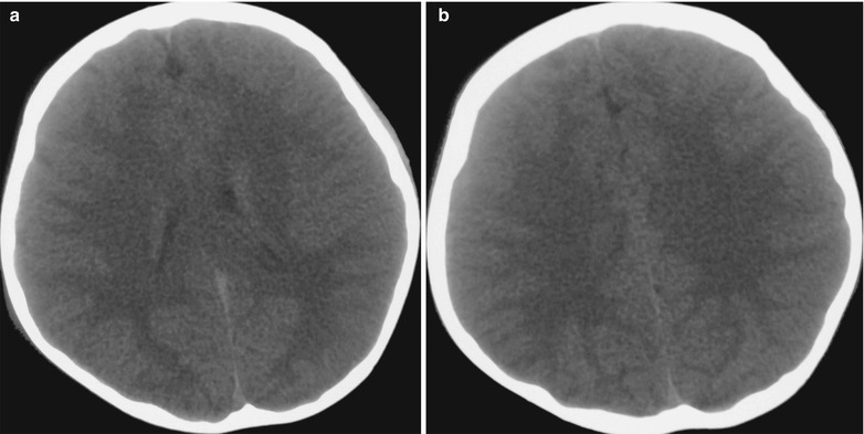
Influenza A (H1N1) complicated by encephalitis. (a, b) CT scanning demonstrated large flakes of low-density foci in the white matter surrounding bilateral cerebral ventricles and in the centrum ovale
Fig. 22.2.
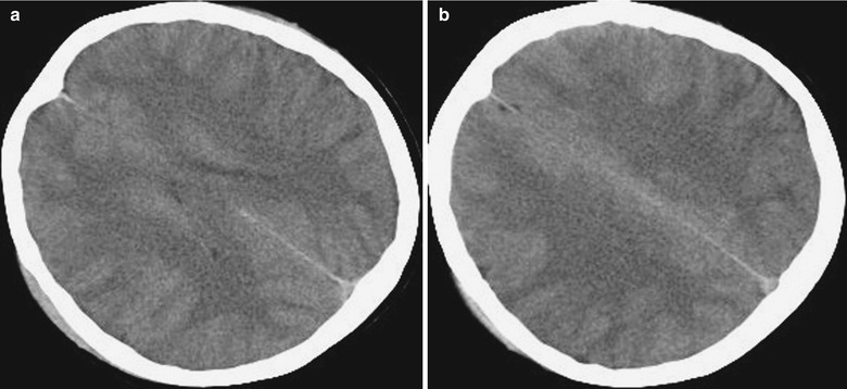
Influenza A (H1N1) complicated by encephalitis. (a, b) CT scanning demonstrates large flakes of low-density foci in the white matter surrounding bilateral cerebral ventricles and in the centrum ovale
Case Study 1
A boy aged 2.5 years complained of fever and cough for 3 days. By physical examinations, T 39.7 °C and bpm 130/min. He had rough breathing sound in both lungs with a large quantity of moist rales. And he had no definitive history of contacts to patients with influenza A (H1N1). Pharyngeal swab by CDC demonstrated positive M gene of influenza A (H1N1) virus, positive NP gene of swine influenza (H1N1) virus, and positive HA gene of influenza A (H1N1) virus. By routine blood tests, WBC 18.7 × 10⁹ /L, GR% 49.34 %, and LY% 41.3 %. By blood biochemical assay, ALT 32 U/L and AST 143 U/L.
Case Study 2
A female patient aged 20 years complained of fever and cough for 4 days as well as headache and conscious disturbance for 1 day. By physical examination, T 40 °C, bpm 128/min, R 25/min, BP 108/58 mmHg, and SPO2 95 %. She was in coma with no response to calling. Her respirations were sobbing-like, with positive cervical resistance and negative meningeal irritation sign. She also had congestive pharynx, occasional moist rales in both lungs, positive bilateral Babinski sign, and positive Chaddock sign. By laboratory tests, she was positive for viral nucleic acid of influenza A (H1N1). By routine blood tests, WBC 39.6 × 10⁹ /L and GR% 92.4 %. By blood biochemistry, UA 676 μmol/L, CK 1482 U/L, CK-MB 347 U/L, and HBDH 1294 U/L. Death occurred due to shock complicated by DIC and multiple organ failure of the brain, lungs, heart, liver, and kidneys as well as critical influenza A (H1N1) complicated by virus meningoencephalitis.
MR Imaging
MR imaging demonstrates diffuse demyelination in the cerebellar hemisphere, brainstem, and white matter of the cerebrum. Contrast imaging demonstrates patches of enhancement and gyrus enhancement in different sizes in the cerebellar hemisphere, brainstem, and parenchyma of the cerebral hemisphere. Haktanir reported one case of meningoencephalitis, with demonstrations of high signal by T2WI in the bilateral frontoparietal interfaces and thalamus. Contrast T1WI demonstrates obvious enhancement of the right frontoparietal interface and enhancement of meninges in bilateral cerebral hemispheres. Susceptibility weighted imaging of T2FFE demonstrates more obvious high signal in the right frontoparietal interface, with no cerebral edema.
Symmetric cerebral multiple foci are a characteristic manifestation of ANE, which can be well demonstrated by CT scanning and MR imaging. The foci are commonly found in the thalamus, tegmentum of the brainstem, white matter surrounding the lateral ventricles, and cerebellar medulla. CT scanning demonstrates symmetric low density in the bilateral thalamus, posterior limb of internal capsule, brainstem, and white matter of bilateral frontal, parietal, and occipital lobes. There are diffuse cerebral edema, absence of the sulcus and the fourth ventricle, and possible brainstem edema. Bilateral basal ganglia infarction has been also reported. MR imaging demonstrates symmetric long T1 and long T2 signals in the bilateral thalamus, basal ganglia, brainstem, splenium of the corpus callosum, bilateral centrum ovale, and paraventricular white matter, which are also high signals by FLAIR. Contrast imaging demonstrates linear and ring-shaped enhancement of the centrum ovale and thalamus but no abnormal enhancement of meninges. DWI demonstrates high signal in the bilateral thalamus or low central signal, boundary ring-shaped high signal, and high signal of other lesions. ADC image demonstrates slightly higher central signal surrounded by ring-shaped lower signal (low value of ADC). Cerebellar involvement is demonstrated as slightly long T1 and slightly long T2 signal in the bilateral cerebellar medulla and in the right cerebellar middle peduncle, which are in slightly high signal by FLAIR and high signal by DWI. Contrast imaging demonstrates obvious enhancement of cerebellar meninges. Compared to traditional sequences, DWI more favorably demonstrates pathological changes, with complex signal changes. DWI demonstrates low signal at the lesion centers in the bilateral thalamus whose pathological basis is necrosis of nerves and gliocytes and surrounding ring-shaped high signal whose pathological basis is cytotoxic edema and limited movement of water. ADC image demonstrates low signal in the center of the thalamus with surrounding high signal and elevated ACD value due to vasogenic edema (Figs. 22.3, 22.4, and 22.5).
Fig. 22.3.
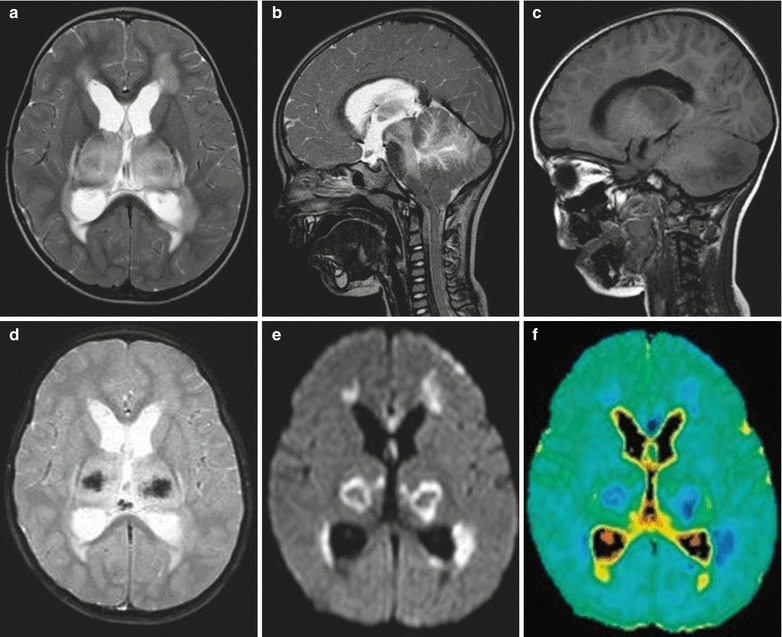
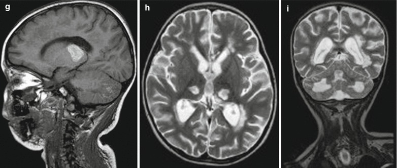
Influenza A (H1N1) complicated by acute necrotizing encephalopathy. (a) In the acute stage, transverse T2WI demonstrates symmetrically increased signal in the bilateral thalamus and white matter surrounding the anterior and posterior horns of lateral ventricles. (b) Sagittal T2WI demonstrates high signals in the cerebellum and brainstem. (c) Sagittal T1WI demonstrates low signal in the brainstem and cerebellar hemisphere. (d) Transverse T2WI and GRE demonstrate low signal at the centers of foci in the thalamus, indicating hemorrhagic necrosis. (e, f) Transverse DWI and ADC image demonstrate limited diffusion of foci in the bilateral thalamus and white matter surrounding the anterior and posterior horns of lateral ventricles. (g) At day 10 after hospitalization, sagittal T1WI demonstrates high signals in the thalamus and cerebellum, in consistency with subacute hemorrhage. (h, i) In the chronic stage, at day 40 after hospitalization, MR imaging demonstrates severe sequelae. Transverse and sagittal T2WI demonstrate atrophy of the bilateral thalamus, with well-defined cavities inside the bilateral thalamus (Note: The case and images are from Ormitti F, et al. AJNR. 2010;31(3):396)
Fig. 22.4.
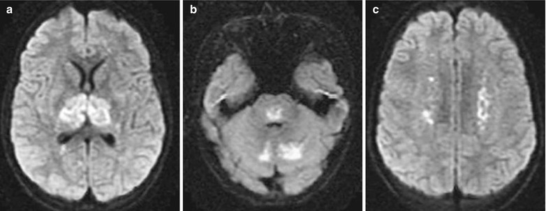
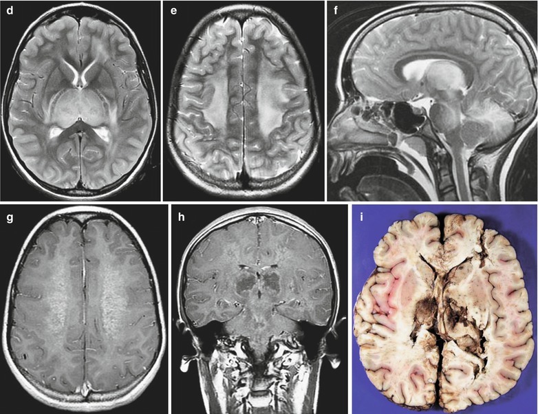
Influenza A (H1N1) complicated by acute necrotizing encephalopathy. (a–c) Transverse DWI demonstrates limited diffusion of the bilateral thalamus, cerebellum, brainstem, and centrum ovale. (d, e) Transverse T2WI demonstrates slightly high or high signal in the thalamus and large flakes of high signals in the bilateral centrum ovale. (f) Sagittal T2WI demonstrates abnormal high signal in the thalamus, mesencephalon, pons, and cerebellum. (g) Contrast transverse T1WI demonstrates slight enhancement of the centrum ovale. (h) Contrast coronal T1WI demonstrates abnormal enhancement of the brainstem and centrum ovale as well as ring-shaped enhancement foci in the bilateral thalamus. (i) Autopsy demonstrates hemorrhagic necrosis of the bilateral thalamus (Note: The case and images are from Lyon JB, et al. Pediatr Radiol. 2010;40(2):200)
Fig. 22.5.
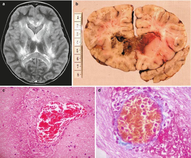
Influenza A (H1N1) complicated by acute necrotizing encephalopathy. (a) MR imaging demonstrates bilateral symmetric abnormalities and abnormal high T2WI signal in the deep white matter of the cerebral hemisphere, caudate nucleus, lenticular nucleus, internal capsule, lateral nucleus of the thalamus and its posterior area, corpus callosum, and tegmentum of the brainstem. (b) By autopsy, the genu of the corpus callosum, mammillary body, and splenium of the corpus callosum are demonstrated from sagittal perspectives. The pathological changes include diffuse cerebral edema, vascular lesions, and necrosis, without obvious perivascular demyelination. In the necrotic area, the necrotic cells are demonstrated with illusive shadows. In the background of rare cells and swollen nerve fiber network, karyopyknosis or fragments of karyolysis and erythrocytic exudation can be found, with no astrocytes and hyperplasic microglia. Ischemic atrophy and pericellular edema are found in many neurons in the cortex of the cerebrum, hippocampus, cerebellum, and brainstem. Thrombosis is found in some minor blood vessels of the brainstem. Diffuse ischemic change of neurons and thrombosis in the brainstem and cerebellar pyramid are pathological changes secondary to cerebral edema. (c) There is a 110 μmm blood vessel in the head of the left caudate nucleus. In addition, exudation of lymphocytes and neutrophils as well as karyolysis with/without accompanying intraluminal thrombosis can be found in the vascular wall with FVL and perivascular space. (d) Martius scarlet blue staining demonstrates fibrin reservoir in the blood vessels (Note: The case and images are from Ng WF, et al. Brain Pathol. 2010;20(1):261)
Case Study 3
A girl aged 3 years complained of high fever and severe convulsion 2 days after catching a cold and diarrhea, with the highest body temperature reaching 41.2 °C. Her condition progressed into coma 2–3 days after her hospitalization. By laboratory test, severe liver failure was indicated, AST 16,852 U/L, ALT 10,300 mm3/L, LDH 27,878 U/L, and PLT 54,000/μL. By examination of cerebrospinal fluid, pressure 170 mmH2O and protein 232 mg/L. CT scanning demonstrates no abnormalities.
Case Study 4
A girl aged 12 years complained of cough and abdominal upset and, following persistent high fever, frequent diarrhea, asthenia, and general pain. She had no history of vaccination against seasonal influenza and influenza A (H1N1). Fast antigen detection by pharyngeal swab defined the diagnosis of influenza A (H1N1).
Case Study 5
A boy aged 7 years was hospitalized due to mental confusion and incoherent talking. He had experienced upper respiratory tract infection 2 weeks ago and had a past history of bronchial asthma. By laboratory test, ALT 5–25 IU/L and complete blood cell, blood sugar, and liver and lung function levels were normal. By cultures of nasopharyngeal secretion and throat swab, he was positive for influenza A (H1N1) virus antibodies. CT scanning fails to demonstrate any space-occupying lesions and cerebral edema. In 12 h after hospitalization, generalized tonic convulsion occurred which then progressed into decorticate posture. His GCS is 3/15 (E1V1M1) and the pupils are 2.5 mm in orthophoria with sluggish reaction to light. In 34 h after hospitalization, the conditions continued to deteriorate with dilated and fixed pupils. Death occurred at day 4 after hospitalization.
Influenza A (H1N1)-Related Respiratory Complications
Pediatric Influenza A (H1N1) Complicated by Pneumonia
Pediatric influenza A (H1N1) complicated by pneumonia has no characteristic chest radiological demonstrations and dynamic changes in its early stage, with no obvious differences from common lung infections. In the progressing stage, parenchymal lesions are more common, possibly with complications of ARDS and mediastinal emphysema. And in the absorption stage, pulmonary interstitial lesions are more common, with blurry pulmonary markings, poor transparency of bilateral lungs, and heterogeneous lung inflation.
X-Ray
Pulmonary Parenchymal Lesions
Pulmonary parenchymal lesions are characterized by infiltrative shadows of pulmonary parenchyma that are commonly bilateral. Some studies demonstrated more common unilateral involvement than bilateral involvement, with more commonly found involvement of the right lung. The lesions are mainly located in the inner middle zone, with possible involvement of upper, middle, and lower lung fields. The involvement of middle and lower lung fields is more common. However, some scholarly studies demonstrated more commonly found lesions in the basilar part of the lungs. Parenchymal infiltration may be characterized by single or multiple small patches of shadows, which can fuse into large flakes of shadows and/or ground-glass opacity (GGO) (Figs. 22.6, 22.7, 22.8, and 22.9). In children, the lesions are commonly demonstrated as patches of shadows, while in infants and young children, the lesions are commonly demonstrated as cotton-wool opacity.
Fig. 22.6.
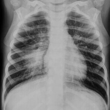
Influenza A (H1N1) complicated by pneumonia. X-ray demonstrates increased and thickened pulmonary markings in both lungs, a large flake of high-density blurry shadow in the right lower lung, as well as enlarged and densely colored hilum
Fig. 22.7.
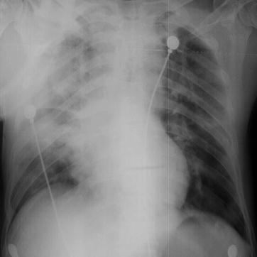
Influenza A (H1N1) complicated by pneumonia. X-ray demonstrates a large flake of high-density blurry shadow in the right upper lung, cloudy shadow in the right middle and upper lung, as well as enlarged and blurry hilum
Fig. 22.8.
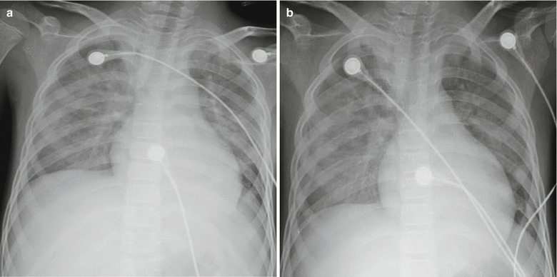
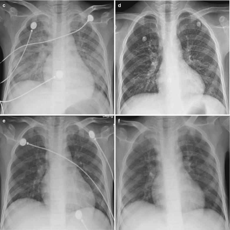
Influenza A (H1N1) complicated by pneumonia. (a) X-ray demonstrates diffuse cotton-woollike shadows in both lungs, decreased transparency of lung fields, and blurry structure of both hili. (b) By reexamination after treatment for 1 day, X-ray demonstrates a rapid progress of the conditions, with cloudy high-density shadows in both lung fields. (c) By reexamination after treatment for 5 days, X-ray demonstrates a progress of the conditions, with diffuse small spots of shadows in both lungs and decreased transparency of lung fields. (d) By reexamination after treatment for 7 days, X-ray demonstrates no obvious changes. (e) By reexamination after treatment for 10 days, X-ray demonstrates improved conditions, with lesion absorption, increased pulmonary markings in both lungs, and blurry pulmonary markings in the right lower lung. (f) By reexamination after treatment for 17 days, X-ray demonstrates no obvious abnormalities in the septum between the heart and lung
Fig. 22.9.
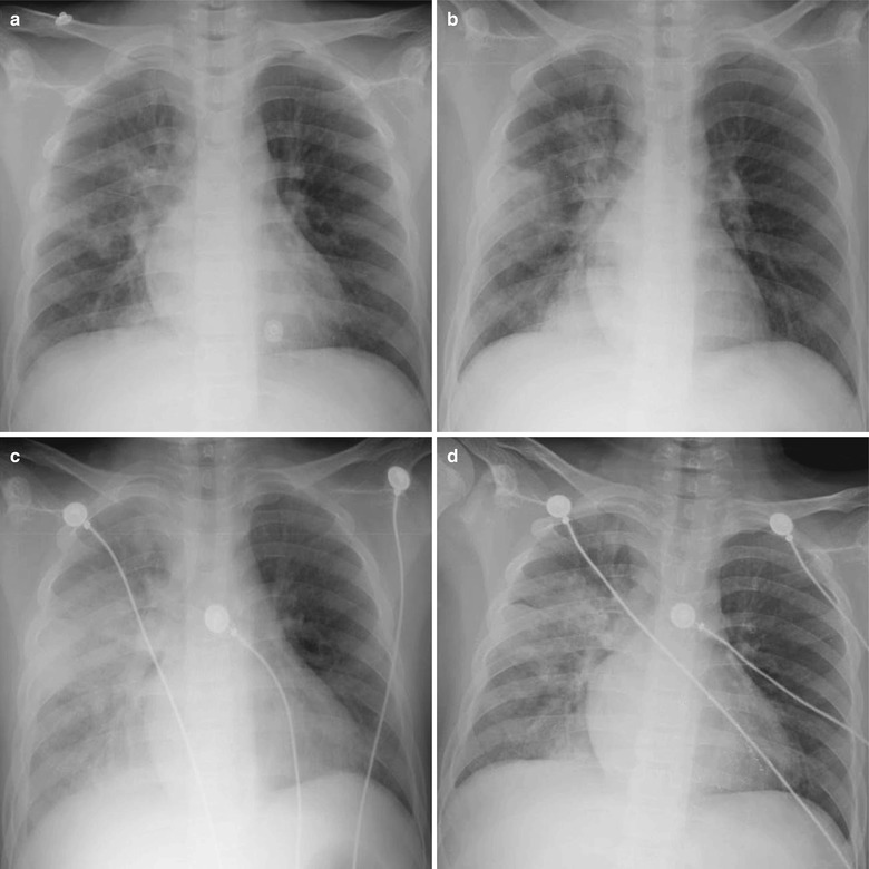
Influenza A (H1N1) complicated by pneumonia. (a) By examination at day 1 after hospitalization, X-ray demonstrates multiple flakes of blurry shadows in the right lung as well as enlarged and densely colored hilar shadow. (b) By reexamination at day 2 after hospitalization, X-ray demonstrates a progress of the conditions, with right pulmonary consolidation. (c) By reexamination at day 6 after hospitalization, X-ray demonstrates lesions with increased density and enlarged range in the right lung as well as diffuse cloudy shadows with increased density in the left lung. (d) By reexamination at day 9 after hospitalization, X-ray demonstrates improved conditions, with lesions absorbed and patches of shadows in the right lung
Pulmonary Interstitial Lesions
Pulmonary interstitial lesions are characterized by increased, thickened, and blurry pulmonary vascular markings, with grids shaped and nodular shadow of different degrees (Fig. 22.10). Some scholars put forward that in the early stage, X-ray of children demonstrates no interstitial changes, which might be related to the limitations of X-ray and the course of illness.
Fig. 22.10.
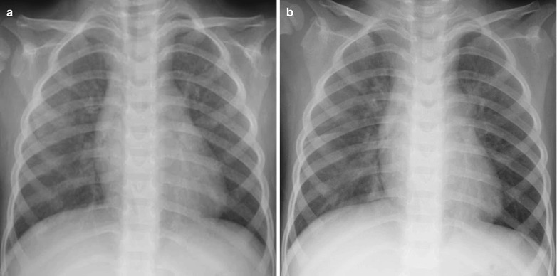
Influenza A (H1N1) complicated by pneumonia. (a) X-ray demonstrates enhanced bilateral pulmonary markings, spots of blurry shadows in bilateral lung fields, as well as enlarged and densely colored hili. (b) By reexamination after treatment for 6 days, X-ray demonstrates clearly defined bilateral pulmonary markings and no abnormal density shadows in both lungs
X-Ray of Severe Cases
In the severe cases, there are also demonstrations of increased pulmonary markings and excessive inflation. The causes may be the involved major airway due to perivascular lesions of the bronchi. The following necrosis of the bronchial wall and the infiltration of neutrophils cause obstruction of the small airway, leading to excessive alveolar inflation.
Others
In some cases, there are also demonstrations of involvement of the pleura, pleural effusion, mediastinal lymphadenectasis, and hilar lymphadenectasis.
Case Study 6
A boy aged 3 years complained of fever and wheezing for 6 days. He also suffered from cough, expectoration, and wheezing after physical activities. Shuanghuanglian mixture was given for 1 day, with following exacerbation of the above symptoms and accompanying dyspnea. At day 6 after the onset, pharyngeal swab was positive. It was reported that several children experienced fever in the kindergarten the boy went. By physical examination, T 39 °C, bpm 118/min, and tonsillar enlargement in first degree was found. The respiration sound of both lungs was rough, with fine moist rales in the left lower lung and large quantity wheezing sound in the right lung. Pharyngeal swab by CDC demonstrated positive M gene of influenza A (H1N1) virus, positive NP gene of swine influenza virus, and positive HA gene of influenza A (H1N1) virus. By routine blood test, WBC 9.73 × 10⁹ /L, GR% 30.64 %, and LY% 31.14 %. By blood biochemistry, AST 42 U/L, ALT 21 U/L, and LDH 274 U/L.
Case Study 7
A foreigner girl aged 12 years complained of fever and cough for 2 days. She also complained of accompanying chills, aversion to cold, and pharyngalgia. She had a history of contact to patients with influenza A (H1N1). By physical examination, T 38.7 °C and tonsillar enlargement in first degree was found. Pharyngeal swab by CDC demonstrated positive M gene of influenza A (H1N1) virus, positive NP gene of swine influenza virus, and positive HA gene of influenza A (H1N1) virus. By routine blood test, WBC 5.7 × 10⁹ /L, GR% 42.1 %, and LY% 40.3 %.
Case Study 8
A boy aged 10 years complained of fever for 3 days. He also complained of mild cough with no sputum, vomiting, no chills, and no fatigue. By physical examination, T 39 °C, the pharynx was congested, and tonsillar enlargement in first degree was found. He had a history of contact to patients with influenza A (H1N1). Pharyngeal swab demonstrated positive M gene of influenza A (H1N1) virus, positive NP gene of swine influenza virus, and positive HA gene of influenza A (H1N1) virus. By routine blood test, WBC 13.39 × 10⁹ /L and GR% 84.1 %.
Case Study 9
A boy aged 12 years complained of cough and expectoration for 2 weeks that progresses with accompanying fever for 3 days. He also complained of fatigue, poor appetite, rhinorrhea, and muscular soreness and pain. By physical examinations, T 39.8 °C, the pharynx was congested, and no tonsillar enlargement was present. He had a history of contact to patients with influenza A (H1N1). Pharyngeal swab by CDC demonstrated positive M gene of influenza A (H1N1) virus, positive NP gene of swine influenza virus, and positive HA gene of influenza A (H1N1) virus. By routine blood test, WBC 3.8 × 10⁹ /L, GR% 23.4 %, and LY% 54.1 %. By blood gas analysis, pH 7.46, PCO2 26 mmHg, and PO2 56.9 mmHg.
Case Study 10
A girl aged 2 years complained of fever, cough, and rhinorrhea for 1 day and a small quantity of tasteless thin whitish sputum. By physical examination, T 39 °C, with no clearly defined route of infection. Pharyngeal swab by CDC demonstrated positive M gene of influenza A (H1N1) virus, positive NP gene of swine influenza virus, and negative HA gene of influenza A (H1N1) virus. By routine blood test, WBC 11.2 × 10⁹ /L, GR% 23.8 %, and LY% 65.4 %.
CT Scanning
Demonstrations of CT scanning are basically the same as those by chest X-ray, including increased, thickened, and blurry pulmonary markings and small flakes of parenchymal shadows distributing along the bronchial tree in the lungs. Sometimes, the small flakes of parenchymal shadows may be large flakes of consolidations, with ground-glass opacity around the lesions. In addition, heterogeneous inflation and spots of interstitial changes can be found (Figs. 22.11, 22.12, and 22.13). In the severe pediatric cases, excessive pulmonary inflation may be found. CT scanning has advantages in clearly defining diseased pulmonary segments and lobes, slight pleural reaction, and small quantities of pleural effusion.
Fig. 22.11.
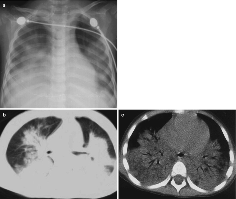
Influenza A (H1N1) complicated by pneumonia. (a) X-ray demonstrates flakes of blurry shadow in both lungs as well as increased and thickened pulmonary markings. (b, c) CT scanning demonstrates large flake of consolidation in both lungs with air bronchus sign
Fig. 22.12.
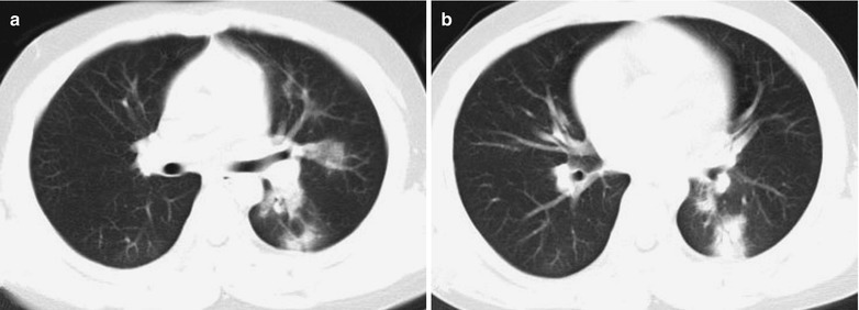
Influenza A (H1N1) complicated by pneumonia. (a, b) CT scanning demonstrates multiple patches of shadows in the superior lingular segment of the left upper lobe and in the dorsal segment of the left lower lobe, with unclearly defined boundaries
Fig. 22.13.
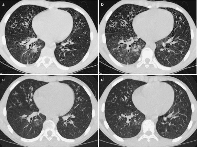
Influenza A (H1N1) complicated by pneumonia and bronchial dilation. (a, b) CT scanning demonstrates small flake of parenchymal shadow and ground-glass opacity in the anterior lower lobe of the right lung and in the inner basal segment of the right lung, widened bronchial canals, and thickened bronchial wall of both lungs, with signet ring sign. (c, d) By reexamination after treatment for 7 days, CT scanning demonstrates the lesions improved and absorbed
Case Study 11
A boy aged 2.5 years complained of fever and cough for 8 days. He received medications of cephalosporin antibiotics and Shuanghuanglian mixture for 2 days, with no improvement and with poor appetite, exacerbated cough, depression, and perleche. Pharyngeal swab was positive at day 9 after the onset. He had an unclearly defined history of contact to patients with influenza A (H1N1). By physical examinations, T 38.7 °C, bpm 13/min, and perleche and scattering white spots were found in the oral mucosa. He had rough breathing sound in both lungs and a large quantity of moist rales in both lungs. Pharyngeal swab by CDC demonstrated positive M gene of influenza A (H1N1) virus, positive NP gene of swine influenza virus, and positive HA gene of influenza A (H1N1) virus. By routine blood test, WBC 1.5 × 10⁹ /L, GR% 47.34 %, and LY% 41.3 %. By blood biochemistry, ALT 32 U/L, AST 143 U/L, CK 301 U/L, and LDH 629 U/L.
Case Study 12
A boy aged 12 years complained of fever and cough for 5 days with whitish phlegm and a body temperature of 39.7 °C. After anti-infection therapy for 4 days, his conditions failed to improve, with a body temperature above 39 °C. Pharyngeal swab at day 5 after the onset was positive. By physical examination, tonsillar enlargement in first degree was found and rough breathing sound was heard in both lungs. He had a history of close contact to patients with influenza A (H1N1). Pharyngeal swab by CDC demonstrated positive M gene of influenza A (H1N1) virus, positive NP gene of swine influenza virus, and positive HA gene of influenza A (H1N1) virus. By routine blood test, WBC 3.47 × 10⁹/L, GR% 24.72 %, and LY% 63.41 %. By blood biochemistry, ALP 400 U/L.
Case Study 13
A boy aged 15 years complained of fever for 4 days as well as cough, shortness of breath, and dyspnea for 3 days. He also complained of dizziness, pharyngalgia, nasal obstruction, rhinorrhea, and cough with yellowish thick sputum difficult to expectorate. By physical examination, T 39.3 °C and grayish complexion caused by acute disease with moderate anemia, exhaustion, normal consciousness, tachypnea, pale conjunctiva, and dry lips with no cyanochroia were observed. He also had symptoms of congested pharynx, decreased breath sounds in the right lower lung, and a large quantity of fine moist rales in both middle and lower lungs that are more obvious in the right lung. He had a history of contact to patients with influenza A (H1N1). Pharyngeal swab demonstrated positive M gene of influenza A (H1N1) virus, positive NP gene of swine influenza virus, and positive HA gene of influenza A (H1N1) virus. By routine blood test, WBC 27.33 × 10⁹/L, GR% 80.7 %, and HGB 92 g/L. By blood gas analysis, PCO2 34 mmHg, PO2 48 mmHg, SPO2 85 %, and Na 132.2 mmol/L. By myocardial enzyme analysis, CK 259 U/L, HBDH 283 U/L, LDH 283 U/L, and CK-MB 26 U/L. By blood biochemistry, AST 81 U/L, ALT 27 U/L, TP 67.2 g/L, ALB 40 g/L, TBIL 3.9 μmol/L, BUN 4 mmol/L, Cr 54 μmol/L, UA 222 μmol/L, K 4.54 mmol/L, Na 131.4 mmol/L, Cl 97.5 mmol/L, and ESR 62 mm/h. The clinical diagnosis was (1) influenza A (H1N1) complicated by severe pneumonia, respiratory failure of type I, and infectious shock, (2) moderate anemia, (3) hypoproteinemia, (4) hemoptysis of unknown causes, and (5) mediastinal emphysema.
Adult Influenza A (H1N1) Complicated by Pneumonia
Influenza A (H1N1) complicated by pneumonia can be primary viral pneumonia or secondary bacterial pneumonia. In the initial stage, viral pneumonia occurs, which may develop into viral and bacterial mixed pneumonia or secondary bacterial pneumonia. Mild cases are characterized by interstitial pneumonia including lesions of intrapulmonary ground-glass opacity (GGO) distributing in lobes and segments. HRCT demonstrates thickened interlobular septa in reticular appearance. Severe cases demonstrate intrapulmonary diffusive GGO that is obvious in the middle and lower lung field as well as obvious consolidation of gas cavity. HRCT demonstrations indicate interstitial changes and alveolar consolidation.
Chest X-Ray
In the early stage (the first 3 days after the onset), chest X-ray demonstrates thickened and blurry pulmonary markings and small patches of shadows, with the foci mostly located in the lower lung field and around the hilum. Due to accompanying bronchiolitis, excessive pulmonary inflation occurs. The progressing stage usually begins at day 4 after the onset, which is characterized by GGO and flakes of parenchymal shadows. The multiple scattering foci integrate rapidly and progress into extensive lesions to involve multiple segments and lobes (Figs. 22.14, 22.15, 22.16, 22.17, 22.18, and 22.19). In the convalescence stage, most of the foci are absorbed, possibly with residual cords or grid-shaped shadows and localized pulmonary emphysema. Bao et al. reported the findings of residual pulmonary giant bullae in the convalescence stage, which may be caused by excessive ventilation of local lungs due to involved terminal bronchioles by inflammation.
Fig. 22.14.
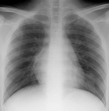
Influenza A (H1N1) complicated by pneumonia. X-ray demonstrates flakes and cotton-woollike shadows in the cardiophrenic angle of the left lower lung and increased pulmonary markings in both lungs
Fig. 22.15.
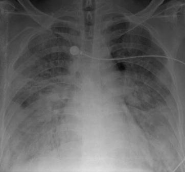
Influenza A (H1N1) complicated by pneumonia. X-ray demonstrates flakes and cotton-woollike blurry shadow with high density in both lungs that is more obvious in both lower lung fields. Blurry pulmonary markings can be found in the lesion area, with enlarged and blurry hilar shadow
Fig. 22.16.
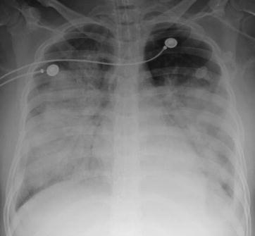
Influenza A (H1N1) complicated by pneumonia. X-ray demonstrates large areas of high-density shadow in both middle and lower lung fields that is more obvious in both lower lung fields, with unclearly defined bilateral costophrenic angles
Fig. 22.17.
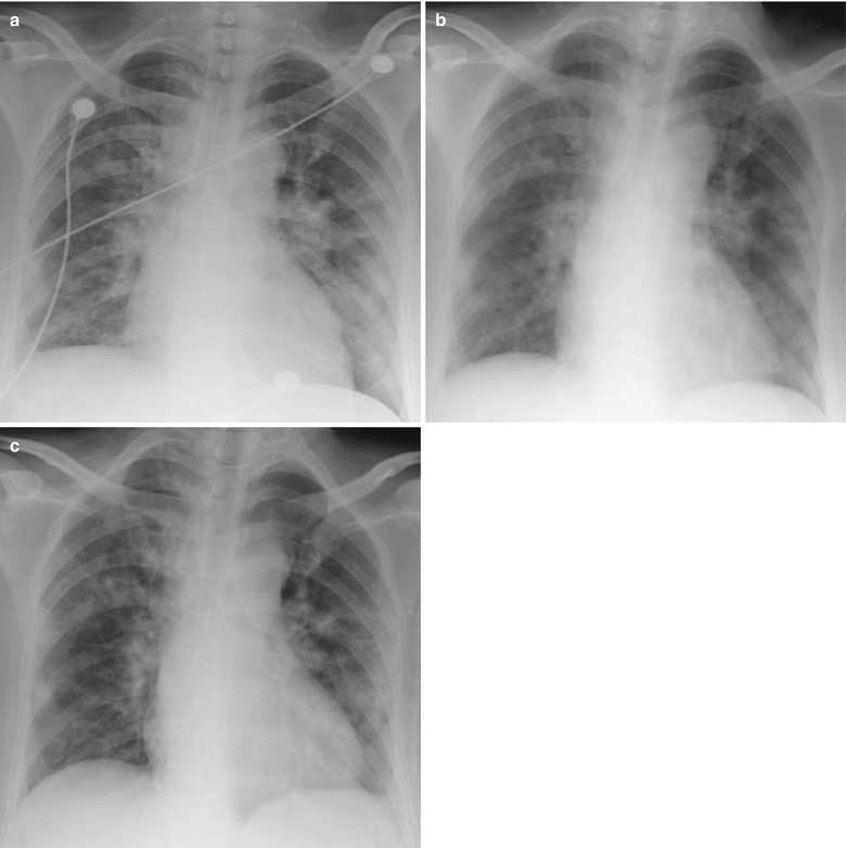
Influenza A (H1N1) complicated by pneumonia. (a) X-ray demonstrates increased pulmonary markings in both lungs, accompanying flakes and cotton-woollike shadow in both lower lungs, and enlarged and densely colored hilar shadows. (b) By reexamination after treatment for 2 days, the conditions are demonstrated to progress, with X-ray demonstrations of multiple flakes and cotton-woollike shadows in both lungs with unclearly defined boundaries, as well as unclearly demonstrated hilar structure and slightly blunt right costophrenic angle. (c) By reexamination after treatment for 4 days, the conditions are demonstrated to be absorbed and improved. X-ray demonstrates increased pulmonary markings of both lungs and accompanying flakes of blurry shadows
Fig. 22.18.
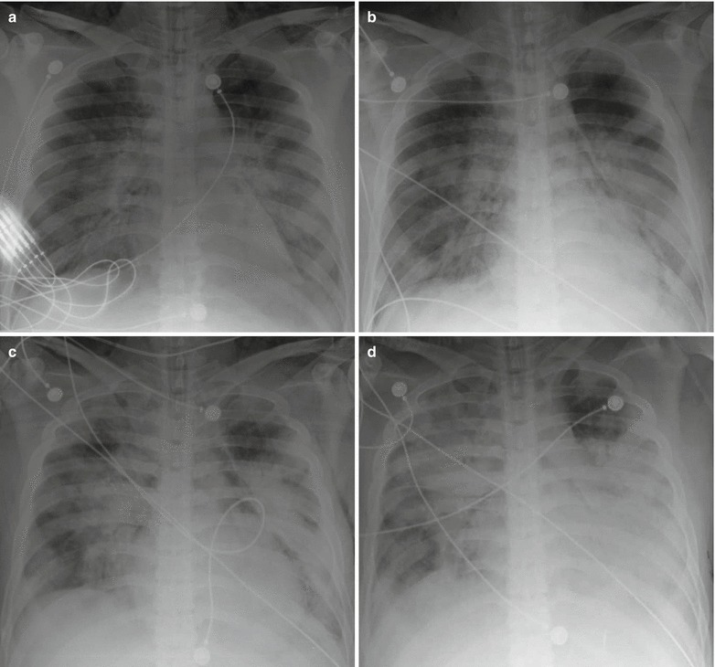
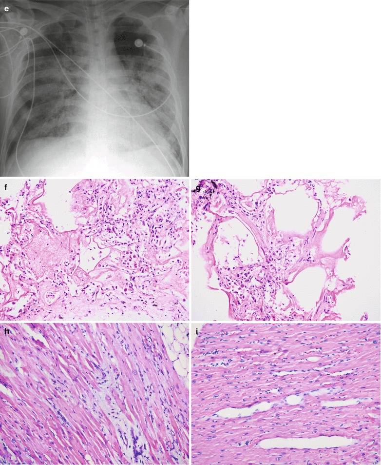
Influenza A (H1N1) complicated by pneumonia. (a) X-ray demonstrates multiple flakes of blurry shadows in both middle and lower lungs, enlarged blurry hilar shadows, and unclearly demonstrated bilateral costophrenic angles. (b) By reexamination after treatment for 1 day, the conditions are demonstrated to progress. X-ray demonstrates more extensive range with flakes of blurry shadows with higher density in bilateral middle and lower lungs. (c) By reexamination after treatment for 2 days, X-ray demonstrates more extensive range with consolidation lesions in both lungs. (d) By reexamination after treatment for 3 days, the conditions are demonstrated with obvious exacerbation. (e) By reexamination after treatment for 9 days, the conditions are demonstrated with obvious improvement. X-ray demonstrates flakes and cotton-woollike blurry shadows in both lungs and increased transparency of both lungs. (f, g) H&E staining demonstrates widened interalveolar space, congested alveolar walls, infiltration of neutrophils and plasma cells that are predominantly mononuclear cells, and exudation of alveolar edema fluid and celluloses (H&E ×20). (h, i) H&E staining demonstrates many inflammatory cells in spaces between myocardial tissues (H&E ×20)
Fig. 22.19.
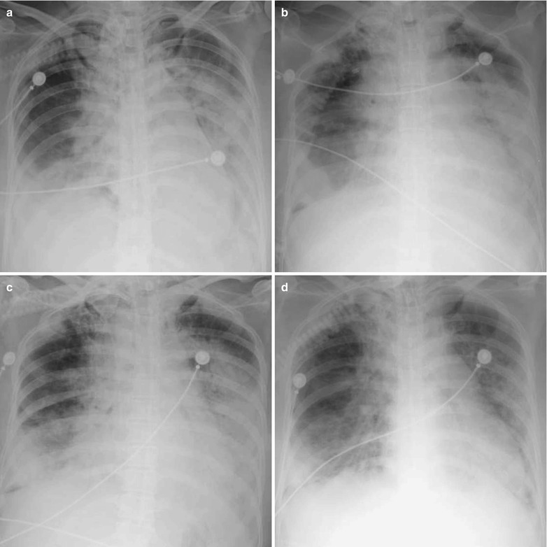
Influenza A (H1N1) complicated by pneumonia. (a) X-ray demonstrates diffusive shadow with increased density in the right lower lung and left middle and lower lung field, enlarged blurry hilar shadow, and unclearly demonstrated upper diaphragm. (b) By reexamination after treatment for 1 day, the conditions are demonstrated to obviously progress. (c) By reexamination after treatment for 2 days, the lesions are demonstrated to have no obvious changes. (d) By reexamination after treatment for 7 days, the lesions are demonstrated to have slight absorption and improvement
Case Study 14
A male patient aged 18 years complained of fever for 3 days, cough for 2 days, and the highest body temperature of 38.5 °C. He also complained of headache, muscular pain, and mild cough with small quantity of yellowish purulent sputum. By physical examination, the pharynx was congested, tonsils were not enlarged, and moist rales were heard in both lungs. He had a history of contact to patients with influenza A (H1N1). Pharyngeal swab demonstrated positive M gene of influenza A (H1N1) virus, positive NP gene of swine influenza virus, and positive HA gene of influenza A (H1N1) virus.
Case Study 15
A male patient aged 37 years complained of fever and cough for 1 week. He also complained of coughing up whitish thick sputum and chest distress, with progressing conditions. Blood gas analysis indicated respiratory failure of type I. After admission, he was given an oxygen inhalation mask. By physical examination, T 39 °C, the pharynx was congested, and moist rales were heard in both lungs. He had a history of contact to patients with influenza A (H1N1). Pharyngeal swab by CDC demonstrated positive M gene of influenza A (H1N1) virus, negative NP gene of swine influenza virus, and negative HA gene of influenza A (H1N1) virus.
Case Study 16
A female patient aged 22 years complained of fever for 1 week and cough with sputum for 3 days. By physical examination, T 40 °C, bpm 180–190/min, and SpO2 65–77 %. She denied history of contact to patients with influenza A (H1N1). Pharyngeal swab demonstrated positive M gene of influenza A (H1N1) virus, positive NP gene of swine influenza virus, and positive HA gene of influenza A (H1N1) virus. By blood gas analysis, pH 7.45, PCO2 39 mmHg, PO2 478 mmHg, and SpO2 69 %. By blood gas analysis following tracheal intubation, pH 7.36–7.26, PCO2 41–56 mmHg, and PO2 37–59 mmHg. By routine blood test, WBC 3.5 × 10⁹/L, GR% 83.6 %, and stab nucleus 8–14 %, with toxic granulation. By blood biochemistry, ALT 109 U/L and AST 507 U/L. The conditions exacerbated at day 2 after the hospitalization, with a sudden slower heart rate, decreased blood pressure, and decreased SpO2 to 35 %. Death occurred after emergency rescuing.
Case Study 17
A male patient aged 27 years complained of fever and cough with sputum for 1 week. He also complained of chills, aversion to cold, rhinorrhea, and nasal obstruction. By physical examination, T 39.5 °C, the pharynx was congested, and moist rales were heard in both lungs. He had a history of contact to patients with influenza A (H1N1). Pharyngeal swab by CDC demonstrated positive M gene of influenza A (H1N1) virus, positive NP gene of swine influenza virus, and negative HA gene of influenza A (H1N1) virus. By routine blood test, WBC 1.93 × 10⁹/L, GR% 59.3 %, and LY% 33.7 %. By blood gas analysis, pH 7.62, PCO2 18 mmHg, and PO2 186 mmHg. By blood biochemistry, ALT 25.7 U/L and AST 84.1 U/L.
Case Study 18
A male patient aged 29 years, with complaints of cough for 7 days and fever for 6 days. He also complained of following wheezing and coughing up reddish foam liked sputum. By physical examination, T 40 °C, pharynx is congested, and moist rales were heard in both lungs. He denied history of contact to patients with influenza A (H1N1). Pharyngeal swab demonstrated positive M gene of influenza A (H1N1) virus, positive NP gene of swine influenza virus, and positive HA gene of influenza A (H1N1) virus. By routine blood test, WBC 17.89 × 10⁹/L, GR% 90.7 %, and LY% 5.5 %. By blood gas analysis, pH 7.33, PCO2 54 mmHg, and PO2 85 mmHg. By blood biochemistry, ALT 40 U/L, AST 32 U/L, Cr 115.9 μmol/L, and urea 16.73 mmol/L.
Case Study 19
A female patient aged 45 years complained of chill, fever, eye upset, and dry cough rarely with sputum for 5 days. She also complained of chest distress and suffocation. She was diagnosed with bronchitis by the physician from another hospital, with following medication of anti-inflammatory drugs. After that, she developed chest distress and shortness of breath and was admitted to our hospital. By physical examination, T 39.5 °C and pharynx was congested. She had a history of contact to patients with influenza A (H1N1). Pharyngeal swab demonstrated positive M gene of influenza A (H1N1) virus, positive NP gene of swine influenza virus, and negative HA gene of influenza A (H1N1) virus. By routine blood test, WBC 3.69 × 10⁹/L, GR% 67.7 %, and LY% 24.7 %. By blood gas analysis, pH 7.488, PCO2 33.2 mmHg, and PO2 46.9 mmHg. By blood biochemistry, ALT 62.9 U/L, AST 144.4 U/L, Cr 46.3 μmol/L, and GLU 7.45 mmol/L.
CT Scanning
In the early stage, HRCT demonstrates thickened bronchial vascular bundles and lobular consolidations that gradually fuse into large flakes (Figs. 22.20 and 22.21). In the progressing stage, HRCT demonstrates nodular shadows of lobular consolidation of central air cavities and multilobular or extensive GGO, possibly accompanied by localized consolidation shadow. With the continued progress of the conditions, GGO foci rapidly fuse and extend, possibly with increased density and flakes of consolidation foci in or out of the GGO (Figs. 22.22 and 22.23). In some cases, a small quantity of pleural effusion may be found. Agarwal et al. reported that 36 % of cases with severe influenza A (H1N1) influenza develop acute pulmonary embolism during hospitalization, which may be caused by high blood coagulation secondary to ARDS.
Fig. 22.20.
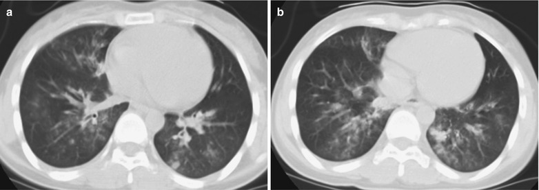
Influenza A (H1N1) complicated by pneumonia. (a, b) CT scanning demonstrates multiple patches of shadows in both lower lobes and the right middle lobe, in different sizes and with uneven density
Fig. 22.21.
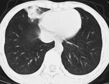
Influenza A (H1N1) complicated by pneumonia. CT scanning demonstrates flake s of high-density shadow in the right middle lobe with uneven density
Fig. 22.22.
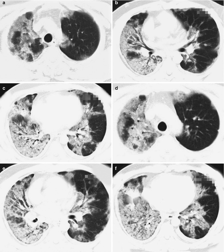
Influenza A (H1N1) complicated by pneumonia. (a–c) CT scanning demonstrates consolidation of both lungs, especially the right lung, and air bronchogram. (d–f) By reexamination after treatment for 4 days, the conditions of both lungs are demonstrated to progress
Fig. 22.23.
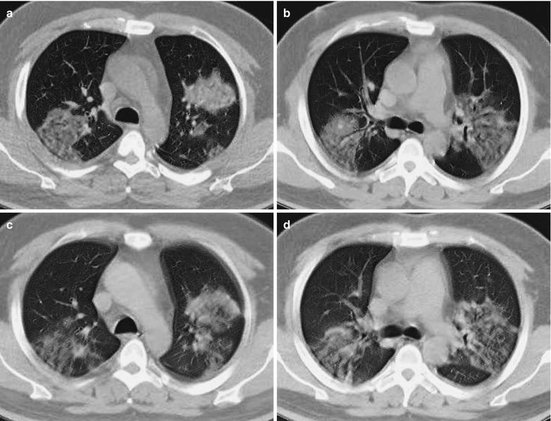
Influenza A (H1N1) complicated by pneumonia. (a, b) CT scanning demonstrates multiple ground-glass opacities in both lungs. (c, d) By reexamination after treatment for 6 days, the foci in both lungs are demonstrated to be absorbed and improved
Case Study 20
A female patient aged 18 years complained of fever and cough for 2 days, dyspnea, and vomiting for 1 day. By physical examination, the pharynx was congested and rough breathing sounds were heard in both lungs with moist rales. She had a history of close contact to patients with influenza A (H1N1). Pharyngeal swab demonstrated negative M gene of influenza A (H1N1) virus, negative NP gene of swine influenza virus, and positive HA gene of influenza A (H1N1) virus. By routine blood test, WBC 11.1 × 10⁹/L and GR% 82 %. By blood gas analysis, pH 7.43, PCO2 35.2 mmHg, PO2 66 mmHg, HCO3 − 23.4 mmol/L, and SO2 93 %.
Case Study 21
A male patient aged 16 years complained of fever for 4 days and right chest pain for 3 days. He also complained of chill, rhinorrhea, coughing up whitish thick phlegm with blood, and shortness of breath. She had a history of close contact to patients with influenza A (H1N1). Pharyngeal swab demonstrated negative M gene of influenza A (H1N1) virus, negative NP gene of swine influenza virus, and positive HA gene of influenza A (H1N1) virus. By routine blood test, WBC 7.9 × 10⁹/L, GR% 82.7 %, and LY% 12.2 %.
Case Study 22
A male patient aged 42 years complained of fever and cough for 5 days. By physical examination, T 39.5 °C, tonsils were enlarged in first degree, and rough breathing sounds were heard in both lungs. He had no definitive history of contact to patients with influenza A (H1N1). Pharyngeal swab demonstrated positive M gene of influenza A (H1N1) virus, positive NP gene of swine influenza virus, and positive HA gene of influenza A (H1N1) virus. By routine blood test, WBC 3.24 × 10⁹/L, GR% 57.74 %, and LY% 32.74 %. By blood biochemistry, TP 55 g/L, AST 100 U/L, ALT 69 U/L, CK 621 U/L, and LDH 511 U/L. During his hospitalization, he had persistent fever, coughed up sputum, and had chest distress. By auscultation, moist rales of both lungs can be heard that are more extensive in the right lung.
Case Study 23
A male patient aged 32 years complained of fever, cough, and pharyngalgia for 6 days, shortness of breath for 2 days, and the highest body temperature of 41 °C. He also reported cough with occasional small quantity of whitish foamy sputum and denied any history of contact to patients with influenza A (H1N1). After admission, his symptoms exacerbated, with severe cough and small quantity of pink foamy sputum. By physical examination, T 36.7 °C, bpm 107/min, R 25/min, BP 119/79 mmHg, SPO2 88 %, tonsils were enlarged in first degree, and many moist rales in both lungs and dry rales in the right middle and lower lung were heard. Pharyngeal swab demonstrated positive M gene of influenza A (H1N1) virus, positive NP gene of swine influenza virus, and positive HA gene of influenza A (H1N1) virus. By routine blood test, WBC 8.2 × 10⁹/L and GR% 84.8 %. By blood gas analysis, pH 7.4, PCO2 24.6 mmHg, PO2 51 mmHg, BE −10 mmol/L and SO2 87 %. By blood biochemistry, ALT 58 U/L, AST 97 U/L, BUN 12 mmol/L, UA 630 U/L, ALP 224 U/L, CRP 128.9 mg/L, CK 2232 U/L, CK-MB 58 U/L, LDH 582 U/L, and HBDH 758 U/L.
In both early and progressing stages, ground-glass opacity is the most common manifestation. In the early stage, chest radiology demonstrates round-like ground-glass opacities distributing under the pleura or around the bronchial blood vessels, which is believed to be the characteristic demonstration of influenza A (H1N1). Ground-glass opacity is commonly complicated by pulmonary consolidation which may be located in the ground-glass opacity or independent of the ground-glass opacity. The ground-glass opacity can be morphological in patches and round-like nodular, localized, multifocal, and large flakes of shadows, with uneven density and unclearly defined boundaries. The occurrence rate of ground-glass opacity is high, which is a rare sign in cases of other pneumonia.
At the center of the ground-glass opacity in the cases of influenza A (H1N1), lobular central nodules can also be found with empty cavities. The mechanism underlying the occurrence of empty cavities may be excessive pulmonary ventilation due to bronchostenosis caused by immune responses. These immune responses may be triggered by spreading of the virus into peribronchial tissues after its invasion into bronchial epithelial tissues.
The chest radiology of cases with severe influenza A (H1N1) complicated by ARDS is characterized by:
Various shapes of the lesions, including patches and large flakes of consolidation shadows, fused patches of shadows or cloudy cotton-woollike shadows, and large flakes of ground-glass opacity; in the cases with one lobe or multiple lobes involved, the density of pulmonary consolidation is even and uniform in white lung sign.
Multiple lobar infiltration and migratory changes in the location and range of the lesions.
Both pulmonary parenchymal and interstitial involvements.
Rapid changes of imaging demonstrations.
Common occurrence of complications, including pneumothorax, mediastinal and subdermal edema (Figs. 22.24 and 22.25), and even retroperitoneal pneumatosis and secondary fungal infection. In rare cases, local destruction and fibrosis of pulmonary tissues can be found, with honeycomb-like lung and insufficient pneumothoracic recruitment.
Fig. 22.24.
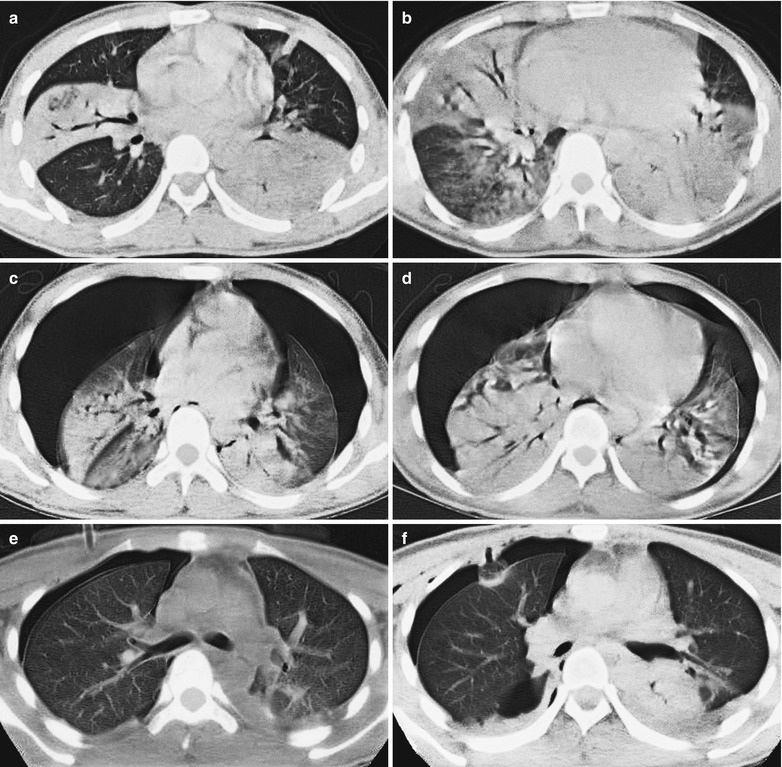
Influenza A (H1N1) complicated by pneumonia and pneumothorax. (a, b) CT scanning demonstrates consolidations in the right middle lobe and both lower lobes with air bronchogram inside the consolidations as well as patches of shadow in the left upper lobe and in the lingual segment of the left lower lobe. (c, d) By reexamination after treatment for 3 days, CT scanning demonstrates stripes of transparent areas with no pulmonary markings in both lateral lungs, compression of both lungs with decreased volumes, and accumulated pulmonary markings. There are also multiple large flakes of consolidations in both lungs, with air bronchogram inside the consolidations. (e, f) By reexamination after treatment for 5 days, CT scanning demonstrates improved pneumothorax of both lungs with absorbed lesions and consolidations in both lower lobes
Fig. 22.25.
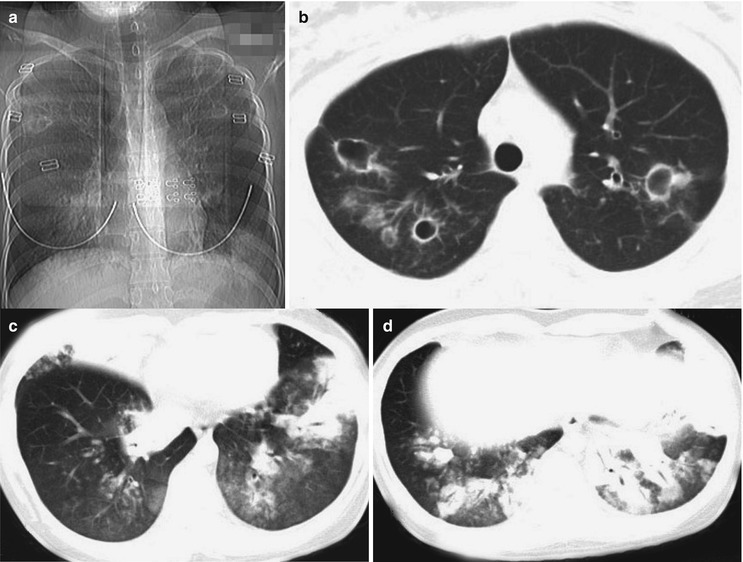
Influenza A (H1N1) complicated by pneumonia and bronchiectasis. (a) X-ray demonstrates flakes of shadows with blurry boundaries in the inner zone of the right middle and lower lung field and the left lower lung field and multiple cavities in both lungs with unclearly defined boundaries. (b) CT scanning demonstrates multiple irregular and thin-walled empty cavities in both upper lobes and surrounding scattering patches of shadows. There are also thickened bronchial wall in the left upper lobe, with widened lumen. (c, d) Multiple consolidations and ground-glass opacities are demonstrated in the right middle lobe, right lower lobe, and inferior lingual segment of the left upper lobe and left lower lobe
Case Study 24
A male patient aged 19 years complained of fever for 6 days, dyspnea, and coughing up sputum for 5 days and the highest body temperature of 38 °C. He also had symptoms of coughing up yellowish thick sputum that is in small quantity, difficult to expectorate, and occasionally with blood and slight difficulty in breathing. By physical examination, T 39 °C and the pharynx was congested. Pharyngeal swab demonstrated positive M gene of influenza A (H1N1) virus, positive NP gene of swine influenza virus, and positive HA gene of influenza A (H1N1) virus. He denied any history of contact to patients with influenza A (H1N1) or patients with influenza-like symptoms. By blood biochemistry, Myo 131 ng/L and CK-MB 7.68 ng/L. By blood gas analysis, pH 7.53, PCO2 27.1 mmHg, and PO2 52 mmHg. He was hospitalized due to the diagnosis of (1) severe and critical influenza A (H1N1), (2) severe pneumonia, (3) respiratory failure of type II and ARDS, and (4) multiple organ dysfunction.
Case Study 25
A female patient aged 21 years complained of fever and cough for 9 days. She received anti-inflammatory therapies, with conditions not improved. Her body temperature fluctuated from 38 to 38.7 °C, with accompanying symptoms of chest distress, shortness of breath, and coughing up yellowish sputum. By physical examination, T 39.7 °C, the pharynx was congested, and tonsils were enlarged in first degree. Moist rales can be heard in both lungs, which are absent after coughing. She had no definitive history of contact to patients with influenza A (H1N1). Pharyngeal swab demonstrated positive M gene of influenza A (H1N1) virus, negative NP gene of swine influenza virus, and positive HA gene of influenza A (H1N1) virus. By routine blood test, WBC 8.64 × 10⁹/L, GR% 76.14 %, and LY% 19.34 %. By blood biochemistry, CK 140 U/L and LDH 547 U/L.
Influenza A (H1N1) Complicated by Pneumonia in Perinatal Women
During pregnancy, pathophysiological changes of many acute or chronic pulmonary diseases are dramatic, leading to intolerance of pregnant women to obviously decreased pulmonary ventilation caused by pneumonia. During the gestational period, decreased total count of T helper lymphocytes and decreased activity of natural killer cells may result in susceptibility of pregnant women to viral and fungal pneumonia.
Mild cases are characterized by ground-glass opacities and small flakes and cotton-woollike high-density shadows in single or multiple pulmonary lobes. Severe cases are characterized by masses of cotton-woollike shadows and ground-glass opacity in multiple pulmonary lobes, with air bronchogram. Within 24 h, the conditions progress into diffuse high-density foci in both lungs, partially accompanied by atelectasis with possible complications of unilateral or bilateral pleuritis or pleural effusion and pericardial effusion (Figs. 22.26 and 22.27). The mechanism underlying pleuritis remains elusive, which may be related to hypoproteinemia, direct viral actions, and endocrinological factors. Most of mild cases have favorable prognosis. But the conditions of severe cases may progress rapidly to cause ARDS, respiratory failure, cardiac failure and renal failure, septic shock, and even death from multiple organ failure.
Fig. 22.26.
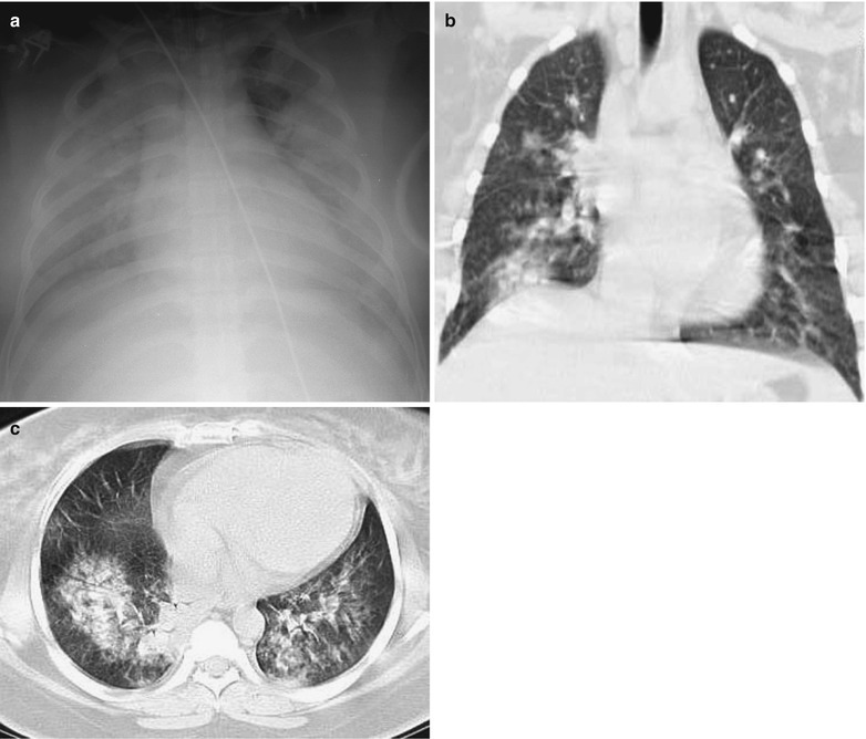
Influenza A (H1N1) complicated by pneumonia. (a) X-ray demonstrates large flakes of high-density shadows in both lung fields. (b, c) CT scanning demonstrates multiple flakes of high-density shadows in both lungs, especially in the right lung
Fig. 22.27.
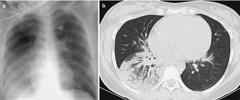
Influenza A (H1N1) complicated by pneumonia. (a) X-ray demonstrates large flake of shadow in the right lower lung, cloudy high-density shadow in the left lower lung, and enlarged and densely colored hilum. (b) CT scanning demonstrates consolidation in the right lower lobe
Case Study 26
A female patient aged 31 years complained of high fever, suffocation, and progressive difficulty in breathing for 2 days which occurred at day 4 after Cesarean section. She also complained of coughing up pink foamy sputum and progressive dyspnea. After symptomatic therapies, her conditions failed to improve, but with rapid deterioration. By physical examination, T 40 °C and bpm 136/min, with oral lip cyanosis, rough breathing sound in both lungs, and overwhelming dry and moist rales in both lungs. She had a history of close contact to patients with influenza A (H1N1). Pharyngeal swab demonstrated negative M gene of influenza A (H1N1) virus, negative NP gene of swine influenza virus, and positive HA gene of influenza A (H1N1) virus. By routine blood test, WBC 12.97 × 10⁹/L and GR% 91.1 %.
Case Study 27
A female patient aged 27 years, at 37+5 gestation, complained of fever for 7 days. She also complained of pharyngeal upset, general soreness and pain, and highest body temperature of 39.5 °C. In addition, frequent cough, pharyngalgia, difficulty lying in supine position, and fluctuation of body temperature between 38 and 39.5 °C occurred 6 days ago. She reported frequent fetal movement for 5 days, vaginal bleeding for 2 days, and decreased fetal movement for 1 day. She had a history of close contact to patients with influenza A (H1N1). Pharyngeal swab demonstrated positive M gene of influenza A (H1N1) virus, negative NP gene of swine influenza virus, and positive HA gene of influenza A (H1N1) virus. By routine blood test, WBC 5.25 × 10⁹/L, HGB 86 g/L, GR% 82.1 %, and LY% 16.4 %. By blood biochemistry, ALB 16 g/L and GLB 22.47 g/L. By blood gas analysis, pH 7.42, PCO2 19.13 mmHg, PO2 63 mmHg, and SO2 95 %.
Appendix: Case Reports by Autopsy
Case Study 28
A male patient aged 74 years complained of fever and cough for 4 days. By physical examination, T 39.4 °C, bpm 120/min, and R 24/min, with mental confusion and deep coma without any response to sound. The breathing sound of both lungs is low, with small quantity of moist rales in both basal lungs. After admission, he was given tracheal intubation connected to a respirator to assist his ventilation. He had a past history of hypertension and chronic bronchitis. Pharyngeal swab demonstrated positive viral nucleic acid of influenza A (H1N1). By routine blood test, WBC 9.51 × 10⁹/L and GR% 80.6 %. By blood gas analysis, pH 7.3, PO2 144 mmHg, PCO2 70 mmHg, HCO3 − 33.4 mmol/L, BE 5.4 mmol/L, and SaO2 98.8 %. By blood biochemistry, ALT 46.6 U/L, AST 56.3 U/L, TBIL 6.9 μmol/L, and Cr 85.6 μmol/L.
Fig. 22.28.
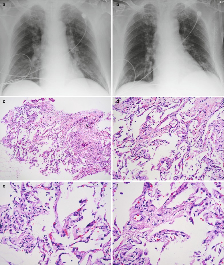
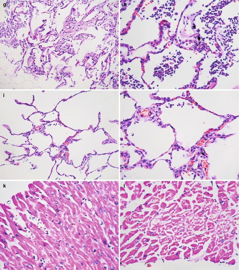
Influenza A (H1N1) complicated by pneumonia. (a, b) X-ray demonstrates multiple blurry shadows in both lungs and enlarged blurry hilar shadows of both lungs. (c–j) The autopsy demonstrates widened pulmonary interstitium, fibrosis of interalveolar space, infiltration of inflammatory cells that are mostly neutrophils, and exudations of mononuclear cells, lymphocytes, and macrophage (H&E ×10). (k, l) There are myocardial interstitial edema and minor vascular dilation (H&E ×40)
Case Study 29
A female patient aged 53 years complained of cough for 7 days and fever for 6 days with the highest body temperature of 40 °C. She also complained of wheezing and suffocating and coughing up pink foamy sputum. By physical examination, the pharynx was congested and moist rales were heard in both lungs. She denied a history of contact to patients with influenza A (H1N1). Pharyngeal swab demonstrated positive M gene of influenza A (H1N1) virus, positive NP gene of swine influenza virus, and positive HA gene of influenza A (H1N1) virus. By routine blood test, WBC 17.89 × 10⁹/L, GR% 90.7 %, and LY% 5.5 %. By blood gas analysis, pH 7.33, PCO2 54 mmHg, and PO2 85 mmHg. By blood biochemistry, ALT 81.3 U/L and AST 166.6 U/L.
Fig. 22.29.
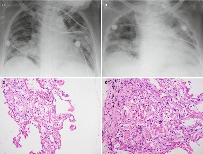
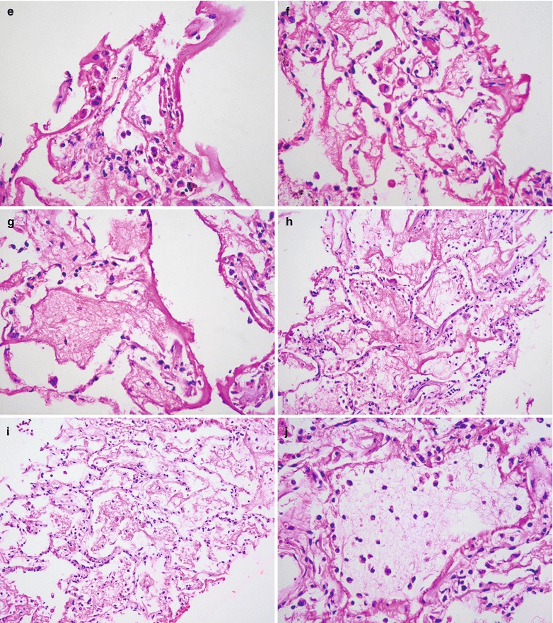
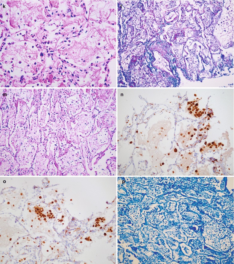
Influenza A (H1N1) complicated by pneumonia. (a) X-ray demonstrates flakes of shadows with increased density in both lung fields that are more obvious in both lower lungs and enlarged blurry hilar shadows. (b) X-ray demonstrates progress of the conditions, with extended range of shadows in both lung fields with high density that are more obvious in the right lower lung and the left lung and enlarged blurry hilar shadows. (c) There are widened interalveolar space, congested alveolar walls, infiltration of neutrophils and plasma cells, and exudations of mononuclear cells, alveolar edema fluid, and celluloses (H&E ×20). (d) Formation of intra-alveolar transparent membrane (H&E ×20). (e) Shedding of alveolar epithelium and exudation of celluloses from some alveolar cavities (H&E ×40). (f) A large quantity of intra-alveolar transparent membranes forms (H&E ×40). (g) Vascular congestion of alveolar walls (H&E ×40). (h) Thinner alveolar walls, occluded vascular vessels, and exudation of celluloses in a large quantity from alveoli (H&E ×20). (i) Following shedding and necrosis of type I epithelial cells, slight hyperplasia of type II epithelial cells occurs (H&E ×20). (j) Exudation of loose celluloses within alveolar cavities (H&E ×40). (k) Exudation of dense celluloses within alveolar cavities (H&E ×40). (l) Exudation of celluloses in a large quantity within alveoli, with no growth of bacteria (Masson staining, ×20). (m) Exudation of celluloses in a large quantity within alveolar cavities (PAS staining × 20). (n, o) Accumulation of macrophages into masses (H&E ×20). (p) A large quantity of inflammatory cell infiltration, with no findings of acid-fast bacilli (H&E ×20)
Case Study 30
A female patient aged 53 years complained of cough for 7 days and fever for 6 days with the highest body temperature of 39.8 °C. By physical examination, the pharynx was congested and moist rales were heard in both lungs. She denied a history of contact to patients with influenza A (H1N1). Pharyngeal swab demonstrated positive M gene of influenza A (H1N1) virus, positive NP gene of swine influenza virus, and positive HA gene of influenza A (H1N1) virus. By routine blood test, WBC 17.89 × 10⁹/L, GR% 90.7 %, and LY% 5.5 %. By blood gas analysis, pH 7.33, PCO2 54 mmHg, and PO2 85 mmHg. By blood biochemistry, ALT 40 U/L, AST 32 U/L, Cr 115.9 μmol/L, and urea 16.73 mmol/L. The clinical diagnosis was influenza A (H1N1) with hepatic lesions.
Fig. 22.30.
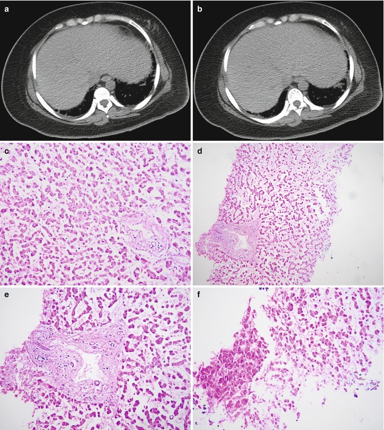
Influenza A (H1N1) complicated by hepatic lesions. (a, b) CT scanning demonstrates enlarged liver with full exterior boundary. (c–f) Pathology demonstrates dissociation of hepatic cords, visible structures in portal areas, no obvious hepatocytic destruction, blurry hepatocytic nucleus, and autolytic changes of hepatocytes
Case Study 31
A male patient aged 29 years complained of cough for 3 days and fever for 2 days. By physical examination, the pharynx was congested and moist rales were heard in both lungs. He denied a history of contact to patients with influenza A (H1N1). Pharyngeal swab demonstrated positive M gene of influenza A (H1N1) virus, positive NP gene of swine influenza virus, and negative HA gene of influenza A (H1N1) virus. By routine blood test, WBC 2.54 × 10⁹/L, GR% 75.1 %, and LY% 15.4 %. By blood gas analysis, pH 7.512, PO2 48.8 mmHg, and PCO2 32.16 mmHg. By blood biochemistry, ALT 166.8 IU/L and AST 270.5 IU/L.
Fig. 22.31.
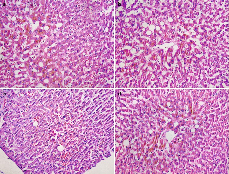
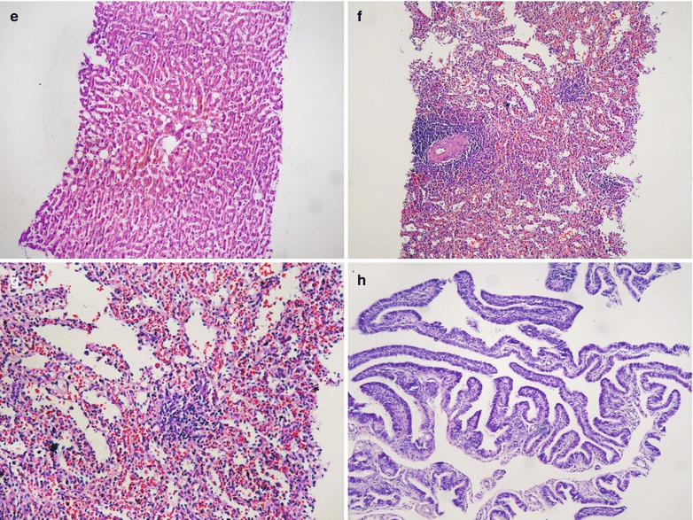
Pathology of influenza A (H1N1). (a–e) Extended and congested hepatic sinus, cholestasis of central hepatocytes, bullous fatty degeneration of hepatocytes, and infiltration of inflammatory cells around portal areas (H&E (a–d) ×10, (e) ×20). (f, g) Dilated and congested splenic sinus, atrophic splenic corpuscles, and decreased lymphocytes (H&E ×10, ×20). (h) Vascular hemorrhage of intestinal mucosa (H&E ×10)
Case Study 32
A female patient aged 22 years complained of fever for 6 days and tachypnea for 2 days with the highest body temperature of 39 °C. She also complained of chills, aversion to cold, cough with sputum, and cyanosis. She experienced spontaneous delivery of a baby boy 2 days ago. By physical examination, the pharynx was congested and diminished breath sounds in both lungs and densely distributed moist rales in both lower lungs were heard. She denied a history of contact to patients with influenza A (H1N1) and patients with influenza-like symptoms. By routine blood test, WBC 14.37 × 10⁹/L, HGB 73 g/L, and GR% 82 %. By blood gas analysis, pH 7.38, PCO2 33 mmHg, and PO2 66 mmHg. By blood biochemistry, ALT 330 U/L, AST 2650 U/L, ALB 23.7 g/L, Cr 58.9 μmol/L, and urea 2.49 mmol/L. She was given anti-infection therapy after her admission and noninvasive use of respirator to assist ventilation. However, her conditions exacerbated and death occurred due to respiratory failure.
Fig. 22.32.
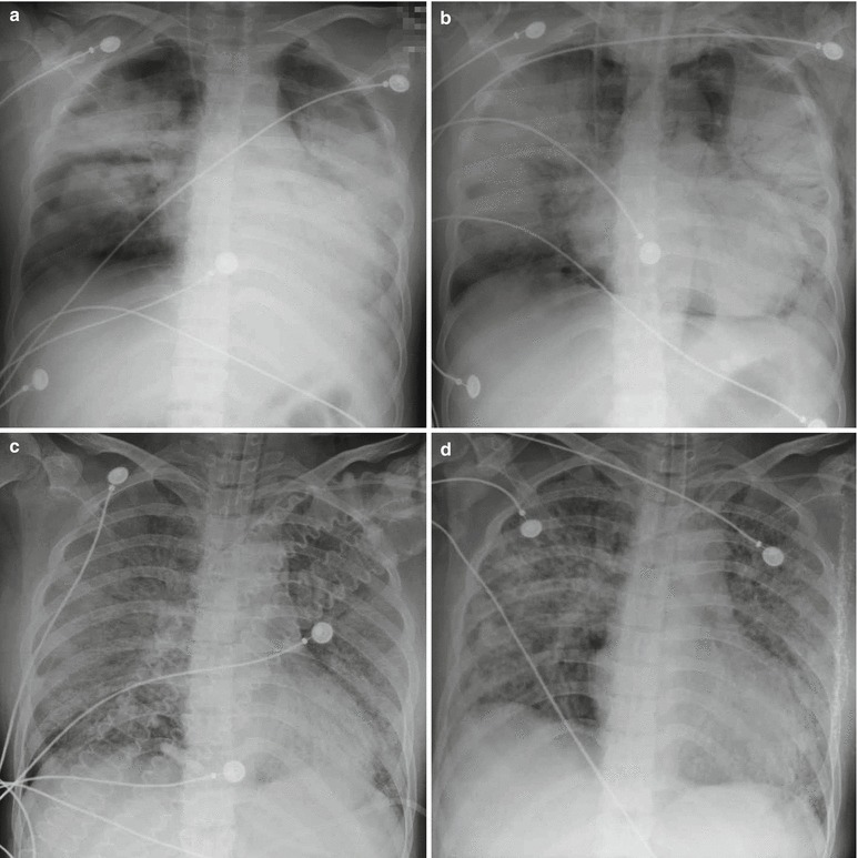
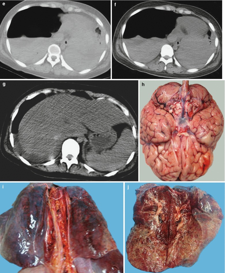
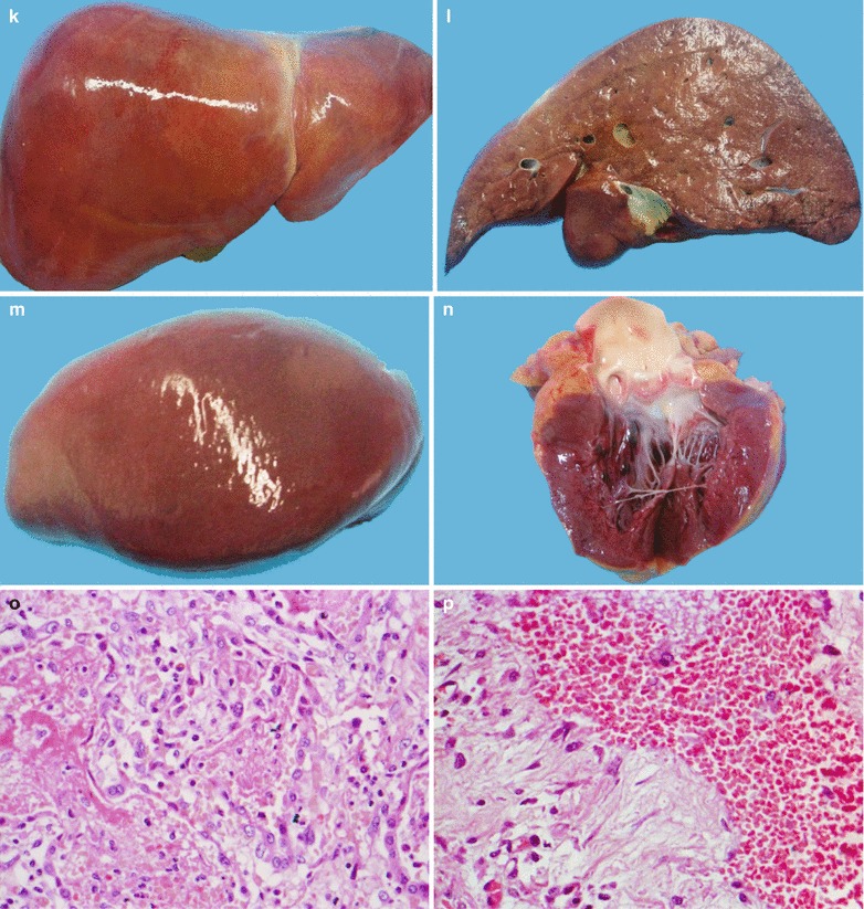
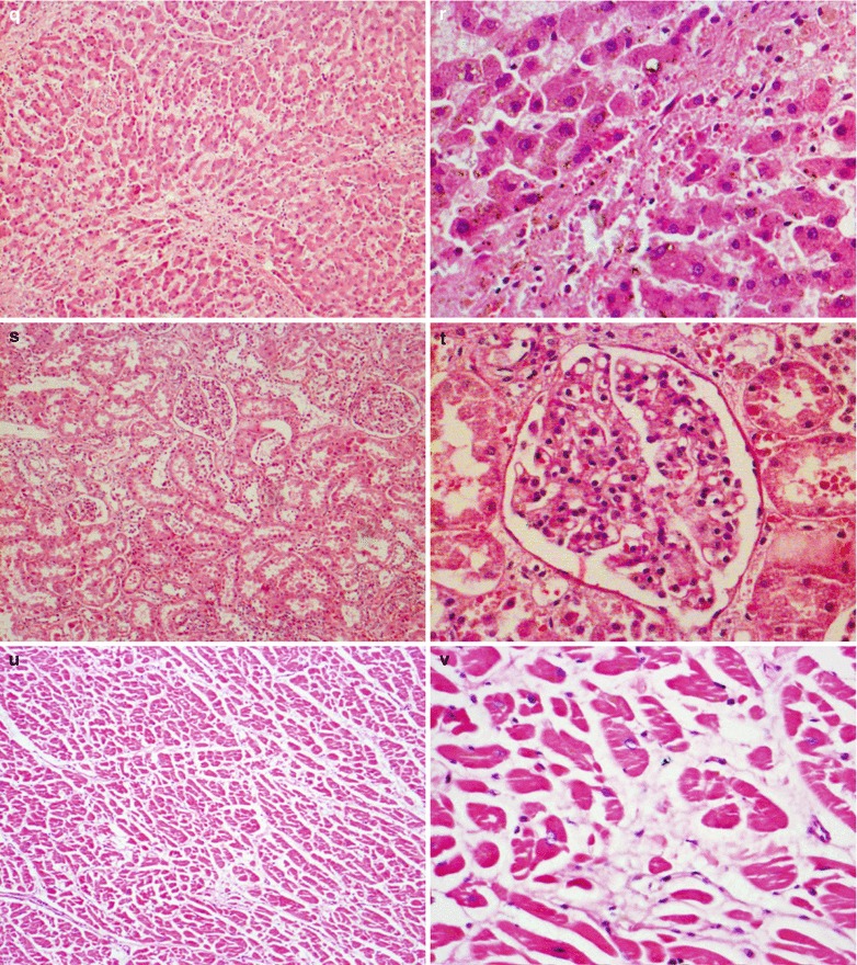
Radiological and pathological changes of influenza A (H1N1). (a) X-ray demonstrates large flakes of shadows with high density in both lung fields with unclearly defined boundaries as well as enlarged heart shadow and blurry bilateral diaphragmatic surface and costophrenic angles. (b) By reexamination after treatment for 4 days, the conditions are demonstrated to progress. X-ray demonstrates extended range with high-density shadows in both lung fields and strips of gas shadows in the bilateral necks and under the skin of the left chest. (c) By reexamination after treatment for 14 days, X-ray demonstrates diffuse high-density shadows in both lung fields. (d) By reexamination after treatment for 23 days, the lesions are absorbed and improved, with increased transparency of both lungs. (e, f) In 2 h after death, chest CT scanning demonstrates a large quantity of pleural effusion in bilateral thoracic cavities and hydropneumothorax in the right thoracic cavity. (g) Abdominal CT scanning demonstrates even density of the liver and liquid density shadow surrounding the posterior liver. (h) The gross specimen demonstrates slight cerebral swelling. (i, j) Shrinkage of lung volumes, yellowish white exudates on the surface and cross sections, extensive intrapulmonary hemorrhage and pulmonary congestion, and accompanying flakes and cords of pulmonary interstitial fibrosis. (k, l) Slight liver swelling and tense liver capsule. (m) Kidneys. (n) Heart. (o) Microscopy demonstrates a small quantity of alveolar cells, fibrosis of most pulmonary tissues, absence of bronchiolar epithelial cells, and a large quantity of inflammatory cells that are predominantly macrophages, in consistency with diffuse alveolar lesions due to necrotic bronchitis and alveolar hemorrhage (H&E ×40). (p) Fibrosis of pulmonary tissues and alveolar hemorrhage (H&E ×40). (q) More inflammatory cells in the interhepatocytic space, dilated hepatic sinus, and a large quantity of erythrocytes in macrophages (H&E ×10). (r) More inflammatory cells in the interhepatocytic space and fragmented necrotic hepatocytes (H&E ×40). (s) Dilated renal sinus and many inflammatory cells in the renal parenchymal space (H&E ×40). (t) A small quantity of inflammatory cells in the space between myocardial cells (H&E ×40). (u) A small quantity of inflammatory cells in the space between myocardical cells (H&E ×10). (v) A small quantity of inflammatory cells in the space between myocardial cells (H&E ×40)
Diagnostic Basis
The diagnosis can be defined based on epidemiological history, clinical manifestations, and etiological examinations. Early detection and early diagnosis play critical role in controlling and curing influenza A (H1N1).
Suspected Cases
A case can be suspected with influenza A (H1N1) with at least one of the following conditions:
A history of close contact to definitive case of influenza A (H1N1) that is in the infective stage within 7 days before the onset of flu-like symptoms; a close contact refers to diagnosing, treating, and attending patients with influenza A (H1N1) in the infective stage with no effective protection, living together with the patient, and contact to respiratory secretion or body fluid of the patient.
Onset of influenza-like clinical manifestations, with influenza A (H1N1) virus positive but no further examination to define the viral subtype.
For the cases with at least one of the above two conditions, etiological examinations should be performed to define the diagnosis.
Clinically Diagnosed Cases
Clinical diagnosis can be made based only on the following conditions. The cases have influenza-like symptoms but with no definitive diagnosis by laboratory tests and examinations in one the same prevailing event of influenza A (H1N1). When other diseases causing influenza-like symptoms are excluded, the cases can be clinically diagnosed with influenza A (H1N1).
Definitively Diagnosed Cases
Influenza A (H1N1) can be defined as the presence of influenza-like clinical manifestations, along with one or more of the following laboratory findings: (1) viral nucleic acid of influenza A (H1N1) virus positive by real-time RT-PCR and RT-PCR; (2) successful isolation of influenza A (H1N1) virus; and (3) at least four times increase of specific antibody against influenza A (H1N1) virus in double sera.
Severe and Critical Cases
The cases with one of the following conditions can be defined as severe: (1) high fever persisting for at least 3 days and accompanying severe cough with thick sputum, bloody sputum, or chest pain; (2) increased frequency of respiration, dyspnea, and cyanosis of oral lips; (3) conscious changes with sluggish response, lethargy, irritation, and convulsion; (4) severe vomiting and diarrhea with dehydration; (5) complication of pneumonia; and (6) obviously exacerbated previous basic diseases.
The cases with one of the following conditions can be defined as critical: (1) respiratory failure, (2) infective toxic shock, (3) multiple organ dysfunction, and (4) other severe clinical conditions requiring intensive care.
Diagnostic Imaging
Pneumonia
X-ray and CT scanning demonstrate patches of exudative shadow, network of shadow, and nodular shadow in both lungs that are more common in the lower lungs. The conditions may progress, with demonstrations of large flakes of consolidation shadow.
Acute Necrotizing Encephalitis
There are multifocal and symmetric cerebral lesions, involving the bilateral thalamus, tegmentum of the brainstem, white matter around the cerebral ventricles, and medulla of the cerebellum. CT scanning demonstrates low-density shadow of the lesions. MR imaging demonstrates long T1 and long T2 signals.
Differential Diagnosis
Differentiation from Diseases of the Central Nervous System
Reye Syndrome
Reye syndrome is an acute encephalopathy accompanied by hepatic fatty degeneration. After viral infection, symptoms of encephalopathy commonly occur, including acute intracranial hypertension, conscious disturbance, and convulsion. Accompanying hepatic dysfunction also occurs. By biochemistry, serum transaminase increases in different degrees, as well as increased blood ammonia and decreased blood sugar. By diagnostic imaging, the main demonstration is the sign of diffuse cerebral edema, rarely with brain parenchymal lesions. Reye syndrome is so dangerous that it can cause death within 24 h. But most survivors experience improved conditions after 2–3 days.
Epidemic Encephalitis B
Epidemic encephalitis B is transmitted by mosquitoes, with common occurrence in summers and in children under the age of 10 years. Clinically, it is characterized by high fever, spasm, conscious disturbance, and meningeal irritation sign. By laboratory examinations, increased WBC count can be found in both cerebrospinal fluid examination and routine blood test that are mainly neutrophilic granulocytes. Serological specific IgM antibody can be detected 3–4 days after the onset. By diagnostic imaging, the lesions can involve the thalamus, cerebral cortex, and spinal cord, with an asymmetric distribution and rare involvement of the brainstem.
Autosomal Dominant Acute Necrotizing Encephalopathy
Autosomal dominant acute necrotizing encephalopathy is a disease with incomplete genetic penetrance, with its genes being located in 2q12.1–2q13. Its pathological foundation is loss of coupling due to mitochondrial oxidative phosphorylation. Autosomal dominant acute necrotizing encephalopathy commonly occurs in children and is characterized by sudden encephalopathy symptoms 2–3 days after the onset of febrile illness. By diagnostic imaging, lesions are symmetrically distributed in the thalamus and brainstem, being similar to those of ANE. Autosomal dominant acute necrotizing encephalopathy tends to relapse which can cause more serious lesions.
Differentiation from Respiratory Diseases
Influenza A (H1N1) complicated by pneumonia has similar imaging demonstrations to other pneumonia, especially viral pneumonia. The definitive diagnosis is based on epidemiological history and laboratory detection for specific virus. Meanwhile, some patients develop pneumonia before their infection of influenza A (H1N1). For these cases, differential diagnosis should be made for definitive diagnosis.
Adenoviral Pneumonia
Adenoviral pneumonia, a common viral pneumonia, commonly occurs in children. It is characterized by peritracheitis, peribronchitis, and alveolitis. X-ray demonstrates thickened and blurry pulmonary markings and pulmonary exudative shadow, commonly with accompanying emphysema. Adenoviral pneumonia rarely develops into respiratory failure and can be effectively treated by antiviral therapies. Its course lasts for a short period of time.
Mycoplasma Pneumonia
Mycoplasma pneumonia is pneumonia with interstitial changes caused by mycoplasma. In the early stage, mycoplasma pneumonia is characterized by imaging demonstrations of increased and blurry pulmonary markings and network of markings. During the progressing stage, the markings are demonstrated as localized or extensive flakes of blurry shadows and large flakes of fanlike shadows stretching from the hilum to exterior lung fields, which are distributed in pulmonary lobes or segments. Such a distribution can also be absent. CT scanning demonstrates early pulmonary interstitial inflammation, network of shadow, and thickened interlobular septum.
Allergic Pneumonia
Allergic pneumonia is characterized by rapid changes of pulmonary demonstrations and foci, which is the same as severe and critical cases of influenza A (H1N1) complicated by pneumonia. However, the main pathological changes of allergic pneumonia are exudative alveolitis and interstitial pneumonia with light and thin pulmonary shadows. After clinical treatment, the foci are rapidly absorbed and their onset is related to allergens, which should be taken into account for differentiation.
Severe Acute Respiratory Syndrome, SARS
SARS, severe acute respiratory syndrome, is also known as infectious atypical pneumonia and is caused by coronavirus. In the early stage, X-ray and CT scanning demonstrate small flakes of shadows in the lungs. In the progressing stage, the small flakes of shadows progress into large flakes or diffuse shadows, possibly complicated by pulmonary consolidation. Being the same as severe influenza A (H1N1) complicated by pneumonia, SARS has a long course, whose differentiation should be based on etiological examinations. However, the imaging demonstrations are different between the two diseases due to different therapies. Influenza A (H1N1) complicated by pneumonia is commonly treated by antiviral therapies, with rare occurrence of intrapulmonary interstitial hyperplasia during the course and in the prognostic outcomes. However, the use of hormone in treating SARS causes obvious intrapulmonary interstitial hyperplasia in patients, with prognostic interstitial hyperplasia or interstitial fibrosis.
Contributor Information
Hongjun Li, Email: lihongjun00113@126.com.
HongJun Li, Email: lihongjun00113@126.com.
Further Reading
- Agarwal PP, Cinti S, Kazerooni EA. Chest radiographic and CT findings in novel swine-origin influenza A(H1N1)virus infection. AJR Am J Roentgenol. 2009;193(6):1488–93. doi: 10.2214/AJR.09.3599. [DOI] [PubMed] [Google Scholar]
- Chen F, Zhao DW, Wen S, et al. Image demonstrations of severe and critical influenza A (H1N1) complicated by pneumonia. Chin J Radiol. 2010;44(2):123–6. [Google Scholar]
- Clinical guideline for influenza A (H1N1) (2010 edition). China: Administrative Office of the Ministry of Health; 2010.
- Finn BC, Rodríguez Pabón EM, Young P. A new cause of cavitated bilateral pulmonary nodules: influenza A (H1N1) virus. Eur J Intern Med. 2010;21(1):50. doi: 10.1016/j.ejim.2009.10.007. [DOI] [PubMed] [Google Scholar]
- Haktanir A. MR imaging in novel influenza A (H1N1)-associated meningoencephalitis. AJNR Am J Neuroradiol. 2010;31(3):394–5. doi: 10.3174/ajnr.A2037. [DOI] [PMC free article] [PubMed] [Google Scholar]
- Jamieson DJ, Honein MA, Rasmussen SA, et al. H1N1 2009 influenza virus infection during pregnancy in the USA. Lancet. 2009;374:451–8. doi: 10.1016/S0140-6736(09)61304-0. [DOI] [PubMed] [Google Scholar]
- Li HJ, Li N. Radiology of influenza A (H1N1)–basics and clinical practice. Beijing: Tsinghua University Press; 2010. [Google Scholar]
- Li HJ, Li N. Radiology of influenza A(H1N1) [M] Dordrecht/Heidelberg/New York/London: Springer; 2013. [Google Scholar]
- Li HJ, Li N, Jin RH, et al. A case of influenza A (H1N1) complicated by pneumonia. Chin J Radiol. 2009;43(12):1337–8. [Google Scholar]
- Li HJ, Bao DY, Li XQ, et al. Imaging demonstrations and pathological analysis of critical influenza A (H1N1) complicated by pneumonia. J Pract Radiol. 2010;25(9):951–4. [Google Scholar]
- Li HJ, Cheng JL, Li N, et al. Critical influenza (H1N1) pneumonia: imaging manifestations and histopathological findings. Chin Med J (Engl) 2012;125(12):2109–14. [PubMed] [Google Scholar]
- Lyon JB, Remigio C, Milligan T, et al. Acute necrotizing encepH alopathy in a child with H1N1 influenza infection. Pediatr Radiol. 2010;40(2):200–5. doi: 10.1007/s00247-009-1487-z. [DOI] [PubMed] [Google Scholar]
- Martin A, Reade EP. Acute necrotizing encepH alopathy progressing to brain death in a pediatric patient with novel influenza A (H1N1) infection. Clin Infect Dis. 2010;50(8):e50–2. doi: 10.1086/651501. [DOI] [PubMed] [Google Scholar]
- Ormitti F, Ventura E, Summa A, et al. Acute necrotizing encepH alopathy in a child during the 2009 influenza A(H1N1) pandemia: MR imaging in diagnosis and follow-up. AJNR Am J Neuroradiol. 2010;31(3):396–400. doi: 10.3174/ajnr.A2058. [DOI] [PMC free article] [PubMed] [Google Scholar]
- Osterlund P, Pirhonen J, Ikonen N, et al. Pandemic H1N1 2009 influenza a virus induces weak cytokine responses in human macropH ages and dendritic cells and is highly sensitive to the antiviral actions of interferons. J Virol. 2010;84(3):1414–22. doi: 10.1128/JVI.01619-09. [DOI] [PMC free article] [PubMed] [Google Scholar]
- Song LJ, Li HJ, Zhao QX. Imaging demonstrations of influenza A (H1N1) complicated by pneumonia: case report of 56 cases. Beijing Med. 2010;332(3):173–5. [Google Scholar]
- Soto-Abraham MV, Soriano-Rosas J, Díaz-Quiñónez A, et al. Pathological changes associated with the 2009 H1N1 virus. N Engl J Med. 2009;361(20):2001–3. doi: 10.1056/NEJMc0907171. [DOI] [PubMed] [Google Scholar]
- Tang ZZ, He YX, Zheng YJ. Pediactric cases report on influenza A (H1N1) complicated by neurological complications during the pandemic of influenza A (H1N1) in 2009. Chin J Pract Pediatr. 2010;25(2):129–31. [Google Scholar]
- Xie ZP, Dai F, Zhu XQ. Image demonstrations of influenza A (H1N1) complicated by acute respiratory distress syndrome. J Pract Radiol. 2010;1261(11):1571–5. [Google Scholar]
- Zeng HW, Huang WX, Gan YG, et al. Imaging demonstrations of pediatric critical influenza A (H1N1) complicated by neurological complications. J Pract Radiol. 2010;25(9):947–50. [Google Scholar]


