Abstract
Electronic structure engineering lies at the heart of efficient catalyst design. Most previous studies, however, utilize only one technique to modulate the electronic structure, and therefore optimal electronic states are hard to be achieved. In this work, we incorporate both Fe dopants and Co vacancies into atomically thin CoSe2 nanobelts for /coxygen evolution catalysis, and the resulted CoSe2-DFe–VCo exhibits much higher catalytic activity than other defect-activated CoSe2 and previously reported FeCo compounds. Deep characterizations and theoretical calculations identify the most active center of Co2 site that is adjacent to the VCo-nearest surface Fe site. Fe doping and Co vacancy synergistically tune the electronic states of Co2 to a near-optimal value, resulting in greatly decreased binding energy of OH* (ΔEOH) without changing ΔEO, and consequently lowering the catalytic overpotential. The proper combination of multiple defect structures is promising to unlock the catalytic power of different catalysts for various electrochemical reactions.
Subject terms: Electrocatalysis, Renewable energy, Electrocatalysis, Nanoscale materials
While electronic-structure engineering lies at the heart of catalyst design, most previous studies utilize only one technique to tune the electronic states. Here, authors demonstrate that Fe doping and Co vacancy work synergistically to approach the activity limit of CoSe2 for oxygen evolution reaction.
Introduction
Design of highly efficient water splitting electrocatalysts for clean hydrogen production is essential for the sustainable development of modern society1,2. Electronic structure engineering via incorporating dopants, vacancies, strains, heterostructures, etc. lies at the heart of efficient catalyst design, as it effectively tunes the binding energies of reaction intermediates3,4. Most previous studies, however, utilize only one technique to manipulate the electronic structure, and thus the catalytic performance is barely satisfactory5,6. For instance, CoSe2 has moderate catalytic activity toward oxygen evolution reaction (OER), and the doping of Fe has been demonstrated to be an effective approach to improving the performance7,8. However, whether the activity could be further enhanced in a synergetic manner by incorporating other defects, such as anion and cation vacancies, has not been reported. We believe that the proper combination of two or more defect structures is essential to achieve near optimal electronic states and ideal intermediate binding energies, which holds the key for the construction of highly efficient water splitting electrocatalysts.
In this work, we seek to fully excavate the catalytic potential of atomically thin CoSe2 nanobelts for OER by incorporating Fe dopants and Co/Se vacancies. Through both experiments and theoretical calculations, we find that the best catalyst is CoSe2–DFe–VCo and the most active center is the Co2 site adjacent to the VCo-nearest surface Fe site. Fe doping and Co vacancy work synergistically to optimize the electronic states of Co2, and therefore the binding energy of OH* is dramatically decreased and high catalytic activity is achieved. By contrast, Se-derived O vacancy has an obvious impact on the binding energy of O* at Co2 site, which results in relatively high overpotential and low catalytic activity.
Results
Synthesis and structural characterization
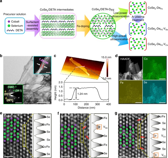
Figure 1a shows the synthetic strategies of different electrocatalysts. Firstly, CoSe2 nuclei were assembled with diethylenetriamine (DETA) under hydrothermal reaction, leading to the formation of CoSe2/DETA lamellar intermediates9. Fe ions were then incorporated into CoSe2/DETA via a wet-impregnation method involving chemical adsorption and cation exchange10. After that, atomically thin CoSe2–DFe nanobelts were obtained by liquid-phase exfoliation using low-power ultrasonication. Se vacancies were created on the surface of exfoliated CoSe2–DFe nanobelts through Ar plasma treatment11, and Co vacancies were extracted by DETA molecules during the exfoliation of CoSe2/DETA under high ultrasonic power12. Figure 1b shows the scanning transmission electron microscopy (STEM) image of the CoSe2–DFe–VCo nanobelts. The selected area electron diffraction (SAED) pattern (left inset) reveals the coexistence of cubic (yellow) and orthorhombic (green) phases of CoSe2 matrix, which is also identified by the X-ray diffraction (XRD) patterns (Supplementary Fig. 1)13. The cross-sectional view of the nanobelts (right inset) suggests an atomic thickness of ~1.20 nm, which is further confirmed by the atomic force microscopy analysis (Fig. 1c). Energy dispersive X-ray spectroscopy (EDS) mapping shows that Co, Se, and Fe dopants are homogeneous distributed throughout the nanobelts (Fig. 1d). The doping ratio of Fe to total cation content is about 18.3% according to the inductively coupled plasma atomic emission spectroscopy (ICP-AES) results (Supplementary Table 1). A certain amount of O was also detected by EDS analysis (Supplementary Fig. 2), attributing to the deviation of Se/Co ratio from the stoichiometry of CoSe2 that results from the low solubility of Na2SeO3 in H2O-DETA mixed solvent13,14. The presence of oxides was also evidenced by Raman spectroscopy (Supplementary Fig. 3), where the peak at 164 cm−1 corresponds to the stretching mode of Se–Se in CoSe212, and the peaks at 190, 515, 612, 472, and 676 cm−1 are assigned to the F2g(1), F2g(2), F2g(3), Eg, and A1g modes of CoOx, respectively15. The created Se and Co vacancies are readily visible via high-angle annular dark-field STEM (HAADF-STEM) imaging (Fig.1e–g). In addition, the intensity profiles along the selected rectangular regions also suggest the missed surface Se and Co atoms in CoSe2–DFe–VSe and CoSe2–DFe–VCo, respectively.
Fig. 1. Synthesis and structural characterization of CoSe2–DFe, CoSe2–DFe–VSe, and CoSe2–DFe–VCo.
a Schematic illustration of the synthetic methods for different catalysts. b Scanning transmission electron microscopy (STEM) image of CoSe2–DFe–VCo nanobelts with selected area electron diffraction pattern (left inset) indicating the mixed cubic and orthorhombic phases and cross-sectional view (right inset) showing the atomic thickness. Scale bar, 200 nm, 5 1/nm (left inset) and 2 nm (right inset). c Atomic force microscopy image of CoSe2–DFe–VCo and height profile along the white line in the image. d Elemental maps of Co, Fe, and Se in CoSe2–DFe–VCo. Scale bar, 300 nm. e–g High-resolution high-angle annular dark-field STEM (HAADF-STEM) images of different catalysts and the intensity profiles along the selected rectangular regions suggest the missed surface Se and Co atoms in CoSe2–DFe–VSe and CoSe2–DFe–VCo, respectively. Scale bar, 0.5 nm.
OER catalytic activity evaluation
To understand how Fe dopants and Co/Se vacancies affect the OER catalytic performance, the prepared catalysts were subjected to systematic electrochemical evaluation via the rotating disk electrode (RDE) method in purified 1 M NaOH (Fig. 2). Cyclic voltammetry (CV) was first conducted to activate the electrodes (Supplementary Fig. 4), and large current densities were recorded during the first few cycles, indicating the instability of CoSe2 in alkaline solution at anodic potentials14. EDS analysis shows that Se signal can hardly be detected after CV test, and SAED pattern indicates the formation of CoOOH phase, which serves as the real host under OER conditions (Supplementary Fig. 5). The thickness of the nanobelts increases from 1.24 to 1.35 nm after CV activation (Supplementary Fig. 6), which is resulted from the significantly increased surface roughness due to CV-induced structural reconstruction.
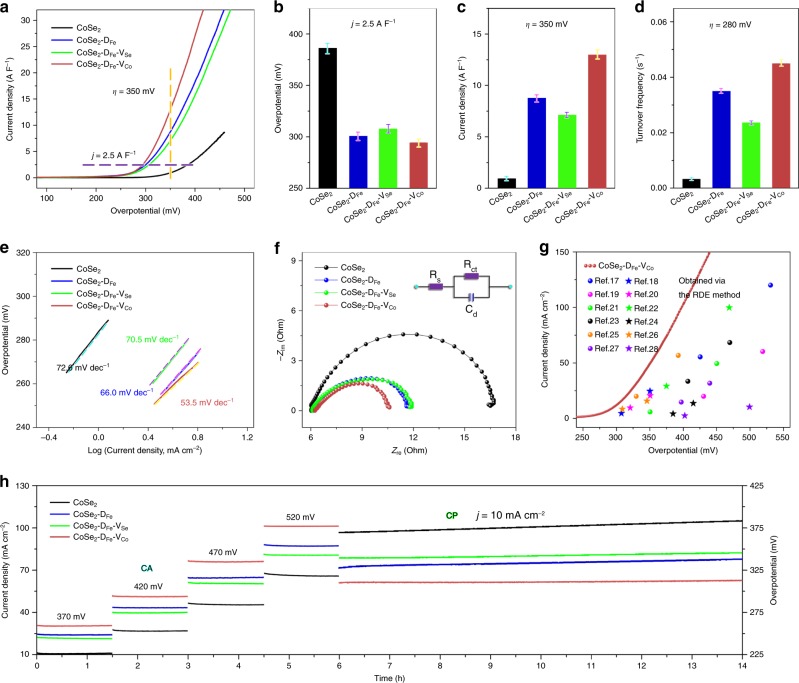
Fig. 2. Catalytic performance for the oxygen evolution reaction.
a Linear sweep voltammetry curves normalized by electrochemical double-layer capacitance. b Overpotentials (η) required to reach a current density (j) of 2.5 A F−1. c Current densities at η = 350 mV. d Turnover frequencies calculated at η = 280 mV. e Tafel plots derived from the polarization curves in low overpotential regions. f Nyquist plots at η = 370 mV with inset showing the equivalent circuit model. g Comparison of the catalytic performance between CoSe2–DFe–VCo and previously reported CoSe2 and FeCo compounds evaluated via the rotating disk electrode (RDE) method. h Durability evaluation via chronoamperometry test at stepwise η of 370, 420, 470, and 520 mV for 6 h, and chronopotentiometry test at j = 10 mA cm−2 for 8 h. The error bars in b–d denote standard deviation of five technical replicates.
The catalytic activities were then assessed by linear sweep voltammetry (LSV, Fig. 2a). The polarization curves were corrected with 95% iR-compensation and then normalized by the electrochemical double-layer capacitance. As shown in Fig. 2b, c, to reach a current density (j) of 2.5 A F−1, the required overpotentials (η) for CoSe2, CoSe2–DFe, CoSe2–DFe–VSe, and CoSe2–DFe–VCo are 385.7, 300.4, 307.3, and 294.2 mV, respectively, and at η = 350 mV, the recorded j are 0.92, 8.73, 7.06, and 12.95 A F−1, respectively. Therefore, Fe doping, in accordance with previous reports, enhances the OER catalytic performance of CoSe2, and further incorporation of Se and Co vacancies leads to slightly decreased and dramatically increased catalytic activity, respectively. It is important to note that the current density achieved by CoSe2–DFe–VCo at η = 350 mV is 30.2% higher than the sum of the current densities of CoSe2–DFe and CoSe2–VCo (Supplementary Fig. 7), indicating a synergistic effect between Fe dopants and Co vacancies. This synergistic effect was further confirmed by changing the sequence of Fe doping and Co vacancy. As shown in Supplementary Fig. 8, the CoSe2–VCo–DFe exhibits higher overpotential and lower current density than CoSe2–DFe–VCo, probably due to that the afterward doping fills the vacancy sites and weakens the role of Co vacancies16.
The high catalytic activity of CoSe2–DFe–VCo was further evidenced by analyzing the turnover frequency (TOF, assuming all cations to be catalytically active) and Tafel slope (derived from the polarization curve in low overpotential region). As shown in Fig. 2d, the apparent TOF of CoSe2–DFe–VCo at η = 280 mV reaches 0.045 s−1, exceeding 0.003, 0.035, and 0.024 s−1 for CoSe2, CoSe2–DFe, and CoSe2–DFe–VSe, respectively, indicative of the high intrinsic catalytic activity. In addition, CoSe2–DFe–VCo exhibits a small Tafel slope of 53.5 mV dec−1, much lower than 72.6, 66.0, and 70.5 mV dec−1 for CoSe2, CoSe2–DFe and CoSe2–DFe–VSe, respectively (Fig. 2e), suggesting the remarkably enhanced reaction kinetics6. The Nyquist plots at η = 370 mV display typical semicircles for different catalysts (Fig. 2f), corresponding to the charge transfer resistances (Rct). Using a relevant equivalent circuit (inset), the fitted results indicate the smallest Rct (4.2 Ω) of CoSe2–DFe–VCo compared with those of CoSe2 (10.5 Ω), CoSe2–DFe (5.3 Ω) and CoSe2–DFe–VSe (5.4 Ω), indicating the efficient electron transfer and the fast ion diffusion8. As a result, the incorporation of Fe dopants and Co vacancies creates highly active catalytic sites with excellent mass transport properties for OER catalysis.
It is also noteworthy that CoSe2–DFe–VCo outperforms commercial IrO2 and RuO2 electrodes (Supplementary Fig. 9), and previously reported CoSe2 and FeCo compounds evaluated via the RDE method (Fig. 2g)17–28. The outstanding performance is firstly attributable to the atomic thickness of the nanobelts, which provides abundant surface active sites for the catalytic reaction (Supplementary Fig. 10). In addition, the proper doping level of Fe optimizes the electrode composition and improves the catalytic performance (Supplementary Fig. 11). More importantly, Fe doping and Co vacancies work synergistically to create highly active centers and dramatically enhance the catalytic activity.
The catalytic stability was further assessed via chronoamperometry (CA) for 6 h and chronopotentiometry (CP) for 8 h. As shown in Fig. 2h, CA test shows that the current density increases with increasing overpotential. More specifically, CoSe2–DFe–VCo delivers average current densities of 30.5, 51.6, 76.3, 101.6 mA cm−2 at η = 370, 420, 470, 520 mV, respectively, exceeding those of CoSe2 (10.8, 26.9, 45.5, 66.3 mA cm−2), CoSe2–DFe (24.6, 43.5, 64.5, 87.0 mA cm−2), and CoSe2–DFe–VSe (21.6, 39.8, 60.5, 80.5 mA cm−2). CP test at j = 10 mA cm−2 shows that the overpotential of CoSe2 suffers an obvious increase of 13.6 mV, while those of CoSe2–DFe, CoSe2–DFe–VSe, and CoSe2–DFe–VCo see slight rises of 8.6, 7.0, and 3.7 mV, respectively, which should be attributed to the enhanced electrical conductivity caused by Fe doping29. Generally, the trend in the activity of different catalysts does not change after long-term durability test.
Understanding defect structures before and after catalysis
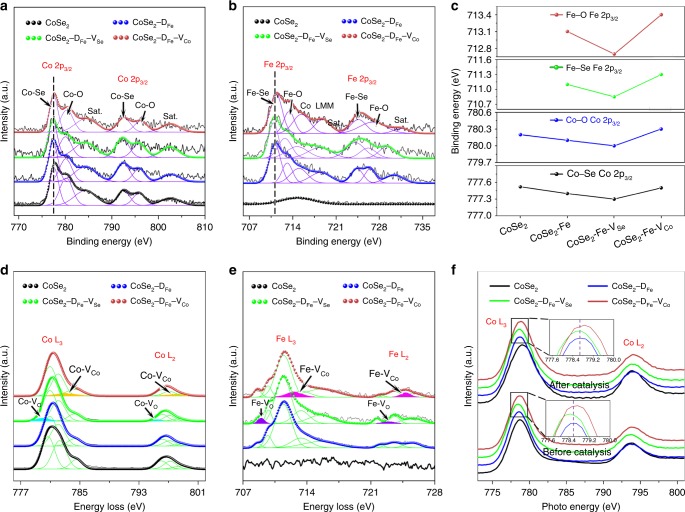
Prior to the mechanism investigation, it is essential to study the stability of dopants and vacancies and probe the evolution of electronic structures during catalysis as they are directly relevant to the catalytic activity30. X-ray photoelectron spectroscopy (XPS) was first performed to understand the primary defects before catalysis. As shown in Fig. 3a, b and Supplementary Fig. 12, Co, Fe, and Se signals are clearly detected, indicating the successful doping of Fe into CoSe2 matrix, and the Co/Fe 2p spectra show the existence of Co/Fe–O bonds due to oxide impurities. The Co/Fe 2p3/2 peaks of Co/Fe–Se and Co/Fe–O in CoSe2–DFe–VSe exhibit negative shifts to lower binding energies, and those in CoSe2–DFe–VCo show positive shifts to higher binding energies, indicating the reduction and oxidation states of metal sites caused by Se/O and Co vacancies, respectively (Fig. 3c)31. The defect structures after CA test at η = 370 mV for 1.5 h were then investigated via electron energy loss spectroscopy (EELS). As shown in Fig. 3d, e, both Co and Fe signals can be detected after catalysis, and no significant changes in their atom ratios were found due to fully purified electrolyte (Supplementary Table 1). For better analysis, the L2,3-edge spectra of Co and Fe were fitted by multiple Gaussian functions, which represent different oxidation states and coordination environments32. Compared with CoSe2−DFe, there are extra Gaussian peaks centered at 780.0 and 795.4 eV (cyan) in Co L2,3-edge spectra, and 709.0 and 723.0 eV (purple) in Fe L2,3-edge spectra of CoSe2–DFe–VSe, which most likely result from the Se-derived O vacancies. In comparison, extra peaks located at 783.0 and 797.9 eV (orange) in Co L2,3-edge spectra, and 712.6 and 724.8 eV (magenta) in Fe L2,3-edge spectra of CoSe2–DFe–VCo, which should be ascribed to the local chemical environment of Co vacancies. Therefore, the surface reconstruction induced by OER catalysis has little impact on the defect structures. To provide further evidence, X-ray absorption near-edge structure (XANES) spectra at the L-edges of Co and Fe before and after catalysis were conducted (Fig. 3f and Supplementary Fig. 13). Obviously, the L3-edge centroids of Co before catalysis shift by 0.10 and 0.12 eV to lower and higher energy positions after the incorporation of Se/O and Co vacancies, respectively33,34. After catalysis, slight broadenings of the white line peaks were observed due to OER-induced complex coordination environment35. Despite the electronic-structure changes, the Co L3-edge centroids of CoSe2–DFe–VSe and CoSe2–DFe–VCo still exhibit 0.06 and 0.11 eV shifts to lower and higher energy positions, respectively. The same trends of energy shift could be obtained from the L2,3-edge XANES spectra of Fe (Supplementary Fig. 13). As a result, both dopants and vacancies are well preserved during OER catalysis, and they consequently determine the catalytic performance of different catalysts.
Fig. 3. Defect structure identification before and after catalysis.
a, b Co 2p and Fe 2p X-ray photoelectron spectroscopy spectra of primary catalysts. c Binding energy shifts of Co–Se, Co–O, Fe–Se, and Fe–O peaks due to doping and vacancies. d, e Co and Fe L-edge electron energy loss spectra after chronoamperometry test at 370 mV for 1.5 h showing the existence of extra peaks in CoSe2–DFe–VSe and CoSe2–DFe–VCo. f Co L-edge X-ray absorption near-edge structure spectra before and after catalysis showing the energy shifts caused by doping and vacancies.
Discussion
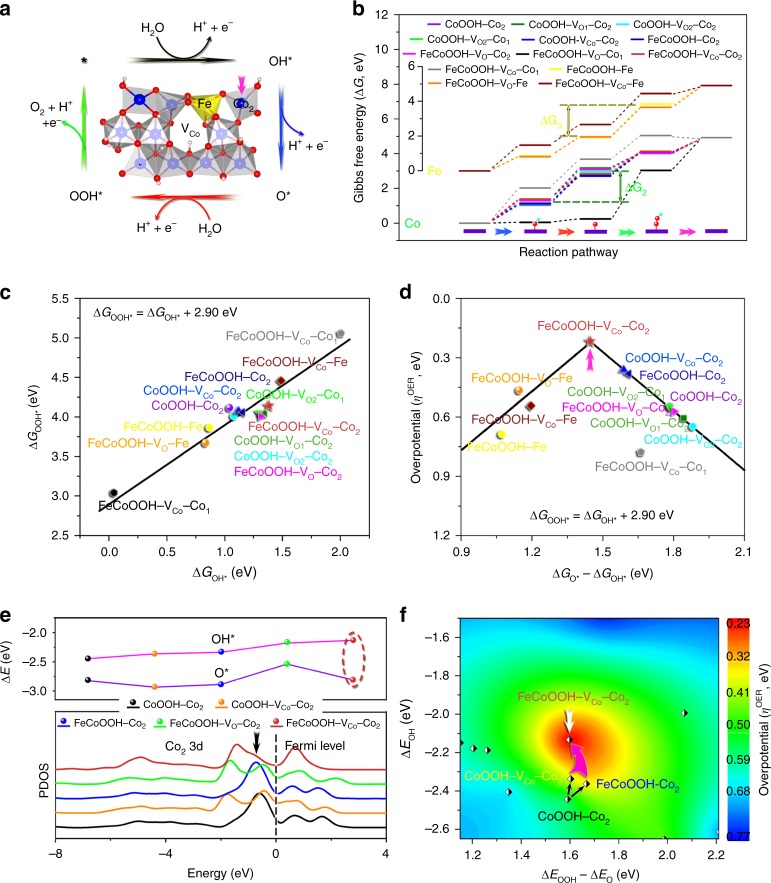
To provide clear insight into the effects of dopants and vacancies on the catalytic activities, we performed Hubbard-corrected density functional theory (DFT + U) calculations. The crystal structure of CoOOH was utilized to build up the periodical surface models as it serves as the real host under OER conditions (Supplementary Figs. 4 and 5). The high-index (01-12) facet was selected as the surface termination due to the proved good coincidence between theoretical and experimental results36, and all potential active sites on (01-12) facets of models with different atomistic arrangements of dopants and vacancies were considered to gain insight into the catalytic mechanism (Supplementary Fig. 14). Figure 4a shows the FeCoOOH–VCo structure and the OER pathway, which involves four proton coupled electron transfer steps and three intermediates of OH*, O*, and OOH*37. The corresponding Gibbs free energies (ΔGi, i = OH*, O*, and OOH*) were calculated and provided in Fig. 4b. As can be seen, the catalytic activities of Co sites are restricted by the transformation of OH* to O*, due to the relatively large difference between ΔGO* and ΔGOH* (namely ΔG2). In comparison, the potential-limiting step of Fe sites is assigned to the transformation of O* to OOH*, owing to the relatively large difference between ΔGOOH* and ΔGO* (i.e., ΔG3). Therefore, Co and Fe sites in CoOOH matrix exhibit different catalytic behaviors for OER. The ΔGi of FeCoOOH–VO–Co1 and FeCoOOH–VCo–Co1 deviate from the general principle probably due to that they are adjacent to the vacancies and their electronic states are greatly altered.
Fig. 4. Hubbard-corrected density functional theory calculations of catalytic activities of different surface metal sites.
a Crystal structure of FeCoOOH–VCo and oxygen evolution reaction reaction pathway on (01-12) facet. b Gibbs free energy profiles along the reaction pathway. c Scaling relation between ΔGOOH* and ΔGOH*. d Volcano plot of overpotential as a function of ΔGO* − ΔGOH* based on the scaling relation. e Calculated partial density of states (PDOS) of Co2 sites and binding energies of OH* and O*. f Contour plot of theoretical overpotential as a function of ΔEOOH − ΔEO and ΔEOH, indicating the near-optimal intermediate binding energies achieved by Fe doping and Co vacancy.
Scaling relations exist between energetics of OH* and OOH* over the above considered metal sites36–38, which are expressed as ΔGOOH* = ΔGOH* + 2.90 eV (Gibbs free energies, Fig. 4c), ΔEOOH* = ΔEOH* + 2.87 eV (adsorption energies, Supplementary Fig. 15a), and ΔEOOH = ΔEOH + 1.03 eV (binding energies, Supplementary Fig. 15b) (Supplementary Table 2). Based on the scaling relation, a universal volcano relationship can be constructed by plotting ηOER as a function of ΔGO* − ΔGOH*38. As shown in Fig. 4d, Fe sites are distributed around the left leg of the volcano which is restricted by the transformation of O* to OOH*, and Co sites are located on the right leg that is determined by the transformation of OH* to O*. Both Fe and Co could serve as OER active centers by analyzing their ηOER, which have been confirmed by previous studies26,39–41. Among all considered metal sites in different models, however, the most active center is the Co2 site that is adjacent to the vacancy-nearest surface Fe site in FeCoOOH–VCo. By comparing the Co2 of CoOOH, FeCoOOH, CoOOH–VCo and FeCoOOH–VCo, we can see that Fe doping and Co vacancy individually decrease the overpotential from 549 to 380 and 360 mV, respectively, while they jointly push Co2 to the top of the volcano, leading to the lowest overpotential of 218 mV among all investigated metal sites. Therefore, the theoretical simulations successfully identify the most active catalytic sites and verify the synergistic effect between Fe dopants and Co vacancies.
To deeply understand how Fe doping and Co vacancy modulate the electronic states of Co2 and consequently tune the intermediate binding energy, the partial density of states of Co2 3d in CoOOH, CoOOH–VCo, FeCoOOH, FeCoOOH–VO, and FeCoOOH–VCo were calculated. As shown in Fig. 4e, an intense negative peak near the Fermi level was observed in CoOOH. Fe doping causes a small shift of the peak away from the Fermi level and Co vacancy induces the splitting of the peak, both of which lead to slightly decreased binding energy of OH*42. Remarkably, the combination of Fe doping and Co vacancy greatly decreases the peak intensity near the Fermi level, and therefore results in dramatically weakened binding with OH*. It is important to note that, the low overpotential is achieved due to that Fe doping and Co vacancy jointly have little impact on the binding energy of O* (dashed ellipse in Fig. 4e). In comparison, even though the combination of Fe doping and O vacancy decreases the binding energy of OH* as well, they have a more obvious influence on the binding energy of O*, which leads to increased overpotential and declined catalytic activity.
A contour plot of 3D overpotential surface as a function of ΔEOOH − ΔEO and ΔEOH can be constructed based on the calculated activities of different metal sites. As shown in Fig. 4f, the overpotential decreases along the direction: blue→cyan→green→yellow→red. CoOOH–Co2 with a moderate overpotential locates in the green region, and the incorporation of Fe doping and Co vacancy individually moves the point to the green–yellow boundary. FeCoOOH–VCo–Co2, sitting close to the center of the red region, possesses near-optimal intermediate binding energies, and near-minimum catalytic overpotential that different systems can research in this study. Therefore, Fe doping and Co vacancy work synergistically to approach the activity limit of Co-based catalysts. This contour map provides valuable guidance for the design of efficient OER catalysts via modulating the electronic structures and intermediate binding energies.
In conclusion, different defect structures were incorporated into atomically thin CoSe2 nanobelts for OER catalysis. We found that the combination of Fe doping and Co vacancy synergistically optimize the electronic states and intermediate binding energies at Co2 site, which is responsible for the dramatically enhanced catalytic activity. This electronic-structure modulation strategy with proper combination of two or more defect structures could efficiently unlock the catalytic power, showing great promise for the rational design of advanced catalysts for various electrochemical reactions.
Methods
Material synthesis
Atomically thin CoSe2 nanobelts were synthesized via a lamellar intermediate-assisted exfoliation approach. In detail, 14.0 ml DETA was slowly poured into 7.0 ml H2O under stirring as severe exothermic reaction occurs. Then, 0.125 g Co(Ac)2·4H2O and 0.130 g Na2SeO3 were added and stirred until completely dissolved. After that, hydrothermal reaction was performed at 160 °C for 10 h, and CoSe2/DETA lamellar intermediates were finally obtained after reaction. Fe doping was performed by adding 1.0 ml of 0.025 mol L−1 Fe(NO3)3·9H2O aqueous solution into the CoSe2/DETA intermediates solution, and atomically thin nanobelts were exfoliated from the intermediates in ethanol by using an ultrasonic homogenizer (on time: 2 s, off time: 1 s, output power: 12%, process time: 5 h). Se vacancies were created on the exfoliated Fe-doped CoSe2 nanobelts via Ar plasma engraving with a irradiation time of 5 min, a power of 100 W, and a gas flow rate of 50 sccm. Co vacancies were incorporated during the exfoliation of Fe-doped CoSe2/DETA intermediates under high-power ultrasonication (on time: 2 s, off time: 1 s, output power: 30%, process time: 2 h). The resulted dispersions were centrifuged at 1000 rmp for 10 min to remove the unexfoliated powders.
Characterizations
The morphology of nanobelts was observed via STEM (JEOL JEM-ARM200F), and the composition elements were identified by atomic-resolution elemental maps via EDS (Oxford XMax100TLE EDS spectrometer). The element contents before catalysis and after CA test at η = 370 mV for 1.5 h were determined by an Optima 2000 DV ICP-emission spectrometer. SAED, XRD (MMA, GBC Scientific Equipment LLC, Hampshire, IL, USA) and Raman scattering (Renishaw 100, 632.8 nm He–Ne laser) were employed to determine the phase structure. The missing lattice atoms were observed by high-resolution HAADF-STEM and analyzed via intensity profile. The electronic states of Co and Fe before catalysis were studied by XPS (PHOIBOS 100 Analyzer from SPECS, Berlin, Germany; Al Kα X-rays), and those after CA test at η = 370 mV for 1.5 h were investigated by EELS (Gatan GIF Quantum ER EELS spectrometer). The EELS characterization was performed at liquid nitrogen temperature to avoid sample damaging and electronic-state changes, and the spectrometer was calibrated against the zero-loss peak at the start of each session. The deconvolution and curve-fitting of the spectra were performed by employing nonlinear least squares fitting tools written in DigitalMicrograph software. The coordination environments of Co and Fe before and after catalysis (CA test at η = 320 mV for 0.5 h) were also studied by XANES (National Synchrotron Radiation Laboratory (NSRL), Hefei, China). The Co and Fe L-edges XANES spectra of catalysts were measured at the photoemission end-station at beamline BL10B in the NSRL, Hefei, China. A bending magnet is connected to the beamline, which is equipped with three gratings covering photon energies from 100 to 1000 eV. The samples were kept in the total-electron-yield mode under an ultrahigh vacuum at 5 × 10⁻¹⁰ mbar. The resolving power of the grating was typically E/ΔE = 1000, and the photon flux was 1 × 10¹⁰ photons per s. Spectra were collected at energies from 770 to 814 eV for Co and 695 to 740 eV for Fe in 0.2 eV energy steps. The XANES raw data were normalized by a procedure consisting of several steps.
Electrochemical measurements
The catalytic performances were evaluated via the RDE method with continuous rotation at 1200 rpm (CHI 760, Shanghai Chenhua Instruments Co. Ltd.), where O2-saturated 1 M NaOH, graphite rod and Hg/HgO electrode were utilized as the electrolyte, counter electrode and reference electrode, respectively. The working electrode was prepared by placing 12 μl of catalyst suspension onto glassy carbon electrode which was dried in a fume cupboard. The catalyst ink was prepared by suspending 2 mg catalyst in 1 ml water/isopropanol/Nafion® mixed solution (3:1:0.2, v/v) under ultrasonication. Before catalytic evaluation, the electrolyte was fully purified with the working electrode via CV cycles between 0.30 and 0.75 V vs. Hg/HgO at 10 mV s−1 until a stable response was achieved. New working electrodes were then activated by six CV cycles, during which CoSe2 was fully converted into CoOOH. LSV was then recorded within the voltage range of 0.30–1.00 V vs. Hg/HgO at 10 mV s−1, and the polarization curves were corrected with 95% iR-compensation and then normalized by electrochemical double-layer capacitance that was estimated from the slope of the difference between anodic and cathodic current densities at 0.35 V vs. Hg/HgO against the scan rate (10, 20, 30, 40, 50, and 60 mV s−1). After that, the electrodes were stabilized at 0.70 V vs. Hg/HgO for 3 min, and then electrochemical impedance spectroscopy was performed at the same potential with an amplitude of 10 mV and a frequency range of 200 kHz–100 mHz. The catalytic durability was finally assessed by CA test at stepwise potentials of 0.70, 0.75, 0.80, and 0.85 V vs. Hg/HgO for 6 h, and CP test at a constant current density of 10 mA cm−2 for 8 h.
Overpotentials were determined by ηOER = EHg/HgO + (0.098 + 0.059 pH − 1.23) V, where EHg/HgO is the recorded potential vs. Hg/HgO. TOF values were calculated based on TOF = jS/4Fn, where j (A cm−2) is the current density at η = 280 mV, S (cm2) is the surface area of glassy carbon electrode, F is the Faraday constant (96,485 C mol−1), and n is the number of moles of the cations assuming all of them are catalytically active. Tafel plots were derived from the polarization curves in low overpotential regions by plotting overpotential against log(current density). The error bars of reported overpotentials, current densities and TOFs were obtained from five replicates after discarding the maximum and minimum values.
Computational methods
In this work, the crystal structure of CoOOH was chosen to build up the periodical surfaces including Fe doping and Co/O vacancy models. The high-index (01-12) facet was adopted as the surface termination36,37, as it exhibits theoretical activities in good agreement with the experimental results. The periodic surface was 14.79 Ǻ × 15.22 Ǻ with a vacuum slab of 18 Å in thickness to separate the layer from its periodic images. All the calculations were performed by spin-polarized plane-wave DFT, as implemented in the CASTEP program43. The geometry optimizations were performed using the BFGS algorithm. The Perdew–Burke–Ernzerhof exchange-correlation functional within the generalized gradient approximation as well as ultrasoft pseudopotentials was selected44,45. The cut-off energy for the plane-wave basis and the Brilliouin zone k-points were chosen as 340.0 eV and 0.04 Ǻ−1 spacing in the Monkhorst–Pack scheme46, respectively, which causes little difference in the calculation results when further increase. To sufficiently consider the on-site Columbic repulsion between the d electrons, the Hubbard U corrections were applied to transition metal d-electrons and the values of U–J parameters for Co (3.42) and Fe (3.29) atoms were taken from the reference47. The convergence criteria for the total energy, forces, stress, atomic displacement, and self-consistent field iterations were set to 1 × 10⁻⁵ eV atom−1, 3 × 10⁻² eV Å−1, 5 × 10⁻² GPa, 1 × 10⁻³ Å, and 1 × 10⁻⁶ eV atom−1, respectively.
The OER reaction steps and Gibbs free energy changes can be expressed by37
| 1 |
| 2 |
| 3 |
| 4 |
where * represents an active site on (01-12) facet, and ΔGi (i = OH*, O*, and OOH*) are Gibbs free energies of OER intermediates. The theoretical overpotential under standard conditions (T = 298.15 K, p = 1 bar, pH = 0) is then given by37
| 5 |
The Gibbs free energies, ΔGi, were determined by the adsorption energies combined with corrections for zero-point energy and entropy, according to ΔGi = ΔEi + ΔZPEi − TΔSi. The ZPE and TS were calculated using DFT calculations of vibrational frequencies and using standard tables for gas-phase molecules48. The ZPEs for H2, H2O, *OH, *O, and *OOH are 0.27, 0.56, 0.35, 0.05, and 0.41 eV, respectively, and the TS corrections for H2 and H2O are 0.41 and 0.67 eV, respectively (we assume S = 0 for the adsorbates on coordinatively unsaturated sites). The adsorption energies of OER intermediates, ΔEi (i = OH*, O*, and OOH*), were calculated relative to H2O and H2 (at U = 0 and pH = 0)
| 6 |
| 7 |
| 8 |
The binding energies, ΔEj (j = OH, O, and OOH), were calculated directly by ΔEj = Ej* − Ej − E*,
where * represents an active site on (01-12) facet, and E is the total energy calculated by using the spin polarization DFT method.
Supplementary information
Acknowledgements
This work is financially supported by an Australian Research Council (ARC) Discovery Projects (DP200100965) and a Griffith University Postdoctoral Fellowship (YUDOU 036 Research Internal). The authors are grateful to Dr David Mitchell and Dr Ningyan Cheng from UOW Electron Microscopy Centre for their assistance in STEM work.
Author contributions
Y.D. conceived the idea, carried out the experiment, and wrote the paper. H.Z. provided research facilities, financial support, and valuable suggestions for the experiment and paper writing. C.T.H. performed the DFT calculations. L.Z. and H.Y. participated in the data analysis and discussion. M.A. and J.M. provided assistance in XRD, XPS, and STEM characterizations.
Data availability
The data that support the findings of this study are available from the corresponding author upon reasonable request.
Competing interests
The authors declare no competing interests.
Footnotes
Peer review information Nature Communications thanks the anonymous reviewers for their contributions to the peer review of this work.
Publisher’s note Springer Nature remains neutral with regard to jurisdictional claims in published maps and institutional affiliations.
Supplementary information
Supplementary information is available for this paper at 10.1038/s41467-020-15498-0.
References
- 1.Suen NT, et al. Electrocatalysis for the oxygen evolution reaction: recent development and future perspectives. Chem. Soc. Rev. 2017;46:337–365. doi: 10.1039/C6CS00328A. [DOI] [PubMed] [Google Scholar]
- 2.Wu T, et al. Iron-facilitated dynamic active-site generation on spinel CoAl2O4 with self-termination of surface reconstruction for water oxidation. Nat. Catal. 2019;2:763–772. doi: 10.1038/s41929-019-0325-4. [DOI] [Google Scholar]
- 3.Jin H, et al. Emerging two-dimensional nanomaterials for electrocatalysis. Chem. Rev. 2018;118:6337–6408. doi: 10.1021/acs.chemrev.7b00689. [DOI] [PubMed] [Google Scholar]
- 4.Pan X, Yang MQ, Fu X, Zhang N, Xu YJ. Defective TiO2 with oxygen vacancies: synthesis, properties and photocatalytic applications. Nanoscale. 2013;5:3601–3614. doi: 10.1039/c3nr00476g. [DOI] [PubMed] [Google Scholar]
- 5.Yan J, et al. Single atom tungsten doped ultrathin α-Ni(OH)2 for enhanced electrocatalytic water oxidation. Nat. Commun. 2019;10:2149. doi: 10.1038/s41467-019-09845-z. [DOI] [PMC free article] [PubMed] [Google Scholar]
- 6.Peng S, et al. Necklace-like multishelled hollow spinel oxides with oxygen vacancies for efficient water electrolysis. J. Am. Chem. Soc. 2018;140:13644–13653. doi: 10.1021/jacs.8b05134. [DOI] [PubMed] [Google Scholar]
- 7.Li H, et al. Fe-doped CoSe2 nanoparticles encapsulated in N-doped bamboo-like carbon nanotubes as an efficient electrocatalyst for oxygen evolution reaction. Electrochim. Acta. 2015;174:297–301. doi: 10.1016/j.electacta.2015.05.186. [DOI] [Google Scholar]
- 8.Zhang J-Y, et al. Rational design of cobalt-iron selenides for highly efficient electrochemical water oxidation. ACS Appl. Mater. Interfaces. 2017;9:33833–33840. doi: 10.1021/acsami.7b08917. [DOI] [PubMed] [Google Scholar]
- 9.Gao MR, Yao WT, Yao HB, Yu SH. Synthesis of unique ultrathin lamellar mesostructured CoSe2-amine (protonated) nanobelts in a binary solution. J. Am. Chem. Soc. 2009;131:7486–7487. doi: 10.1021/ja900506x. [DOI] [PubMed] [Google Scholar]
- 10.Stevens MB, et al. Reactive Fe-sites in Ni/Fe (oxy)hydroxide are responsible for exceptional oxygen electrocatalysis activity. J. Am. Chem. Soc. 2017;139:11361–11364. doi: 10.1021/jacs.7b07117. [DOI] [PubMed] [Google Scholar]
- 11.Chang A, Zhang C, Yu Y, Yu Y, Zhang B. Plasma-assisted synthesis of NiSe2 ultrathin porous nanosheets with selenium vacancies for supercapacitor. ACS Appl. Mater. Interfaces. 2018;10:41861–41865. doi: 10.1021/acsami.8b16072. [DOI] [PubMed] [Google Scholar]
- 12.Liu Y, et al. Low overpotential in vacancy-rich ultrathin CoSe2 nanosheets for water oxidation. J. Am. Chem. Soc. 2014;136:15670–15675. doi: 10.1021/ja5085157. [DOI] [PubMed] [Google Scholar]
- 13.Zheng YR, et al. Doping-induced structural phase transition in cobalt diselenide enables enhanced hydrogen evolution catalysis. Nat. Commun. 2018;9:2533. doi: 10.1038/s41467-018-04954-7. [DOI] [PMC free article] [PubMed] [Google Scholar]
- 14.Gui Y, et al. Manipulating the assembled structure of atomically thin CoSe2 nanomaterials for enhanced water oxidation catalysis. Nano Energy. 2019;57:371–378. doi: 10.1016/j.nanoen.2018.12.063. [DOI] [Google Scholar]
- 15.Dou Y, et al. Atomic layer-by-layer Co3O4/graphene composite for high performance lithium-ion batteries. Adv. Energy Mater. 2016;6:1501835. doi: 10.1002/aenm.201501835. [DOI] [Google Scholar]
- 16.Lesnyak V, Brescia R, Messina GC, Manna L. Cu vacancies boost cation exchange reactions in copper selenide nanocrystals. J. Am. Chem. Soc. 2015;137:9315–9323. doi: 10.1021/jacs.5b03868. [DOI] [PMC free article] [PubMed] [Google Scholar]
- 17.Zhou Q, et al. Active-site-enriched iron-doped nickel/cobalt hydroxide nanosheets for enhanced oxygen evolution reaction. ACS Catal. 2018;8:5382–5390. doi: 10.1021/acscatal.8b01332. [DOI] [Google Scholar]
- 18.Zheng YR, et al. An efficient CeO2/CoSe2 nanobelt composite for electrochemical water oxidation. Small. 2015;11:182–188. doi: 10.1002/smll.201401423. [DOI] [PubMed] [Google Scholar]
- 19.Zhou Y, et al. Co3O4@(Fe-doped)Co(OH)2 microfibers: facile synthesis, oriented-assembly, formation mechanism, and high electrocatalytic activity. ACS Appl. Mater. Interfaces. 2017;9:30880–30890. doi: 10.1021/acsami.7b10402. [DOI] [PubMed] [Google Scholar]
- 20.Zhao X, et al. Engineering the electrical conductivity of lamellar silver-doped cobalt(II) selenide nanobelts for enhanced oxygen evolution. Angew. Chem. Int. Ed. 2017;56:328–332. doi: 10.1002/anie.201609080. [DOI] [PubMed] [Google Scholar]
- 21.Jin H, et al. Fe incorporated α-Co(OH)2 nanosheets with remarkably improved activity towards the oxygen evolution reaction. J. Mater. Chem. A. 2017;5:1078–1084. doi: 10.1039/C6TA09959A. [DOI] [Google Scholar]
- 22.Li J, et al. Fe-doped CoSe2 nanoparticles encapsulated in N-doped bamboo-like carbon nanotubes as an efficient electrocatalyst for oxygen evolution reaction. Electrochim. Acta. 2018;265:577–585. doi: 10.1016/j.electacta.2018.01.211. [DOI] [Google Scholar]
- 23.Liang L, et al. Metallic single-unit-cell orthorhombic cobalt diselenide atomic layers: robust water-electrolysis catalysts. Angew. Chem. Int. Ed. 2015;54:12004–12008. doi: 10.1002/anie.201505245. [DOI] [PubMed] [Google Scholar]
- 24.Gao MR, et al. CoSe2 nanobelts composite catalyst for efficient water oxidation. ACS Nano. 2014;8:3970–3978. doi: 10.1021/nn500880v. [DOI] [PubMed] [Google Scholar]
- 25.Xu H, et al. Phosphorus-doped cobalt-iron oxyhydroxide with untrafine nanosheet structure enable efficient oxygen evolution electrocatalysis. J. Colloid Interface Sci. 2018;530:58–66. doi: 10.1016/j.jcis.2018.06.061. [DOI] [PubMed] [Google Scholar]
- 26.Zhang L, et al. Ultrathin iron-cobalt oxide nanosheets with abundant oxygen vacancies for the oxygen evolution reaction. Adv. Mater. 2017;29:1606793. doi: 10.1002/adma.201606793. [DOI] [PubMed] [Google Scholar]
- 27.Xiao C, Lu X, Zhao C. Unusual synergistic effects upon incorporation of Fe and/or Ni into mesoporous Co3O4 for enhanced oxygen evolution. Chem. Commun. 2014;50:10122–10125. doi: 10.1039/C4CC04922E. [DOI] [PubMed] [Google Scholar]
- 28.Gao MR, Xu YF, Jiang J, Zheng YR, Yu SH. Water oxidation electrocatalyzed by an efficient Mn3O4/CoSe2 nanocomposite. J. Am. Chem. Soc. 2012;134:2930–2933. doi: 10.1021/ja211526y. [DOI] [PubMed] [Google Scholar]
- 29.Li N, et al. Influence of iron doping on tetravalent nickel content in catalytic oxygen evolving films. Proc. Natl Acad. Sci. USA. 2017;114:1486–1491. doi: 10.1073/pnas.1620787114. [DOI] [PMC free article] [PubMed] [Google Scholar]
- 30.Jiang H, He Q, Zhang Y, Song L. Structural self-reconstruction of catalysts in electrocatalysis. Acc. Chem. Res. 2018;51:2968–2977. doi: 10.1021/acs.accounts.8b00449. [DOI] [PubMed] [Google Scholar]
- 31.Guo C, et al. Engineering high-energy interfacial structures for high-performance oxygen-involving electrocatalysis. Angew. Chem. Int. Ed. 2017;56:8539–8543. doi: 10.1002/anie.201701531. [DOI] [PubMed] [Google Scholar]
- 32.Zhang S, et al. Determination of manganese valence states in (Mn3+, Mn4+) minerals by electron energy-loss spectroscopy. Am. Mineral. 2010;95:1741–1746. doi: 10.2138/am.2010.3468. [DOI] [Google Scholar]
- 33.Ling T, et al. Activating cobalt(II) oxide nanorods for efficient electrocatalysis by strain engineering. Nat. Commun. 2017;8:1509. doi: 10.1038/s41467-017-01872-y. [DOI] [PMC free article] [PubMed] [Google Scholar]
- 34.Cheng J, et al. Enhanced insulating behavior in the Ir-vacant Sr2Ir1-xO4 system dominated by the local structure distortion. J. Synchrotron Rad. 2018;25:1123. doi: 10.1107/S1600577518005799. [DOI] [PubMed] [Google Scholar]
- 35.Cao L, et al. Identification of single-atom active sites in carbon-based cobalt catalysts during electrocatalytic hydrogen evolution. Nat. Catal. 2019;2:134. doi: 10.1038/s41929-018-0203-5. [DOI] [Google Scholar]
- 36.Bajdich M, García-Mota M, Vojvodic A, Nørskov JK, Bell AT. Theoretical investigation of the activity of cobalt oxides for the electrochemical oxidation of water. J. Am. Chem. Soc. 2013;135:13521–13530. doi: 10.1021/ja405997s. [DOI] [PubMed] [Google Scholar]
- 37.Friebel D, et al. Identification of highly active Fe sites in (Ni,Fe)OOH for electrocatalytic water splitting. J. Am. Chem. Soc. 2015;137:1305–1313. doi: 10.1021/ja511559d. [DOI] [PubMed] [Google Scholar]
- 38.Man IC, et al. Universality in oxygen evolution electrocatalysis on oxide surfaces. ChemCatChem. 2011;3:1159–1165. doi: 10.1002/cctc.201000397. [DOI] [Google Scholar]
- 39.Burke MS, et al. Cobalt-iron (oxy)hydroxide oxygen evolution electrocatalysts: the role of structure and composition on activity, stability, and mechanism. J. Am. Chem. Soc. 2015;137:3638–3648. doi: 10.1021/jacs.5b00281. [DOI] [PubMed] [Google Scholar]
- 40.Smith RDL, et al. Spectroscopic identification of active sites for the oxygen evolution reaction on iron-cobalt oxides. Nat. Commun. 2017;8:2022. doi: 10.1038/s41467-017-01949-8. [DOI] [PMC free article] [PubMed] [Google Scholar]
- 41.Zhang B, et al. Homogeneously dispersed multimetal oxygen-evolving catalysts. Science. 2016;352:333–337. doi: 10.1126/science.aaf1525. [DOI] [PubMed] [Google Scholar]
- 42.Qiu B, et al. Fabrication of nickel-cobalt bimetal phosphide nanocages for enhanced oxygen evolution catalysis. Adv. Funct. Mater. 2018;28:1706008. doi: 10.1002/adfm.201706008. [DOI] [Google Scholar]
- 43.Clark SJ, et al. First principles methods using CASTEP. Z. Kristallogr. 2005;220:567–570. [Google Scholar]
- 44.Vanderbilt D, et al. Soft self-consistent pseudopotentials in a generalized eigenvalue formalism. Phys. Rev. B. 1990;41:7892–7895. doi: 10.1103/PhysRevB.41.7892. [DOI] [PubMed] [Google Scholar]
- 45.Perdew JP, Burke K, Ernzerhof M. Generalized gradient approximation made simple. Phys. Rev. Lett. 1996;77:3865–3868. doi: 10.1103/PhysRevLett.77.3865. [DOI] [PubMed] [Google Scholar]
- 46.Monkhorst HJ, Park JD. Special points for Brillouin-zone intergrations. Phys. Rev. B. 1976;13:5188–5192. doi: 10.1103/PhysRevB.13.5188. [DOI] [Google Scholar]
- 47.Xu H, Cheng D, Cao D, Zeng XC. A universal principle for a rational design of single-atom electrocatalysts. Nat. Catal. 2018;1:339–348. doi: 10.1038/s41929-018-0063-z. [DOI] [Google Scholar]
- 48.Valdés Á, Qu ZW, Kroes GJ, Rossmeisl J, Nørskov JK. Oxidation and photo-oxidation of water on TiO2 surface. J. Phys. Chem. C. 2008;112:9872–9879. doi: 10.1021/jp711929d. [DOI] [Google Scholar]
Associated Data
This section collects any data citations, data availability statements, or supplementary materials included in this article.
Supplementary Materials
Data Availability Statement
The data that support the findings of this study are available from the corresponding author upon reasonable request.


