

Highlights
-
•
Organoids are excellent platforms to discover measures against emerging pathogens.
-
•
Organoid models of ZIKV, C. difficle, and H. pylori infection have been established.
-
•
Microinjection, co-cultures and monolayers are used for host–microbe studies.
-
•
Triple co-cultures of immune cells, microbes and organoids are futuristic next steps.
-
•
Organoids grown in synthetic matrices have more reproducibility and applicability.
Abstract
Recent advances in host–microbe interaction studies in organoid cultures have shown great promise and have laid the foundation for much more refined future studies using these systems. Modeling of Zika virus (ZIKV) infection in cerebral organoids have helped us understand its association with microcephaly. Similarly, the pathogenesis of bacterial (Helicobacter pylori, Clostridium difficile) and viral (Norovirus, Rotaviruses) infections have been precisely dissected in organoid cultures. Additionally, direct associations between microbial colonization of tissues and diseases like cancer have also been deciphered. Here we discuss the most recent and striking studies on host–microbe interactions in organoid cultures, highlighting various methods which can be used for developing microbe-organoid co-culture systems.
Current Opinion in Immunology 2017, 48:15–22
This review comes from a themed issue on Host pathogens
Edited by Marc Pellegrini and Liz Hartland
For a complete overview see the Issue and the Editorial
Available online 9th August 2017
http://dx.doi.org/10.1016/j.coi.2017.07.012
0952-7915/© 2017 Elsevier Ltd. All rights reserved.
Introduction
The “germ theory of diseases”, which hypothesizes that diseases are caused due to the action of microorganisms, was the crowning achievement of a French scientist Louis Pasteur, who in 1860s refuted the theory of spontaneous generation [1]. Ever since, various hypothesis on the microbial pathogenicity have been proposed and established [2, 3]. Initially believed to primarily be assailants leading to disease states i.e. pathogenic (pathos is the Greek word for disease and genes means “born of”), scientists now recognize that host–microbe crosstalk is not always detrimental but also beneficial as in the case of gut symbionts (derived from symbiōsis, meaning “state of living together” in Greek) [4]. More so, disease states are a result of a two-way interaction that occurs between the host cells or tissues and the microorganisms. As per the chain of infection model, host–pathogen interactions can lead to either host immunity or an aggravated immune response due to infection, depending on six factors including the susceptibility of the host, route of entry and colonization potential of the microbe (Figure 1 ) [5, 6]. Recent years have seen a surge in interest in understanding this complex interplay between the microbes and the host organism. According to the World Health Organization (WHO), at least 12% of all human pathogens are considered as Emerging Infectious Diseases (EID) including malaria, Severe acute respiratory syndrome (SARS), Zika virus disease, HIV/AIDS etc., thus making it indispensable for us to understand the mechanism of action of molecular components involved in host–pathogen interaction during infection with EIDs [7].
Figure 1.

Illustration of the “chain of infection” model. Infection results from the interaction between the host, microbe and the environment. Six elements together constitute the chain of infection starting from pathogen, reservoir, portal of exit, means of transmission, portal of entry and ends with the infection of a new host. Organoids can be used to study the various links of the “chain of infection” model and help in the prevention and treatment of infectious diseases.
Model organisms and animal models like fruit fly Drosophila melanogaster, zebrafish Danio rerio and mice have been instrumental in providing valuable insights into host–microbe interactions; however, their limited translation potential to humans due to uncontrollable microbial diversity and significant inter-specie variances proves to be a major disadvantage [8, 9, 10, 11]. Recently developed humanized mice models are more relevant to human diseases, allowing better understanding of microbe interactions, but are expensive and difficult to maintain [12, 13]. Ex vivo two-dimensional (2D) cell cultures of immortalized cell lines grown as monolayers and are functionally closer to the ‘real situation’ in humans, but lack the three-dimensional (3D) in vivo architectural details. In recent years, matrix or scaffold based 3D in vitro culture systems grown as spheroids or aggregates have gained widespread interest [14]. 3D Organoids or “mini organs on a dish” are adult stem cell (ASC) or pluripotent stem cell (hPSC) derived structures that can be grown from resident stem cells and present all organ specific cell types on their surface. Organoids from various tissues have been generated using both adult and pluripotent stem cells [15, 16, 17, 18, 19•]. They recapitulate the composition, diversity and organization of cell types much better than any other existing in vitro system, therefore providing better opportunities to develop more efficacious control measures against emerging pathogens. In this review, we discuss the past, present and future of the use of 3D organoid cultures of various tissues as disease models for host–microbe interaction studies (Table 1 ).
Table 1.
Studying host–microbe interactions in organoid cultures Table shows organoids of different organs being used for studying microbe–host interactions. List shows microbes tested in intestinal, gastric, brain and gall bladder organoids and other organisms which can be potentially studied in the future in organoid cultures
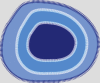
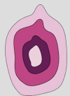
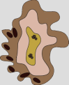
| Intestine/colon | Stomach | Brain | Gall bladder | Liver | Lung | |
|---|---|---|---|---|---|---|
| Organoid |  |
 |
 |
 |
 |
 |
| Microbe/infection modeled | - C. difficle | - H. pylori | - Zika virus | - S. typhi | - P. vivaxa | - Rous sarcoma virusa |
| - S. typhi | - Epstein Barr Virusa | - Chikungunya virusa | (Malaria) | - Influenza virusa | ||
| - Norovirus | - Japanese encephalitis virusa | - Hepatitis virus A, B, C, Ea | - Rhinovirusa | |||
| - Rotavirus | - Venezuelan equine encephalitis virusa | - Corornavirusa | ||||
| - Shigellaa | ||||||
| - Enteric adenovirusa | ||||||
| - Cryptosporidiuma | ||||||
| Source | Human ASCs, iPSCs | Human ASCs, PSCs | Human iPSCs | Mouse ASCs | Human ASCs, PSCsa | Human ASCs, PSCsa |
| Reference | [28, 33••, 34, 35••, 36•, 37, 38••, 39•, 40] | [32•, 33••, 34, 35••, 36•, 37, 3, 39•, 40, 41, 42••, 43, 44••] | [45•, 46, 47••, 48•, 49•, 50, 51••, 52•, 53••] | [55••] | – | – |
Not yet published/potential future studies.
Modeling infectious diseases in organoids
Intestinal organoids model gastrointestinal diseases
In 2009, in the first of its kind model system, ever expanding 3D intestinal organoids were grown from non-transformed mouse adult tissue stem cells [20, 21]. Subsequently, conditions for growing organoids from adult human colon, small intestine and adenocarcinoma were also developed [22]. Intestinal organoids can be maintained in culture for long-term without procuring genetic aberrations or alterations and they retain their apico-basal polarity. Intestinal organoids are Wnt activity dependent, consisting mostly of resident proliferating stem cells which can be directed towards a differentiated cell state by withdrawl of niche factors. They have also been generated from human pluripotent stem cells or hPSCs (including embryonal stem cells — ESCs and induced pluripotent stem cells or iPSCs) [23]. In both these systems, organoids are grown in scaffolds with extracellular matrix like Matrigel® (Corning) or Basement membrane extract (BME) supplemented with a cocktail of growth factors essential for stem cells proliferation. Designer matrices or synthetic hydrogel networks with a well-defined composition have recently been tested to support organoid growth. These will further improve the reproducibility and applicability of organoid culture systems [24••].
Intestinal organoids have been used to model diseases such as colorectal carcinoma (CRC) and Cystic fibrosis (CF) [25, 26, 27]. Another compelling application of intestinal organoids has been their use in studying the pathogenesis of various infectious diseases and in understanding host–microbe dynamics [28, 29]. Organoids can be used to study the various links of the “chain of infection” model (Figure 1). For example, the epithelial cells of the intestinal organoids can be modeled as a reservoir and portal of exit for intracellular parasites like Cryptosporidium etc. Organoids can also be used to study the mechanism of transmission e.g. if a pathogen is airborne and can spread from an infected to an uninfected organoid. The study of portal of entry of pathogens and the role of specific cell types for e.g. modeling the penetration of intestinal epithelium by Shigella via the M-cells is also possible using organoid cultures. Studies using mouse small intestinal organoids with terminally differentiated secretory Paneth cells co-cultured with Escherichia coli or its antigens have given insights into the effects of microbial antigens on the function and changing facets of Paneth cells, identifying IFN-γ as a potent immune component which facilitates release of antimicrobial factors into the gut lumen [30•]. Clostridium difficile (C. difficile) and Salmonella typhi (S. typhi) are the two-major bacterial intestinal pathogens causing diarrhea and gastrointestinal failures in humans. These pathogens have affinity towards the apical side of the epithelium, thus to mimic that interaction in 3D organoids, groups now use two different methods — 1) microinjection, 2) mechanical disruption of organoids and subsequent introduction of the microbe [31•, 32•]. Alternatively, 3D organoids can be dissociated into single cells and grown as a monolayer with the apical side facing upwards. These monolayers can then be exposed to pathogens via their addition to the media (Figure 2 ). However, in this case, assessment of effects on the basolateral surface is not possible. In proof of principle studies, live Salmonella typhimurium was microinjected into the closed iPSC derived intestinal organoid lumen [33••, 34]. Gene expression profiling and biochemical analysis of these organoids revealed massive NF-κB activation and upregulation of cytokine-mediated signaling. Factors like Interleukin (IL)-6, 8 and TNFα were also found to be secreted, consistent with previous findings in animal studies. Likewise, in a model for obligate anaerobe C. difficile infection (CDI), the Spence lab used pluripotent stem cell derived intestinal organoids and microinjected C. difficile toxin A (TcdA) and Toxin B (TcdB) purified from strain VPI 10463 into the lumen. While TcdA had previously been shown to be more potent in mice models, TcdB had a stronger effect in cell lines [35••]. Interestingly, infection in the 3D organoid model was closer to the in vivo situation. Within a few hours of infection, the distribution of tight junctional marker zonula occludens (ZO-1) was altered. Furthermore, cell–cell adhesion marker E-Cadherin and actin cytoskeletal rearrangements were seen in organoids injected with C. difficile toxin A but not C. difficile toxin B. In another study, the Worrell laboratory observed a reduction in NHE3 and MUC2 protein levels in C. difficile infected organoids as compared to normal organoids. This could be a way the microbe creates a favorable environment for its colonization [36•].
Figure 2.

Methods of studying host–microbe interactions. Organoids can be microinjected with the microbe using a fine capillary microinjector. In this case the microbe is in direct contact with the apical side of epithelial cells. Organoids can also be sheared into smaller pieces, mixed with pathogen and re-plated with Matrigel/BME. Alternatively, 3D organoids can be dissociated into single cells by enzymatic treatment and grown as 2D monolayer cultures. Microbes are then introduced into the media.
Viral pathogen Human Norovirus (HuNoV) infection leads to a self-limiting stomach flu or viral gastroenteritis and is one of the most common causes of acute gastroenteritis in the world [37]. Following close behind is Rotavirus, which is the second most common cause of gastric diarrhea in humans. Despite the rampant nature of both these viruses, no proper vaccine has yet been developed against them due to the lack of a good model organism or in vitro system supporting their growth. In striking studies, Ettayebi and group modeled HuNoV infection in an organoid — virus co-culture system, successfully showing that the virus can infect and faithfully replicate inside the absorptive enterocytic cells of the epithelium, with only a specific GII.3 HuNov strain displaying bile requirement [38••]. Furthermore, a differential response of patients with histo-blood group antigen (HBGA) variability was observed towards different HuNoV strains, a fact which was also seen previously in cultured gastrointestinal epithelial cells (Caco-2) upon Norovirus infection. Similarly, researchers have shown that Rotavirus strain (simian SA11) from clinical samples can replicate in iPSC-derived intestinal organoids [39•, 40]. Future research on gastrointestinal viruses, parasites and bacterial pathogens using organoid cultures should help identify therapeutic targets and in developing novel diagnostics and vaccines.
Understanding the pathophysiology of Helicobacter pylori infection in gastric organoids
The occurrence of bacterium Helicobacter pylori (H. pylori) is so common that at any given point, more than 50% of the world's population harbors H. pylori in the upper gastrointestinal tract. [41]. Acute H. pylori infection is associated as a major risk factor for peptic ulcers, gastric adenocarcinoma and gastritis. To study this association further, researchers microinjected H. pylori into the lumen of gastric organoids derived from human gastric mucosa. Schlaermann and group developed a monolayer culture from similar 3D gastric organoids [42••, 43]. Colonization of the bacteria led to an increase in proliferation of Lgr5+ stem cells which was in turn found to be induced by bacterial virulence factor CagA expression [44••]. Furthermore, an inflammatory response was induced and promoted by the differentiated cells of the bacteria infected organoids. It would not be far-fetched to hypothesize that such an inflammatory response could be the bridging factor connecting excessive microbial colonization of H. pylori in the stomach to the occurrence of gastric cancer in humans. In another study, scientists used gastric organoids to determine that a potent chemoattractant — urea which emanates from the epithelial cell wall is essential for H. pylori colonization in the gastric mucosa [45•]. Gastric organoid models would thus not only be beneficial to study H. pylori pathogenesis but also to dissect the implications of such microbial colonizations in the organ and in understanding their role in the causation of diseases like cancer and inflammatory bowel disease (IBD) in humans.
Cerebral organoids as a model of ZIKV infection
A breakthrough in the field of neuroscience came with the development of brain organoids from human iPSCs by the Knoblich group in 2013 [46, 47••]. Around the same time, Zika virus (ZIKV), a mosquito-borne flavivirus came into prominence into the modern world after a public outbreak of the virus in Brazil. ZIKV was first identified in 1947 from the blood of a rhesus monkey found in Uganda and in humans in 1952. By Feb 2016 World Health Organization (WHO) had declared Zika virus infection as a public health emergency. The virus spreads mainly by the Aedes aegypti mosquito and its occurrence is strongly associated with microcephaly. However, the pathogenesis of the viral infection and how it effects the brain neurons was not fully understood until recently. Employing pluripotent stem cell (ESCs and iPSCs) derived cerebral organoids multiple recent studies have now deciphered the sequence of disease progression in Zika virus infection [46, 47••, 48•]. Multiple groups demonstrated that ZIKV infection causes disruption of cerebral organoid cortical layers, abrogating growth and thus halting the process of neurogenesis. Researchers found that the Toll-like receptor 3 (TLR3) activation, which occurs upon ZIKV infection, leads to deregulated neurogenesis and thus decrease in the pool of functional neurons [49•]. Gabriel and group further showed that the pattern of pathogenicity was different when two new ZIKV isolates were used instead of the highly passaged MR766 strain. The new strains infected apical proliferating progenitors, interfering with centrosomal protein assembly, which in turn led to their premature differentiation and apoptosis, giving rise to features of microcephaly [50, 51••, 52•]. In a drug repurposing screen of ∼6000 compounds, caspase-3 activity inhibitors Emricasan and Niclosamide, a category B anthelmintic, were found to be effective in limiting ZIKV induced neural cortical progenitor death and ZIKV replication [53••]. Scientists have now developed innovative cost-effective miniature spinning bioreactors to generate cerebral organoids from human iPSCs [54•]. In light of these recent studies, the United States Centers for Disease Control Prevention (CDC) in April 2016 concluded that ZIKV infection causes microcephaly (CDC, 2016).
Dissecting associations between microbes and cancer — gall bladder organoids
Neefjes and colleagues recently exploited gallbladder organoids derived from Ink4a/Arf (Cdkn2a) tumor suppressor deficient mice for to draw a direct association between chronic Salmonella enterica serovar typhimurium infection and gallbladder carcinoma (GBC). WT Salmonella infection leads to colorectal adenocarcinoma formation in mice. When mouse gallbladder organoids were infected with WT Salmonella, they presented features of loss of polarity, like those seen in the GBC mouse model. Additionally, WT Salmonella pre-exposed organoids were found to have neoplastic transformations via activation of AKT and MAPK signaling and could grow in a growth-factor deficient media [55••]. Organoid and microbe co-cultures would be instrumental in further dissecting the molecular basis of such associations.
Future of organoid — microbe studies
Given that organoid cultures of various other tissues including liver, lung, kidney and ovary have already been established (unpublished data from Clevers lab), it would be exciting to mimic other infectious diseases such as malaria (P. vivax and P. falciparum) and hepatitis (HBV) in liver organoids, Rous sarcoma virus (RSV) in lung organoids and Epstein Barr Virus (EBV) infection in gastric organoids. Hepatitis C (HCV) and Human Immunodeficiency virus (HIV) co-infection studies have been a subject of immense interest [56•]. While HIV is known to enhance HCV infection, the direct alteration of the course of HIV infection and AIDS upon HCV infection remains debated. It would be interesting to study HCV–HIV co-infection in organoid cultures. While most commensal microbes (microbiome) are anaerobic and the organoid lumen is about 5% aerobic, it would be interesting to tweak the current organoid culture systems to understand the host-microbiome interplay that exists in our body. Another important addition would be the inclusion of immune and endothelial cells to fully access how microbial fluctuations modulate immune cell responses, leading to disease states. Future research using organoid models to dissect the pathogenesis of various diseases is bound to open exciting new avenues to tread and lead us towards novel drug discoveries and improved worldwide healthcare.
Conflict of interest
The authors declare no conflict of interest.
References and recommended reading
Papers of particular interest, published within the period of review, have been highlighted as:
• of special interest
•• of outstanding interest
Acknowledgements
We are thankful to Dr. Inha Heo (Clevers lab) and Dr. Antoni Hendrickx (UMC, Utrecht) for scientific discussions and help with illustrations. DD is the recipient of a VENI grant from the Netherlands Organisation for Scientific Research (NWO-ALW, 016.Veni.171.015). H.C. is named as the inventor of several patents related to Lgr5 stem-cell-based organoid technology.
References
- 1.Casanova J.L. Human genetic basis of interindividual variability in the course of infection. Proc Natl Acad Sci U S A. 2015;112:E7118–E7127. doi: 10.1073/pnas.1521644112. [DOI] [PMC free article] [PubMed] [Google Scholar]
- 2.Spanò S., Gao X., Hannemann S., Lara-Tejero M., Galán J.E. A bacterial pathogen targets a host rab-family GTPase defense pathway with a GAP. Cell Host Microbe. 2016;19:216–226. doi: 10.1016/j.chom.2016.01.004. [DOI] [PMC free article] [PubMed] [Google Scholar]
- 3.Sterkel A.K., Lorenzini J.L., Fites J.S., Subramanian Vignesh K., Sullivan T.D., Wuthrich M., Brandhorst T., Hernandez-Santos N., Deepe G.S., Klein B.S. Fungal mimicry of a mammalian aminopeptidase disables innate immunity and promotes pathogenicity. Cell Host Microbe. 2016;19:361–374. doi: 10.1016/j.chom.2016.02.001. [DOI] [PMC free article] [PubMed] [Google Scholar]
- 4.Caballero S., Kim S., Carter R.A., Leiner I.M., Sušac B., Miller L., Kim G.J., Ling L., Pamer E.G. Cooperating commensals restore colonization resistance to vancomycin-resistant Enterococcus faecium. Cell Host Microbe. 2017;21 doi: 10.1016/j.chom.2017.04.002. 592–602.e4. [DOI] [PMC free article] [PubMed] [Google Scholar]
- 5.Liu Y., Liu J., Du S., Shan C., Nie K., Zhang R., Li X.-F., Zhang R., Wang T., Qin C.-F. Evolutionary enhancement of Zika virus infectivity in Aedes aegypti mosquitoes. Nature. 2017 doi: 10.1038/nature22365. [DOI] [PMC free article] [PubMed] [Google Scholar]
- 6.Leffler E.M., Band G., Busby G.B.J., Kivinen K., Le Q.S., Clarke M., Bojang K.A., Conway D.J., Jallow M., Sisay F. Resistance to malaria through structural variation of red blood cell invasion receptors. Science. 2017 doi: 10.1126/science.aam6393. [DOI] [PMC free article] [PubMed] [Google Scholar]
- 7.Bloom D.E., Black S., Rappuoli R. Emerging infectious diseases: a proactive approach. Proc Natl Acad Sci U S A. 2017;114:4055–4059. doi: 10.1073/pnas.1701410114. [DOI] [PMC free article] [PubMed] [Google Scholar]
- 8.Liu X., Hodgson J.J., Buchon N. 2017. Drosophila as a model for homeostatic, antibacterial, and antiviral mechanisms in the gut A conserved midgut structure from fly to human. [DOI] [PMC free article] [PubMed] [Google Scholar]
- 9.Rolig A.S., Parthasarathy R., Burns A.R., Bohannan B.J.M., Guillemin K. Individual members of the microbiota disproportionately modulate host innate immune responses. Cell Host Microbe. 2015;18:613–620. doi: 10.1016/j.chom.2015.10.009. [DOI] [PMC free article] [PubMed] [Google Scholar]
- 10.Thevaranjan N., Puchta A., Schulz C., Naidoo A., Szamosi J.C., Verschoor C.P., Loukov D., Schenck L.P., Jury J., Foley K.P. Age-associated microbial dysbiosis promotes intestinal permeability, systemic inflammation, and macrophage dysfunction. Cell Host Microbe. 2017;21 doi: 10.1016/j.chom.2017.03.002. 455–466.e4. [DOI] [PMC free article] [PubMed] [Google Scholar]
- 11.Kanther M., Rawls J.F. Host–microbe interactions in the developing zebrafish. Curr Opin Immunol. 2010;22:10–19. doi: 10.1016/j.coi.2010.01.006. [DOI] [PMC free article] [PubMed] [Google Scholar]
- 12.Walsh N.C., Kenney L.L., Jangalwe S., Aryee K.-E., Greiner D.L., Brehm M.A., Shultz L.D. Humanized mouse models of clinical disease. Annu Rev Pathol Mech Dis. 2017;12 doi: 10.1146/annurev-pathol-052016-100332. annurev-pathol-052016-100332. [DOI] [PMC free article] [PubMed] [Google Scholar]
- 13.Macpherson A.J., McCoy K.D. Standardised animal models of host microbial mutualism. Mucosal Immunol. 2015;8:476–486. doi: 10.1038/mi.2014.113. [DOI] [PMC free article] [PubMed] [Google Scholar]
- 14.Clevers H. Modeling development and disease with organoids. Cell. 2016;165:1586–1597. doi: 10.1016/j.cell.2016.05.082. [DOI] [PubMed] [Google Scholar]
- 15.Boj S.F., Hwang C., II, Baker L.A., Chio I.I.C., Engle D.D., Corbo V., Jager M., Ponz-Sarvise M., Tiriac H., Spector M.S. Organoid models of human and mouse ductal pancreatic cancer. Cell. 2015;160:324–338. doi: 10.1016/j.cell.2014.12.021. [DOI] [PMC free article] [PubMed] [Google Scholar]
- 16.Dye B.R., Hill D.R., Ferguson M.A., Tsai Y.-H., Nagy M.S., Dyal R., Wells J.M., Mayhew C.N., Nattiv R., Klein O.D. In vitro generation of human pluripotent stem cell derived lung organoids. Elife. 2015;4:1–25. doi: 10.7554/eLife.05098. [DOI] [PMC free article] [PubMed] [Google Scholar]
- 17.Huch M., Gehart H., Van Boxtel R., Hamer K., Blokzijl F., Verstegen M.M.A., Ellis E., Van Wenum M., Fuchs S.A., De Ligt J. Long-term culture of genome-stable bipotent stem cells from adult human liver. Cell. 2015;160:299–312. doi: 10.1016/j.cell.2014.11.050. [DOI] [PMC free article] [PubMed] [Google Scholar]
- 18.Barker N., Huch M., Kujala P., van de Wetering M., Snippert H.J., van Es J.H., Sato T., Stange D.E., Begthel H., van den Born M. Lgr5+ve stem cells drive self-renewal in the stomach and build long-lived gastric units in vitro. Cell Stem Cell. 2010;6:25–36. doi: 10.1016/j.stem.2009.11.013. [DOI] [PubMed] [Google Scholar]
- 19•.Takebe T., Zhang R.-R., Koike H., Kimura M., Yoshizawa E., Enomura M., Koike N., Sekine K., Taniguchi H. Generation of a vascularized and functional human liver from an iPSC-derived organ bud transplant. Nat Protoc. 2014;9:396–409. doi: 10.1038/nprot.2014.020. [DOI] [PubMed] [Google Scholar]; Authors first time developed a triple co-culture incorporating liver hepatocytes, endothelial cells and MSCs to form organoids.
- 20.Ootani A., Li X., Sangiorgi E., Ho Q.T., Ueno H., Toda S., Sugihara H., Fujimoto K., Weissman I.L., Capecchi M.R. Sustained in vitro intestinal epithelial culture within a Wnt-dependent stem cell niche. Nat Med. 2010;15:701–706. doi: 10.1038/nm.1951. [DOI] [PMC free article] [PubMed] [Google Scholar]
- 21.Sato T., Vries R.G., Snippert H.J., van de Wetering M., Barker N., Stange D.E., van Es J.H., Abo A., Kujala P., Peters P.J. Single Lgr5 stem cells build crypt-villus structures in vitro without a mesenchymal niche. Nature. 2009;459:262–265. doi: 10.1038/nature07935. [DOI] [PubMed] [Google Scholar]
- 22.Sato T., Stange D.E., Ferrante M., Vries R.G.J., Van Es J.H., Van Den Brink S., Van Houdt W.J., Pronk A., Van Gorp J., Siersema P.D. Long-term expansion of epithelial organoids from human colon, adenoma, adenocarcinoma, and Barrett's epithelium. Gastroenterology. 2011;141:1762–1772. doi: 10.1053/j.gastro.2011.07.050. [DOI] [PubMed] [Google Scholar]
- 23.Watson C.L., Mahe M.M., Múnera J., Howell J.C., Sundaram N., Poling H.M., Schweitzer J.I., Vallance J.E., Mayhew C.N., Sun Y. An in vivo model of human small intestine using pluripotent stem cells. Nat Med. 2014;20:1310–1314. doi: 10.1038/nm.3737. [DOI] [PMC free article] [PubMed] [Google Scholar]
- 24••.Gjorevski N., Sachs N., Manfrin A., Giger S., Bragina M.E., Ordóñez-Morán P., Clevers H., Lutolf M.P. Designer matrices for intestinal stem cell and organoid culture. Nature. 2016;539:560–564. doi: 10.1038/nature20168. [DOI] [PubMed] [Google Scholar]; Well-defined synthetic matrices were developed which supported intestinal organoid growth. These matrices provide a defined niche and thus will enable more reproducibility in experiments.
- 25.Schwank G., Koo B.K., Sasselli V., Dekkers J.F., Heo I., Demircan T., Sasaki N., Boymans S., Cuppen E., Van Der Ent C.K. Functional repair of CFTR by CRISPR/Cas9 in intestinal stem cell organoids of cystic fibrosis patients. Cell Stem Cell. 2013;13:653–658. doi: 10.1016/j.stem.2013.11.002. [DOI] [PubMed] [Google Scholar]
- 26.Van De Wetering M., Francies H.E., Francis J.M., Bounova G., Iorio F., Pronk A., Van Houdt W., Van Gorp J., Taylor-Weiner A., Kester L. Prospective derivation of a living organoid biobank of colorectal cancer patients. Cell. 2015;161:933–945. doi: 10.1016/j.cell.2015.03.053. [DOI] [PMC free article] [PubMed] [Google Scholar]
- 27.Drost J., van Jaarsveld R.H., Ponsioen B., Zimberlin C., van Boxtel R., Buijs A., Sachs N., Overmeer R.M., Offerhaus G.J., Begthel H. Sequential cancer mutations in cultured human intestinal stem cells. Nature. 2015;521:43–47. doi: 10.1038/nature14415. [DOI] [PubMed] [Google Scholar]
- 28.Klotz C., Aebischer T., Seeber F. Stem cell-derived cell cultures and organoids for protozoan parasite propagation and studying host–parasite interaction. Int J Med Microbiol. 2012;302:203–209. doi: 10.1016/j.ijmm.2012.07.010. [DOI] [PubMed] [Google Scholar]
- 29.Fatehullah A., Tan S.H., Barker N. Organoids as an in vitro model of human development and disease. Nat Cell Biol. 2016;18:246–254. doi: 10.1038/ncb3312. [DOI] [PubMed] [Google Scholar]
- 30•.Farin H.F., Karthaus W.R., Kujala P., Rakhshandehroo M., Schwank G., Vries R.G.J., Kalkhoven E., Nieuwenhuis E.E.S., Clevers H. Paneth cell extrusion and release of antimicrobial products is directly controlled by immune cell-derived IFN-γ. J Exp Med. 2014;211:1393–1405. doi: 10.1084/jem.20130753. [DOI] [PMC free article] [PubMed] [Google Scholar]; The changes in Paneth cell dynamics in response to antimicrobial products were demonstrated for the first time in organoid cultures.
- 31•.Dutta D., Heo I., Clevers H. Disease modelling in stem cell-derived 3D organoid systems. Trends Mol Med. 2017;23:393–410. doi: 10.1016/j.molmed.2017.02.007. [DOI] [PubMed] [Google Scholar]; Reviews the various methods for introducing microbes into organoids.
- 32•.Bartfeld S., Clevers H. Organoids as model for infectious diseases: culture of human and murine stomach organoids and microinjection of Helicobacter pylori. J Vis Exp. 2015 doi: 10.3791/53359. [DOI] [PMC free article] [PubMed] [Google Scholar]; Detailed video protocol of the microinjection procedure.
- 33••.Forbester J.L., Goulding D., Vallier L., Hannan N., Hale C., Pickard D., Mukhopadhyay S., Dougan G. Interaction of Salmonella enterica serovar Typhimurium with intestinal organoids derived from human induced pluripotent stem cells. Infect Immun. 2015;83:2926–2934. doi: 10.1128/IAI.00161-15. [DOI] [PMC free article] [PubMed] [Google Scholar]; Modeled the interaction of S. typhi with intestinal organoids in hIPSC-derived organoids, validating them as promising models for assessing epithelium–pathogen interactions.
- 34.Wilson S.S., Tocchi A., Holly M.K., Parks W.C., Smith J.G. A small intestinal organoid model of non-invasive enteric pathogen–epithelial cell interactions. Mucosal Immunol. 2015;8:352–361. doi: 10.1038/mi.2014.72. [DOI] [PMC free article] [PubMed] [Google Scholar]
- 35••.Leslie J.L., Huang S., Opp J.S., Nagy M.S., Kobayashi M., Young V.B., Spence J.R. Persistence and toxin production by Clostridium difficile within human intestinal organoids result in disruption of epithelial paracellular barrier function. Infect Immun. 2015;83:138–145. doi: 10.1128/IAI.02561-14. [DOI] [PMC free article] [PubMed] [Google Scholar]; Modeled C. difficle infection showing that different toxins of C. difficile have different toxicity effects.
- 36•.Engevik M.A., Engevik K.A., Yacyshyn M.B., Wang J., Hassett D.J., Darien B., Yacyshyn B.R., Worrell R.T. Human Clostridium difficile infection: inhibition of NHE3 and microbiota profile. Am J Physiol Gastrointest Liver Physiol. 2015;308:G497–G509. doi: 10.1152/ajpgi.00090.2014. [DOI] [PMC free article] [PubMed] [Google Scholar]; Studied a potential mechanism of bacterial colonization in organoids by inhibition of NHE3 and modulating microbiota dynamics.
- 37.Zheng D.P., Ando T., Fankhauser R.L., Beard R.S., Glass R.I., Monroe S.S. Norovirus classification and proposed strain nomenclature. Virology. 2006;346:312–323. doi: 10.1016/j.virol.2005.11.015. [DOI] [PubMed] [Google Scholar]
- 38••.Ettayebi K., Crawford S.E., Murakami K., Broughman J.R., Karandikar U., Tenge V.R., Neill F.H., Blutt S.E., Zeng X.-L., Qu L. Replication of human noroviruses in stem cell-derived human enteroids. Science (80) 2016;353:1387–1393. doi: 10.1126/science.aaf5211. 3:1–6. [DOI] [PMC free article] [PubMed] [Google Scholar]; The first ever in vitro system for the culture of Norovirus in human organoid cultures was developed.
- 39•.Yin Y., Bijvelds M., Dang W., Xu L., Van Der Eijk A.A., Knipping K., Tuysuz N., Dekkers J.F., Wang Y., De Jonge J. Modeling rotavirus infection and antiviral therapy using primary intestinal organoids. Antiviral Res. 2015;123:120–131. doi: 10.1016/j.antiviral.2015.09.010. [DOI] [PubMed] [Google Scholar]; Demonstrated that human organoids support infection of patient-derived rotavirus strains and allow for individualized evaluation of the efficacy of antiviral medications.
- 40.Finkbeiner S.R., Zeng X.L., Utama B., Atmar R.L., Shroyer N.F., Estes M.K. Stem cell-derived human intestinal organoids as an infection model for rotaviruses. MBio. 2012;3:1–6. doi: 10.1128/mBio.00159-12. [DOI] [PMC free article] [PubMed] [Google Scholar]
- 41.Amieva M., Peek R.M. Pathobiology of Helicobacter pylori-induced gastric cancer. Gastroenterology. 2016;150:64–78. doi: 10.1053/j.gastro.2015.09.004. [DOI] [PMC free article] [PubMed] [Google Scholar]
- 42••.Bartfeld S., Bayram T., Van De Wetering M., Huch M., Begthel H., Kujala P., Vries R., Peters P.J., Clevers H. In vitro expansion of human gastric epithelial stem cells and their responses to bacterial infection. Gastroenterology. 2015;148 doi: 10.1053/j.gastro.2014.09.042. 126–136.e6. [DOI] [PMC free article] [PubMed] [Google Scholar]; Studied the pathogenesis of H. pylori in human gastric organoids. First study to establish the microinjection protocol for adult stem cell derived cultures.
- 43.Schlaermann P., Toelle B., Berger H., Schmidt S.C., Glanemann M., Ordemann J., Bartfeld S., Mollenkopf H.J., Meyer T.F. A novel human gastric primary cell culture system for modelling Helicobacter pylori infection in vitro. Gut. 2016;65:202–213. doi: 10.1136/gutjnl-2014-307949. [DOI] [PMC free article] [PubMed] [Google Scholar]
- 44••.McCracken K.W., Catá E.M., Crawford C.M., Sinagoga K.L., Schumacher M., Rockich B.E., Tsai Y.-H., Mayhew C.N., Spence J.R., Zavros Y. Modelling human development and disease in pluripotent stem-cell-derived gastric organoids. Nature. 2014;516:400–404. doi: 10.1038/nature13863. [DOI] [PMC free article] [PubMed] [Google Scholar]; Using pluripotent stem cells, gastric organoids were developed which modeled H. pylori infection like the in-vivo situation.
- 45•.Huang J.Y., Sweeney E.G., Sigal M., Zhang H.C., Remington S.J., Cantrell M.A., Kuo C.J., Guillemin K., Amieva M.R. Chemodetection and destruction of host urea allows Helicobacter pylori to locate the epithelium. Cell Host Microbe. 2015;18:147–156. doi: 10.1016/j.chom.2015.07.002. [DOI] [PMC free article] [PubMed] [Google Scholar]; Reported that Urea secreted by epithelial cells attract H. pylori to the epithelial cells, allowing for co-localization which eventually leads to inflammation and gastric cancer.
- 46.Lancaster M.A., Knoblich J.A. Generation of cerebral organoids from human pluripotent stem cells. Nat Protoc. 2014;9:2329–2340. doi: 10.1038/nprot.2014.158. [DOI] [PMC free article] [PubMed] [Google Scholar]
- 47••.Lancaster M.A., Renner M., Martin C.-A., Wenzel D., Bicknell L.S., Hurles M.E., Homfray T., Penninger J.M., Jackson A.P., Knoblich J.A. Cerebral organoids model human brain development and microcephaly. Nature. 2012;501:373–379. doi: 10.1038/nature12517. [DOI] [PMC free article] [PubMed] [Google Scholar]; First ever brain organoids were developed from pluripotent stem cells. These organoids model the fetal brain and can be used for developmental studies and for disease modeling for microcephaly, autism etc.
- 48•.Garcez P.P., Loiola E.C., Madeiro da Costa R., Higa L.M., Trindade P., Delvecchio R., Nascimento J.M., Brindeiro R., Tanuri A., Rehen S.K. Zika virus impairs growth in human neurospheres and brain organoids. Science (80) 2016;352:816–818. doi: 10.1126/science.aaf6116. [DOI] [PubMed] [Google Scholar]; Show that ZIKV abrogates neurogenesis by targeting human brain cells, reducing their viability and growth as neurospheres and brain organoids.
- 49•.Cugola F.R., Fernandes I.R., Russo F.B., Freitas B.C., Dias J.L.M., Guimarães K.P., Benazzato C., Almeida N., Pignatari G.C., Romero S. The Brazilian Zika virus strain causes birth defects in experimental models. Nature. 2016 doi: 10.1038/nature18296. [DOI] [PMC free article] [PubMed] [Google Scholar]; Reported the mechanism of halting of neurogenesis leading to decreased number of neurons and thus microcephaly.
- 50.Wells M.F., Salick M.R., Wiskow O., Ho D.J., Worringer K.A., Ihry R.J., Kommineni S., Bilican B., Klim J.R., Hill E.J. Genetic ablation of AXL does not protect human neural progenitor cells and cerebral organoids from Zika virus infection. Cell Stem Cell. 2016;19:703–708. doi: 10.1016/j.stem.2016.11.011. [DOI] [PubMed] [Google Scholar]
- 51••.Dang J., Tiwari S.K., Lichinchi G., Qin Y., Patil V.S., Eroshkin A.M., Rana T.M. Zika virus depletes neural progenitors in human cerebral organoids through activation of the innate immune receptor TLR3. Cell Stem Cell. 2016;19:258–265. doi: 10.1016/j.stem.2016.04.014. [DOI] [PMC free article] [PubMed] [Google Scholar]; Reported a link between the activation of the immune component TLR3 and its association with Zika virus infection leading to microcephaly.
- 52•.Gabriel E., Ramani A., Karow U., Gottardo M., Natarajan K., Gooi L.M., Goranci-Buzhala G., Krut O., Peters F., Nikolic M. Recent Zika virus isolates induce premature differentiation of neural progenitors in human brain organoids. Cell Stem Cell. 2017;20 doi: 10.1016/j.stem.2016.12.005. 397–406.e5. [DOI] [PubMed] [Google Scholar]; This article showed that newer strains of ZIKV have a different mode of infection as compared to an older strain. Newer strains induce premature differentiation and death as compared to other strains which abrogated neurogenesis.
- 53••.Xu M., Lee E.M., Wen Z., Cheng Y., Huang W.-K., Qian X., Kouznetsova J., TCW J., Ogden S.C., Hammack C. Identification of small-molecule inhibitors of Zika virus infection and induced neural cell death via a drug repurposing screen. Nat Med. 2016;22:1101–1107. doi: 10.1038/nm.4184. [DOI] [PMC free article] [PubMed] [Google Scholar]; In a one of its kind screen, the study used brain organoids to identify drugs/molecules which could potentially be used to control the replication and spread of ZIKV.
- 54•.Qian X., Nguyen H.N., Song M.M., Hadiono C., Ogden S.C., Hammack C., Yao B., Hamersky G.R., Jacob F., Zhong C. Brain-region-specific organoids using mini-bioreactors for modeling ZIKV exposure. Cell. 2016;165:1238–1254. doi: 10.1016/j.cell.2016.04.032. [DOI] [PMC free article] [PubMed] [Google Scholar]; Innovative mini-bioreactors were used to develop cerebral organoids which were cost effective and fully functional, mimicking the phenotypes observed in-vivo upon ZIKV infection.
- 55••.Scanu T., Spaapen R.M., Bakker J.M., Pratap C.B., Wu L.en, Hofland I., Broeks A., Shukla V.K., Kumar M., Janssen H. Salmonella manipulation of host signaling pathways provokes cellular transformation associated with gallbladder carcinoma. Cell Host Microbe. 2015;17:763–774. doi: 10.1016/j.chom.2015.05.002. [DOI] [PubMed] [Google Scholar]; This article showed that Salmonella enterica can promote transformation of genetically predisposed cells, potentially leading to GBC.
- 56•.Graham C.S., Baden L.R., Yu E., Mrus J.M., Carnie J., Heeren T., Koziel M.J. Influence of human immunodeficiency virus infection on the course of Hepatitis C virus infection: a meta-analysis. Clin Infect Dis. 2001;33:562–569. doi: 10.1086/321909. [DOI] [PubMed] [Google Scholar]; A landmark study showing the influence of HIV on expansion of HCV virus infection.